R E S E A R C H A R T I C L E
Open Access
Influence of epigallocatechin-3-gallate in
promoting proliferation and osteogenic
differentiation of human periodontal
ligament cells
Jie Liu
1,2†, Yi Lu
1†, Jin Liu
3, Changxiong Jin
1, Yuchen Meng
1and Dandan Pei
1,2*Abstract
Background:Epigallocatechin-3-gallate (EGCG) was recently proposed to have the potential to regulate bone
metabolism, however, its influence on osteogenesis remains controversial. The present study aimed to investigate the effects of EGCG on the proliferation and osteogenesis of human periodontal ligament cells (hPDLCs).
Methods:Cells were cultured in osteogenic medium and treated with EGCG at various concentrations. Cell
proliferation was analyzed using a CCK-8 assay and acridine orange (AO)/ethidium bromide (EB) staining. Flow cytometry was used to measure the intracellular reactive oxygen species (ROS) potential of hPDLCs. The expression levels of osteogenic marker genes and proteins in hPDLCs, including type I collagen (COL1), runt-related
transcription factor 2 (RUNX2), osteopontin (OPN), and osterix (OSX), were determined by quantitative real-time polymerase chain reaction (qRT-PCR) and western blot analysis. In addition, alkaline phosphatase (ALP) activity was monitored both quantitatively and qualitatively. Extracellular matrix mineralization was further analyzed by alizarin red S staining.
Results:The results showed that EGCG concentrations from 6 to 10μM increased the ROS level and inhibited the
cell proliferation of hPDLCs. EGCG concentrations from 2 to 8μM effectively increased extracellular matrix mineralization, in which 4 and 6μM EGCG generated the most mineralizing nodules. The ALP activity and the mRNA and protein expression levels of the tested osteogenic markers were most strongly up-regulated by treatment with 4 and 6μM EGCG.
Conclusions:The present study demonstrated that EGCG might promote the osteogenesis of hPDLCs in a
dose-dependent manner, with concentrations of 4 and 6μM EGCG showing the strongest osteogenic enhancement without cytotoxicity, indicating a promising role for EGCG in periodontal regeneration in patients with deficient alveolar bone in the future.
Keywords:Epigallocatechin-3-gallate, Human periodontal ligament cells, Proliferation, Osteogenic differentiation
© The Author(s). 2019Open AccessThis article is distributed under the terms of the Creative Commons Attribution 4.0 International License (http://creativecommons.org/licenses/by/4.0/), which permits unrestricted use, distribution, and reproduction in any medium, provided you give appropriate credit to the original author(s) and the source, provide a link to the Creative Commons license, and indicate if changes were made. The Creative Commons Public Domain Dedication waiver (http://creativecommons.org/publicdomain/zero/1.0/) applies to the data made available in this article, unless otherwise stated. * Correspondence:peidandan1986@126.com
†Jie Liu and Yi Lu contributed equally to this work.
1
Key Laboratory of Shaanxi Province for Craniofacial Precision Medicine Research, College of Stomatology, Xi’an Jiaotong University, Xi’an, China
2Department of Prosthodontics, College of Stomatology, Xi’an Jiaotong
University, 98 Xiwu Road, Xi’an 710004, Shaanxi, China
Background
The periodontium, consisting of the alveolar bone, gin-giva, periodontal ligament, and cementum, serves as a po-tent support for tooth stability, nutrition and resistance to stress during mastication movement. Based on an epi-demiological investigation, the major cause of bone loss in the periodontium has been considered to be periodontitis, which is highly prevalent and affects up to 538 million people worldwide, and this number is likely to increase as many populations continue growing [1]. The destruction of periodontal bone tissue due to severe periodontitis may result in gingival recession, uncomfortable tooth mobility, and eventual tooth loss [2]. The restoration of damaged periodontal tissue, especially the alveolar bone, is the ul-timate purpose of periodontal therapy. In routine clinical settings, antimicrobial, non-surgical, and surgical ap-proaches are currently being used, either separately or in combination, to prevent the progression of periodontitis. Although these treatments are effective for controlling periodontal disease, they are not competent for predict-able regeneration. Hence, seeking an effective method for periodontal regeneration is of paramount importance for both dentists and patients.
Epigallocatechin-3-gallate (EGCG), which is the most abundant and functional catechin polyphenol in green tea, has gained great attention in the past decades due to its multiple health benefits [3]. As a natural compound, EGCG has been reported to have antimicrobial, antioxi-dant, antitumor, antiviral, and antimutagenic effects in systemic diseases [3, 4]. Furthermore, several studies have indicated that local drug therapy using green tea could reduce gingival inflammation and improve peri-odontal parameters, such as probing depth, bleeding on probing and clinical attachment loss [5–7]. The bio-logical effects of EGCG with regard to improving peri-odontal health may be achieved through the inhibition of the adherence of Porphyromonas gingivalis and the disruption of the initial step of biofilm formation [8, 9]. These findings indicate that EGCG might be a promising alternative for treating patients with periodontitis.
In addition to the abovementioned biological effects of EGCG, recently, EGCG was reported to have the ability to regulate bone metabolism [10–12]. Bone remodeling is regulated by the coupled actions of osteoblasts and osteo-clasts [13, 14]. The primary pathogenesis of osteopenic diseases, including periodontitis, is the imbalance between osteoblastic activity and osteoclastic activity. Previous studies demonstrated that EGCG could inhibit pro-inflammatory cytokine expression, including tumor necrosis factor-α(TNF-α) and interleukin-6 (IL-6), which are responsible for the reduction on osteoclastic activity [13,15], or even induce the apoptosis of osteoclasts to in-hibit bone loss [16, 17]. Aside from these studies, which have focused on the suppression of osteoclasts, the effects
of EGCG on osteogenesis have not been clearly elucidated thus far. Epidemiological investigations have suggested a close association between bone mineral density and con-sumption of green tea, which is the primary main source of EGCG for humans with osteoporosis [18]. Vali et al. [19] found that EGCG increased alkaline phosphatase (ALP) activity and the formation of mineralized bone nod-ules in SaOS-2 human osteoblast-like cells. Thus, it was hypothesized that EGCG possessed the potential to pro-mote osteogenesis. However, conflicting results reported that EGCG appeared to inhibit the osteogenic differenti-ation of MC3T3-E1 cells [20], and another study reported that ALP activity and calcium content were repressed by EGCG in an ectopic bone formation model [21]. Thus, further studies are required to provide evidence for this controversial topic.
Human periodontal ligament cells (hPDLCs), which are constitutive of several cell types and possess high self-renewal and pluripotency potential, may serve as seed cells in periodontal bone regeneration [22, 23]. Studies that used hPDLCs for bone regeneration indicated that hPDLCs could differentiate into osteoblast-like cells and construct alveolar bone-like tissues [23,24]. In the present study, we aimed to investigate the effects of different con-centrations of EGCG on the cell proliferation and osteo-genesis of hPDLCs to clarify the role played by EGCG in osteogenesis and its potential applications for periodontal bone regeneration.
Materials and methods Cell isolation and culture
The present study was approved by the Ethics Committee of College of Medicine & Hospital of Stomatology, Xi’an Jiaotong University (approval number xjkqll[2016]NO.048). Primary periodontal ligaments were obtained from human premolars that were extracted for orthodontic reasons. Briefly, the periodontal ligament tissues attached to the mid-third of the root surfaces were scraped off with a sharp surgical scalpel, followed by treatment with 1 mg/mL type I collagenase (Gibco, Los Angeles, CA, USA) for 30 min. Then, the tissues were cultured in dishes with growth medium at 37 °C, in air with 95% humidity plus 5% CO2,
and the medium was changed every 3 d. The growth medium consisted of 89% α-minimum essential medium (α-MEM; Invitrogen, USA), 10% (v/v) fetal bovine serum (FBS; Gibco), and 1% antibiotic (100 U/ml streptomycin and 100 U/ml penicillin, both from Gibco). When the cells reached 70–80% confluence, they were digested with 0.25% trypsin (Gibco) for further passaging. Passages 3 to 6 were used in the present study.
EGCG treatment
10 mM stock solution according to the manufacturer’s instructions. Due to light and temperature sensitivity, the stock solution of EGCG was protected from light and maintained at−20 °C until use. Prior to application to cells, the EGCG stock solution was diluted with pre-warmed growth medium at 37 °C. For subsequent exper-iments, hPDLCs were cultured with EGCG at concentrations of 0 (the control group), 2, 4, 6, 8 and 10μM in either growth medium or osteogenic differenti-ation medium (ODM) for the specified times prior to further experiments. The ODM consisted of the growth medium, as described above, with additional supplemen-tation of 50μM L-ascorbic acid, 0.1 μΜdexamethasone and 10 mM β-glycerophosphate (all purchased from Sigma-Aldrich). The medium was renewed every 3 d.
Cell proliferation
The Cell Counting Kit-8, purchased from Dojindo (CCK-8; Kumamoto, Japan), was used to explore the role played by EGCG on the proliferation of hPDLCs, according to the manufacturer’s instructions. Briefly, hPDLCs at a density of 2 × 10³ cells per well were seeded into clear-bottomed 96-well culture plates and cultured for 24 h. Subsequently, the growth medium was replaced by medium in which EGCG had been added at the various concentrations men-tioned above. At time points of 24, 48 or 72 h, 10μL of CCK-8 reagent was added to each tested well and incu-bated at 37 °C for 1 h, protected from light. The absorbance value was recorded by a Thermo Scientific microplate ab-sorbance reader at 450 nm (Shanghai, China).
Acridine orange/ethidium bromide (AO/EB) staining
To estimate the cell viability following EGCG treatment, hPDLCs were stained with a dual-fluorescence solution consisting of AO and EB (Sigma-Aldrich), according to the manufacturer’s instructions. Generally, AO pene-trates living cells with intact membranes and then binds to DNA to generate green fluorescence, while EB only enters dead cells with damaged membranes to generate red fluorescence. The hPDLCs were seeded in 6-well plates at a density of 1 × 10⁵cells per well and incubated in the growth medium for 24 h. After removing the growth medium, fresh medium containing the above-mentioned concentrations of EGCG was added, and the cells were cultured for 24 h. Then, 300μL of the dual-fluorescence staining solution, containing 0.1 g/L AO and 0.1 g/L EB at a proportion of 1:1, was added to each well and incubated for 10 min. Fluorescence was viewed through an Olympus FSX100 fluorescence microscope (Olympus Corporation, Tokyo, Japan).
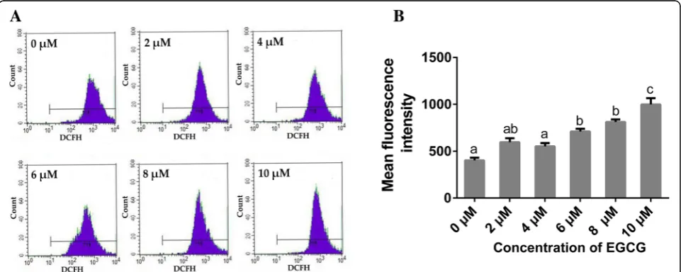
Quantification of reactive oxygen species (ROS)
ROS levels were detected using 2′, 7′ -dichlorofluores-cein diacetate (DCFH-DA; Beyotime, Shanghai, China)
according to the manufacturer’s instructions. The cell culture and EGCG treatment protocols were the same as those described for the AO/EB staining assay. After cul-turing for 72 h, the hPDLCs were incubated with 200μL growth medium supplemented with 10μM DCFH-DA at 37 °C for 30 min. Then the cells were harvested and measured by flow cytometry (BD Biosciences, San Jose, CA, USA) with an excitation at 490 nm and an emission at 530 nm. The generation of intracellular ROS was then quantified.
ALP activity
A Quantichrom Alkaline Phosphatase Assay Kit (Nan-jing Jiancheng Bioengineering Institute, Nan(Nan-jing, China) was used to evaluate the effects of EGCG on the intra-cellular ALP activity of hPDLCs. The cells were seeded into a 24-well plate at a density of 2 × 10⁴cells per well and cultured in growth medium for 24 h. Then the medium was replaced with ODM containing EGCG at the concentrations mentioned above and cultured for 7 d. The cells were scraped off and solubilized in 200μL lysis buffer containing 0.2% Triton-X 100 (Sigma-Al-drich) for 30 min at 37 °C. After that, the cell lysates were centrifuged to remove the cell impurities. ALP ac-tivity was then tested according to the manufacturer’s protocol. Then, 30μL of the obtained supernatant and 100μL of ALP substrate solution were transferred to a new clear-bottomed 96-well plate. After incubation for 15 min at 37 °C, 150μL of coloration reagent was added to each well, and the samples were measured at a 520 nm wavelength with a microplate reader. The ALP activ-ity was calculated against the total protein content, as determined by a Pierce™BCA Assay Kit (Thermo Scien-tific) and was expressed as light units/mg protein. Be-sides, the BCIP/NBT Alkaline Phosphatase Staining Kit (Beyotime) was used according to the manufacturer’s in-structions, on day 7 for qualitative observation.
Alizarin red S assay
determined by comparing the absorbance to a standard curve of known alizarin red S concentrations. The deter-mination was based on the assumption that an alizarin red S - calcium complex was produced by 1 mol of ali-zarin red S binding to 2 mol of calcium [25]. Therefore, the results of extracellular mineralization, as determined through alizarin red S staining, were expressed as the calcium concentration ultimately.
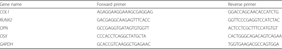
Quantitative real-time polymerase chain reaction (qRT-PCR)
QRT-PCR was used to detect the relative changes in the mRNA expression levels of runt-related transcription factor 2 (RUNX2), osteopontin (OPN), osterix (OSX) and type I collagen (COL1) which are recognized as markers of osteogenic differentiation. Cells were cultured in six-well plates with growth medium at a density of 1 × 10⁵cells per well for 24 h. Then the culture medium was replaced with ODM containing EGCG at the previously mentioned concentrations and renewed every 3 d. After 7 d, the cells were lysed using TRIzol Reagent (Invitro-gen) to extract the total mRNA and then converted to synthesize cDNA using a Primer Script® RT Reagent Kit (TaKaRa, Tokyo, Japan). The sequences of the primers used in the present study are shown in Table 1. Finally, the obtained cDNA was amplified with SYBR Green PCR Master Mix (TaKaRa) in an ABI 7500 Real-Time PCR system (Applied Biosystems, Singapore). The reac-tion condireac-tions for qRT-PCR were set according to the manufacturer’s instructions. The relative mRNA expres-sion levels were calculated using the 2-△△Ct method, in which the mRNA expression levels of the selected osteo-genic genes were normalized to the mRNA expression level of GAPDH. The results were calculated as fold changes.
Western blot
Western blot was used to analyze the protein expression levels of COL1, RUNX2, OPN and OSX. The hPDLCs were treated as described for the qRT-PCR assay. The cells were disrupted in pre-cooled RIPA buffer (Beyo-time) after being treated with EGCG for 14 d. The pro-tein content in the extracted cell lysates was determined using a Pierce™ BCA Assay Kit (Thermo Scientific)
according to the manufacturer’s instructions. The ob-tained proteins (20μg) were separated by 8~12% SDS-polyacrylamide gels. Then, the proteins were trans-ferred onto polyvinylidene difluoride membranes (PVDF; Millipore, MA, USA) with a pore size of 0.45μm, followed by blocking with 5% fat-free dry milk for 3 h. Subsequently, the membranes were incubated with indi-vidual primary antibodies at 4 °C overnight. The follow-ing antibodies were used: anti-COL1 (ab138492, Abcam, MA, USA), anti-RUNX2 (ab23981, Abcam), anti-OPN (ab91655, Abcam) and anti-OSX (ab22552, Abcam). The membranes were then incubated with the secondary antibody (ab205718, Abcam) conjugated with horserad-ish peroxidase for 1 h. The protein bands on the PVDF membranes were then stained using an enhanced chemi-luminescence reagent. The stained bands were visualized using the Image Studio Lite software (Media Cybernetics Inc., Bethesda, MD, USA) and quantified by comparing the band intensities to that of GAPDH.
Statistical analysis
All tests were performed three times which consisted of more than three independent experiments. The results were analyzed using SPSS 18.0 software (IBM, Chicago, IL, USA). For ROS assay, ALP activity assay, alizarin red S assay, qRT-PCR and western blot, one-way ANOVA followed by Tukey’s multiple post hoc tests was used to evaluate the differences among groups after normality (Shapiro–Wilk test) and homoscedasticity (modified Levene test) were assessed. Data were expressed as mean ± standard deviation. For the CCK-8 assay, two-way ANOVA followed by Tukey’s multiple tests was used. Data were expressed as median and interquartile range (IQR). Significant differences were set in advance atp< 0.05.
Results
Cell viability and proliferation
The effects of EGCG on cell proliferation, as determined by the CCK-8 assay, are shown in Fig.1A. Two-way ANOVA revealed that the concentration of EGCG (F= 16.090, p< 0.05) and the culture time (F= 112.040,p< 0.05) both sig-nificantly influenced the proliferation of hPDLCs. The interaction between EGCG concentration and culture time was also significant (F= 3.827,p< 0.05). The groups treated
Table 1Primers for polymerase chain reaction
Gene name Forward primer Reverse primer
COL1 AGAGGAAGGAAAGCGAGGAG GGACCAGCAACACCATCTG
RUNX2 GACGAGGCAAGAGTTTCACC GGTTCCCGAGGTCCATCTAC
OPN GCCGAGGTGATAGTGTGGTT ACTCCTCGCTTTCCATGTGT
OSX CCCACCTCAGGCTATGCTA CACTGGGCAGACAGTCAGAA
GAPDH GCACCGTCAAGGCTGAGAAC TGGTGAAGACGCCAGTGGA
with 0, 2 and 4μM EGCG showed comparable levels of cell proliferation for all of the tested time points, 24, 48 and 72 h, except for the 4μM EGCG group at 24 h, while concen-trations of 8 and 10μM EGCG had adverse effects on the proliferation of hPDLCs. No significant differences were found between the 6μM EGCG group and the control group at 24 or 48 h. A decrease in proliferation was found in the 6μM EGCG group after culturing for 72 h when compared to the control group.
The cell viability results, as detected by AO/EB stain-ing, are shown in Fig.1B. The cells in the groups treated
with 0 to 6μM EGCG primarily exhibited green fluores-cence, indicating that these cells were in a healthy state with a fine form of nucleus. The number of apoptotic cells stained with red fluorescence increased in the groups treated with 8 and 10μM EGCG. Cell apoptosis was more significant in the 10μM EGCG group than that in the 8μM EGCG group.
Intracellular ROS expression
Figure 2 presents the expression levels of intracellular ROS after EGCG treatment. ROS appeared to be Fig. 1Effects of EGCG on cell proliferation of hPDLCs. (A)Cell proliferation was determined using CCK-8 assay after EGCG treatment for 24, 48 or 72 h (n= 5). Boxplots represent median and interquartile range (IQR). For the factor“concentration of EGCG”in each chart, groups at the same time-point labeled with different letters (lower case letters for 24 h, upper case letters for 48 h and numbers for 72 h) are significantly different from each other (p< 0.05) and those with the same letters exhibit no significant difference. For the factor of“culture time”in each chart, groups treated with the same concentration of EGCG that are connected with a horizontal bar are not significant; (B) Representative microscopic fluorescence images of AO/EB stained specimens after EGCG treatment for 72 h (n= 3). Green fluorescence indicates living cells, while red fluorescence indicates apoptotic cells. Bar, 100μm
induced by EGCG in a dose-dependent manner. No sig-nificant differences in the production of ROS were found among the groups treated with 0, 2 or 4μM EGCG. Treatment with 6 to 10μM EGCG induced more ROS when compared to the control group, while no signifi-cant differences were found between the 6 and 8μM EGCG groups. The 10μM EGCG group exhibited the highest level of ROS expression among all the groups.
ALP activity and extracellular matrix mineralization
Images of the hPDLCs after ALP staining are shown in Fig. 3A. The quantitative analysis of ALP activity is shown in Fig. 3C. It was revealed that ALP activities in the 2 and 4μM EGCG groups were significantly higher than those in the other groups, while the 10μM EGCG group showed remarkably lower ALP activity among all of the groups tested. In addition, no significant differ-ences in ALP activity were found among groups treated with 0, 6 or 8μM EGCG.
As shown in Fig.3B, mineralizing nodules were found in all of the groups after cell culture for 21 d. Quantita-tive analysis of the calcium content in the mineralizing deposits is shown in Fig. 3D. Except for the 10μM EGCG group, all of the groups exhibited a higher degree
of mineralization than the control group. The 4 and 6μM EGCG groups showed the highest calcium con-tents in the extracellular matrix among all of the groups, and no significant differences were found between these two groups.
Gene expression
EGCG treatment significantly increased the mRNA levels of COL1, RUNX2, OPN and OSX in most of the treatment groups, as shown in Fig.4. The groups treated with 4 to 8μM EGCG showed a remarkable increase in the expression levels of the osteogenic related genes tested in the present study. ForCOL1, RUNX2andOSX, the gene expression levels peaked in the 6μM EGCG group. For OPN, the gene expression levels peaked in the 8μM EGCG group.
Protein expression
The results of the protein expression analysis using western blot are shown in Fig.5. Compared to the con-trol group, the protein levels of COL1 were significantly increased in all of the EGCG groups. For RUNX2, only the 4μM EGCG treatment up-regulated the protein levels significantly. Treatment with 2 and 4μM EGCG
Fig. 4Relative expression levels ofCOL1,RUNX2,OPNandOSXmeasured by qRT-PCR after treatment with EGCG for 7 d. Semi-quantitative analysis of relative gene expression levels. Data are presented as mean ± standard deviation (n= 5). The bars with different lower case letters are significantly different from each other (p< 0.05), and those with the same letter exhibit no significant difference
had no effect on the expression levels of OPN and OSX. While treatment with 6μM EGCG up-regulated the pro-tein levels of OPN and OSX. The 10μM EGCG treat-ment decreased the protein levels of OSX when compared to the control group.
Discussion
Using bone augmentation procedures to promote peri-odontal bone regeneration is the dominant strategy for reestablishing both function and aesthetics in patients with deficient alveolar bone [26]. Currently, green tea is one of the most popular beverages consumed worldwide. As the most abundant catechin and a major bioactive component of green tea, EGCG has been reported to be associated with bone metabolism [13]. However, the spe-cific effects of EGCG on osteogenic differentiation have been ambiguous. In the present study, the effects of EGCG on the proliferation and osteogenesis of hPDLCs were evaluated to clarify its potential role in periodontal bone regeneration.
Primary-cultured hPDLCs were used in the present study. Cell proliferation was evaluated by CCK-8 assay and AO/EB staining. The results suggested that supple-mentation with 4μM EGCG significantly increased the proliferation of hPDLCs at the early stage, whereas 8 or 10μM EGCG remarkably inhibited proliferation. These results are consistent with those of a recent study, in which Jin et al. [27] reported that 5μM EGCG promoted the cell proliferation of human bone marrow derived mesenchymal cells. Moreover, several studies indicated that EGCG exerted negative effects on the proliferation of osteoblasts in vitro, especially at doses beyond 20μM [28,29]. Kamon et al. [20] reported that EGCG had no impact on the proliferation of murine osteoblastic MC3T3-E1 cells. The discrepancies in results between previous studies and the present study may be attributed to the different experimental conditions, such as the cell types and exposure times used.
The influence of EGCG on the proliferation of cells may be due to its effect on intracellular ROS levels, which can cause DNA damage in the nucleus [28–30]. Under normal conditions, cells can eliminate excess ROS to maintain normal biological functions. If not eliminated, superfluous ROS may impair the host DNA, leading to the permanent loss of cell function or even cell death [30]. EGCG has been reported to possess both antioxidant and pro-oxidant effects [16, 31]. At lower concentrations, the antioxidant effect of EGCG was re-ported to be predominant, leading to its protective effect on DNA. However, EGCG at higher concentrations may possess a relatively stronger ability to exert pro-oxidant action, which may gradually overpower its antioxidant system, resulting in a pro-oxidant effect on DNA. As shown in Fig. 2, compared to the control group, the 2
and 4μM EGCG treatments had no effects on intracel-lular ROS generation, and EGCG treatments exceeding 6μM significantly increased intracellular ROS levels. Simultaneously, after incubation for 72 h with 6 to 10μM EGCG, the proliferation of hPDLCs was inhibited when compared to that of the control group (Fig. 1A), indicating a cytotoxic effect of EGCG at higher concentrations.
The effects of EGCG on osteogenesis were further evaluated in the present study. ALP activity is commonly used as an indicator of the early stage of osteogenesis because it can provide essential phosphoric acid for the deposition of apatite through hydrolyzing phosphate [32]. As a result of osteogenesis, the formation of min-eralizing nodules in the extracellular matrix could be used as an intuitionistic index [33]. Thus, ALP enzyme activity was tested at day 7, which is considered to be an early time point for osteogenesis, and alizarin red S staining was performed after incubation with EGCG for 21 d. The ALP activity assay and alizarin red S staining are considered to be gold standard indicators for the early and late stages of osteogenic differentiation re-spectively. The results in the present study demonstrated that treatment with 2 to 8μM EGCG could promote the mineralization of hPDLCs, with 4 to 6μM EGCG show-ing the most significant impacts, which is consistent with a previous study using human bone marrow mesen-chymal stem cells [27]. However, when using MC3T3-E1 cells, Kamon et al. [20] showed a contradictory result and reported that EGCG at concentrations from 1 to 10μM suppressed osteogenic differentiation. It appears that the osteogenic responses to EGCG may be dependent on the cell type.
by stochastic post-transcription and post-translation, as well as experimental errors and noise interferences [37].
However, the detailed molecular mechanisms involved in EGCG-induced osteogenic capability in human peri-odontal ligament cells remain unclear. Previous studies showed that the activation of the Nrf2, Wnt/β-catenin and p44/p42 MAP kinase signaling pathways might be involved in the cellular osteogenic response to EGCG in osteoblasts [38–40]. With this background, EGGC may induce osteogenic differentiation of hPDLCs via the above mechanisms and we plan to elucidate the molecu-lar mechanisms in a future study. Additionally, there are other limitations of the present study. It should be men-tioned that most cells in living tissues or organs are cap-able of reproducing the paracrine and exocrine signals generated by cell-cell and cell-extracellular matrix inter-actions [41]. These interactions are often critical for guiding and maintaining specific attachment-dependent cellular identities in vivo. Nevertheless, these micro en-vironmental modulators of cell behavior are often miss-ing in dissociated two-dimensional monolayer cell cultures, even in three-dimensional culture models [42]. Thus, further study is needed to clarify the role of EGCG in periodontal bone regeneration in vivo.
Conclusions
The present study demonstrated that EGCG might pro-mote the osteogenesis of hPDLCs in a dose-dependent manner, suggesting a potential therapeutic role of EGCG as a beneficial supplement when treating patients with periodontal bone loss. The concentration of EGCG should be limited to a certain range in future applica-tions because the concentration affects the osteogenic ability of cells.
Abbreviations
ALP:Alkaline phosphatase; AO: Acridine orange; CCK-8: Cell Counting Kit-8; COL1: Type I collagen; DCFH-DA: 2′, 7′-Dichlorofluorescein diacetate; EB: Ethidium bromide staining; EGCG: Epigallocatechin-3-gallate; FBS: Fetal bovine serum; GAPDH: Glyceraldehyde-3-phosphate dehydrogenase; hPDLCs: Human periodontal ligament cells; IL-6: Interleukin-6; IQR: Interquartile range; ODM: Osteogenic differentiation medium; OPN: Osteopontin; OSX: Osterix; PVDF: Polyvinylidene difluoride; qRT-PCR: Quantitative real-time polymerase chain reaction; ROS: Reactive oxygen species; RUNX2: Runt-related transcription factor 2; TNF-α: Tumor necrosis factor-α;α-MEM:α-minimum essential medium
Acknowledgements Not applicable.
Funding
The current work was supported by the grants from the National Natural Science Foundation of China (81870798, 81400551 and 81704128), the Fundamental Research Funds for the Central Universities (xjj2017097), the Innovation Project of Science and Technology of Shaanxi Province (2016KTCL03–10), Xi’an Science and Technology Planning Project (201805100YX8SF34–3) and the Youth Research Fund from College of Medicine & Hospital of Stomatology, Xi’an Jiaotong University (201705). The authors declare that the funding bodies did not contribute to the design of
the study, collection, analysis and interpretation of data or the writing of the manuscript.
Availability of data and materials
The datasets used and/or analyzed during the current study available from the corresponding author on reasonable request.
Authors’contributions
DP designed and directed the experiments, and revised the whole manuscript thoroughly. JL2 (Jie Liu) and YL performed most of the experiments and wrote the manuscript. JL1 (Jin Liu) and CJ participated in some of the experiments and wrote part of the manuscript. YM performed the analysis for all of the results and revised the whole manuscript. All authors have given final approval of this version to be published. All authors agreed to be accountable for all aspects of the work in ensuring that questions related to the accuracy or integrity of any part of the work are appropriately investigated and resolved. All authors have read and approved the final manuscript.
Ethics approval and consent to participate
The present study was approved by the Ethics Committee of College of Medicine & Hospital of Stomatology, Xi’an Jiaotong University. All patients or their parents have signed the informed consent form.
Consent for publication Not applicable.
Competing interests
The authors declare that they have no competing interests.
Publisher’s Note
Springer Nature remains neutral with regard to jurisdictional claims in published maps and institutional affiliations.
Author details
1Key Laboratory of Shaanxi Province for Craniofacial Precision Medicine
Research, College of Stomatology, Xi’an Jiaotong University, Xi’an, China.
2Department of Prosthodontics, College of Stomatology, Xi’an Jiaotong
University, 98 Xiwu Road, Xi’an 710004, Shaanxi, China.3Department of Periodontics, College of Stomatology, Xi’an Jiaotong University, Xi’an, China.
Received: 5 November 2018 Accepted: 16 April 2019
References
1. Kassebaum NJ, Smith AGC, Bernabe E, Fleming TD, Reynolds AE, Vos T, Murray CJL, Marcenes W, Collaborators GBDOH. Global, regional, and national prevalence, incidence, and disability-adjusted life years for oral conditions for 195 countries, 1990-2015: a systematic analysis for the global burden of diseases, injuries, and risk factors. J Dent Res. 2017;96(4):380–7. 2. Han J, Menicanin D, Gronthos S, Bartold PM. Stem cells, tissue engineering
and periodontal regeneration. Aust Dent J. 2014;59(Suppl 1):117–30. 3. Pastoriza S, Mesias M, Cabrera C, Rufian-Henares JA. Healthy properties of
green and white teas: an update. Food Funct. 2017;8(8):2650–62. 4. Afzal M, Safer AM, Menon M. Green tea polyphenols and their potential role
in health and disease. Inflammopharmacology. 2015;23(4):151–61. 5. Kushiyama M, Shimazaki Y, Murakami M, Yamashita Y. Relationship between
intake of green tea and periodontal disease. J Periodontol. 2009;80(3):372–7. 6. Hrishi TS, Kundapur PP, Naha A, Thomas BS, Kamath S, Bhat GS. Effect of
adjunctive use of green tea dentifrice in periodontitis patients - a randomized controlled pilot study. Int J Dent Hyg. 2016;14(3):178–83. 7. Chava VK, Vedula BD. Thermo-reversible green tea catechin gel for local
application in chronic periodontitis: a 4-week clinical trial. J Periodontol. 2013;84(9):1290–6.
8. Fournier-Larente J, Morin MP, Grenier D. Green tea catechins potentiate the effect of antibiotics and modulate adherence and gene expression in Porphyromonas gingivalis. Arch Oral Biol. 2016;65:35–43.
10. Tominari T, Ichimaru R, Yoshinouchi S, Matsumoto C, Watanabe K, Hirata M, Grundler FMW, Inada M, Miyaura C. Effects of O-methylated (−)-epigallocatechin gallate (EGCG) on LPS-induced osteoclastogenesis, bone resorption, and alveolar bone loss in mice. FEBS Open Bio. 2017; 7(12):1972–81.
11. Kaida K, Honda Y, Hashimoto Y, Tanaka M, Baba S. Application of green tea catechin for inducing the osteogenic differentiation of human
dedifferentiated fat cells in vitro. Int J Mol Sci. 2015;16(12):27988–8000. 12. Chu C, Deng J, Hou Y, Xiang L, Wu Y, Qu Y, Man Y. Application of PEG and
EGCG modified collagen-base membrane to promote osteoblasts proliferation. Mater Sci Eng C Mater Biol Appl. 2017;76:31–6.
13. Shen CL, Yeh JK, Cao JJ, Wang JS. Green tea and bone metabolism. Nutr Res. 2009;29(7):437–56.
14. Sims NA, Vrahnas C. Regulation of cortical and trabecular bone mass by communication between osteoblasts, osteocytes and osteoclasts. Arch Biochem Biophys. 2014;561:22–8.
15. Oka Y, Iwai S, Amano H, Irie Y, Yatomi K, Ryu K, Yamada S, Inagaki K, Oguchi K. Tea polyphenols inhibit rat osteoclast formation and differentiation. J Pharmacol Sci. 2012;118(1):55–64.
16. Nakagawa H, Wachi M, Woo JT, Kato M, Kasai S, Takahashi F, Lee IS, Nagai K. Fenton reaction is primarily involved in a mechanism of
(−)-epigallocatechin-3-gallate to induce osteoclastic cell death. Biochem Biophys Res Commun. 2002;292(1):94–101.
17. Nakagawa H, Hasumi K, Takami M, Aida-Hyugaji S, Woo JT, Nagai K, Ishikawa T, Wachi M. Identification of two biologically crucial hydroxyl groups of (−)-epigallocatechin gallate in osteoclast culture. Biochem Pharmacol. 2007; 73(1):34–43.
18. Guo M, Qu H, Xu L, Shi DZ. Tea consumption may decrease the risk of osteoporosis: an updated meta-analysis of observational studies. Nutr Res. 2017;42:1–10.
19. Vali B, Rao LG, El-Sohemy A. Epigallocatechin-3-gallate increases the formation of mineralized bone nodules by human osteoblast-like cells. J Nutr Biochem. 2007;18(5):341–7.
20. Kamon M, Zhao R, Sakamoto K. Green tea polyphenol (−)-epigallocatechin gallate suppressed the differentiation of murine osteoblastic MC3T3-E1 cells. Cell Biol Int. 2009;34(1):109–16.
21. Takita H, Kikuchi M, Sato Y, Kuboki Y. Inhibition of BMP-induced ectopic bone formation by an antiangiogenic agent (epigallocatechin 3-gallate). Connect Tissue Res. 2002;43(2–3):520–3.
22. Tian Y, Bai D, Guo WH, Li J, Zeng J, Yang LQ, Jiang ZT, Feng L, Yu M, Tian WD. Comparison of human dental follicle cells and human periodontal ligament cells for dentin tissue regeneration. Regen Med. 2015;10(4):461–79. 23. Memmert S, Nokhbehsaim M, Damanaki A, Nogueira AVB, Papadopoulou
AK, Piperi C, Basdra EK, Rath-Deschner B, Gotz W, Cirelli JA, et al. Role of cathepsin S in periodontal wound healing-an in vitro study on human PDL cells. BMC Oral Health. 2018;18(1):60.
24. Mu S, Guo S, Wang X, Zhan Y, Li Y, Jiang Y, Zhang R, Zhang B. Effects of deferoxamine on the osteogenic differentiation of human periodontal ligament cells. Mol Med Rep. 2017;16(6):9579–86.
25. Norgaard R, Kassem M, Rattan SI. Heat shock-induced enhancement of osteoblastic differentiation of hTERT-immortalized mesenchymal stem cells. Ann N Acad Sci. 2006;1067:443–7.
26. Daga D, Mehrotra D, Mohammad S, Singh G, Natu SM. Tentpole technique for bone regeneration in vertically deficient alveolar ridges:a review. J Oral Biol Craniofac Res. 2015;5(2):92–7.
27. Jin P, Wu HY, Xu GJ, Zheng L, Zhao JM. Epigallocatechin-3-gallate (EGCG) as a pro-osteogenic agent to enhance osteogenic differentiation of mesenchymal stem cells from human bone marrow: an in vitro study. Cell Tissue Res. 2014;356(2):381–90.
28. Mah YJ, Song JS, Kim SO, Lee JH, Jeon M, Jung UW, Moon SJ, Kim JH, Choi HJ. The effect of epigallocatechin-3-gallate (EGCG) on human alveolar bone cells both in vitro and in vivo. Arch Oral Biol. 2014;59(5):539–49.
29. Jung IH, Lee DE, Yun JH, Cho AR, Kim CS, You YJ, Kim SJ, Choi SH. Anti-inflammatory effect of (−)-epigallocatechin-3-gallate on
Porphyromonas gingivalis lipopolysaccharide-stimulated fibroblasts and stem cells derived from human periodontal ligament. J Periodontal Implant Sci. 2012;42(6):185–95.
30. Chu CY, Deng J, Man Y, Qu YL. Green tea extracts Epigallocatechin-3-gallate for different treatments. Biomed Res Int. 2017.
31. Weisburg JH, Weissman DB, Sedaghat T, Babich H. In vitro cytotoxicity of epigallocatechin gallate and tea extracts to cancerous and normal cells
from the human oral cavity. Basic Clin Pharmacol Toxicol. 2004;95(4): 191–200.
32. Niu LN, Sun JQ, Li QH, Jiao K, Shen LJ, Wu D, Tay F, Chen JH. Intrafibrillar-silicified collagen scaffolds enhance the osteogenic capacity of human dental pulp stem cells. J Dent. 2014;42(7):839–49.
33. Braun J, Hack A, Weis-Klemm M, Conrad S, Treml S, Kohler K, Walliser U, Skutella T, Aicher WK. Evaluation of the osteogenic and chondrogenic differentiation capacities of equine adipose tissue-derived mesenchymal stem cells. Am J Vet Res. 2010;71(10):1228–36.
34. Komori T. Regulation of bone development and extracellular matrix protein genes by RUNX2. Cell Tissue Res. 2010;339(1):189–95.
35. Artigas N, Urena C, Rodriguez-Carballo E, Rosa JL, Ventura F. Mitogen-activated protein kinase (MAPK)-regulated interactions between Osterix and Runx2 are critical for the transcriptional Osteogenic program. J Biol Chem. 2014;289(39):27105–17.
36. Foster BL, Ao M, Salmon CR, Chavez MB, Kolli TN, Tran AB, Chu EY, Kantovitz KR, Yadav M, Narisawa S. Osteopontin regulates dentin and alveolar bone development and mineralization. Bone. 2018;107:196–207.
37. Bortoluzzi EA, Niu LN, Palani CD, El-Awady AR, Hammond BD, Pei DD, Tian FC, Cutler CW, Pashley DH, Tay FR. Cytotoxicity and osteogenic potential of silicate calcium cements as potential protective materials for pulpal revascularization. Dent Mater. 2015;31(12):1510–22.
38. Liu S, Yang L, Mu S, Fu Q. Epigallocatechin-3-gallate ameliorates glucocorticoid-induced osteoporosis of rats in vivo and in vitro. Front Pharmacol. 2018;9:447.
39. Kawabata T, Tokuda H, Sakai G, Fujita K, Matsushima-Nishiwaki R, Otsuka T, Kozawa O. Repression of IGF-I-induced osteoblast migration by
(−)-epigallocatechin gallate through p44/p42 MAP kinase signaling. Biomed Rep. 2018;9(4):318–26.
40. Shen CL, Kwun IS, Wang S, Mo H, Chen L, Jenkins M, Brackee G, Chen CH, Chyu MC. Functions and mechanisms of green tea catechins in regulating bone remodeling. Curr Drug Targets. 2013;14(13):1619–30.
41. Gao G, Cui X. Three-dimensional bioprinting in tissue engineering and regenerative medicine. Biotechnol Lett. 2016;38(2):203–11.