INTERNATIONAL JOURNAL OF PHARMACEUTICAL, CHEMICAL AND BIOLOGICAL SCIENCES
Available online at
www.ijpcbs.comEUDRAGIT POLYMERS IN COLON TARGETED
ORAL DELIVERY OF INSULIN
Rajesh Kumar
1, DK.Majumdar
1, AK. Panda
2and DP. Pathak
31
Department of Pharmaceutics, Delhi Institute of Pharmaceutical Sciences and Research,
Formerly College of Pharmacy (University of Delhi) Pushp Vihar, Sector-III,
New Delhi-110017, India.
2
National Institute of Immunology, Aruna Asaf Ali Marg, New Delhi, India.
3Department of Pharmaceutical chemistry, Delhi Institute of Pharmaceutical Sciences
and Research, Formerly College of Pharmacy (University of Delhi) Pushp Vihar,
Sector-III, New Delhi-110017, India.
INTRODUCTION
Insulin is a 51-amino acid polypeptide. It has 2 chains-A and B. A-chain consists of 21 amino acids while B-chain consists of 30 amino acids. Both chains are linked by disulphide bonds.In mammals, insulin is synthesized in the pancreas within the β-cells1-3 of the islets of Langerhans. 4-9 One million to three million islets of
Langerhans (pancreatic islets) form the endocrine part of the pancreas, which is primarily an exocrinegland. The endocrine portion accounts for only 2% of the total mass of the pancreas. Within the islets of Langerhans, beta cells constitute 65–80% of all the cells. It is however first synthesized as a single polypeptide called preproinsulin10 in pancreatic
β-cells. Preproinsulin contains a 24-residue signal peptide11 which directs the nascent
polypeptide chain to the rough endoplasmic reticulum (RER).12-15The signal peptide is
cleaved as the polypeptide is translocated into lumen of the RER, forming proinsulin. In the RER the proinsulin folds into the correct conformation and 3 disulfide bonds are formed. About 5–10 min after its assembly in the
endoplasmic reticulum, proinsulin is transported to the trans-Golgi network (TGN) where immature granules are formed. Transport to the TGN may take about 30 min.Proinsulin16undergoes maturation into
active insulin through the action of cellular endopeptidases known as prohormone convertases (PC1 and PC2), as well as the exoprotease carboxypeptidase E. The endopeptidases cleave at 2 positions, releasing a fragment called the C-peptide,17-20 and leaving 2
peptide chains, the B- and A- chains, linked by 2 disulfide bonds. The cleavage sites are each located after a pair of basic residues (lysine-64 and arginine-65, and arginine-31 and -32). After cleavage of the C-peptide, these 2 pairs of basic residues are removed by the carboxypeptidase. The C-peptide is the central portion of proinsulin, and the primary sequence of proinsulin goes in the order "B-C-A" (the B and A chains were identified on the basis of mass and the C-peptide was discovered later).The resulting mature insulin is packaged inside mature granules waiting for metabolic signals (such as leucine, arginine, glucose and mannose)
Research Article
ABSTRACT
The colon targeted drug delivery has a number of important implications in the field of pharmacotherapy. Oral colon targeted drug delivery of insulin has recently gained attention. Targeting of insulin to the colon via oral administration protect the insulin from degradation or release in the stomach and small intestine. It also ensures abrupt or controlled release of the insulin in the proximal colon. Various drug delivery systems have been designed that deliver the drug quantitatively to the colon and then trigger the release of drug. This review will cover different types of Eudragit polymers which can be used in formulation of colon targeted drug delivery systems.
and vagal nerve stimulation to be exocytosed from the cell into the circulation.
The endogenous production of insulin is regulated in several steps along the synthesis pathway
• At transcription21-27from the insulin
gene
28-31-• In mRNA stability
• At the mRNA translation
• In the posttranslational modifications. Insulin and its related proteins have been shown to be produced inside the brain, and reduced levels of these proteins are linked to Alzheimer's disease.
Insulin is stored in pancreas as its biological precursor proinsuilin, which is a single chain polypeptide that is cleaved by proteolysis on demand to form insulin, with C-peptide as one of the byproduct. Insulin is stored in granules in β cells of islets of langerhans and consists of two atoms of Zn and six molecules of insulin.
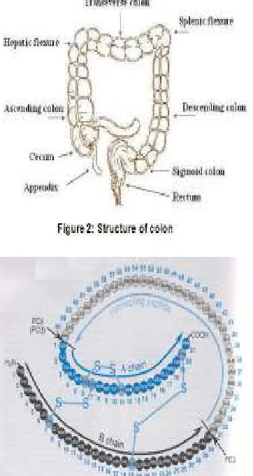
The amino acid sequence of human proinsulin is shown in Fig.3. By proteolytic cleavage, four basic amino acids (residues 31, 32, 64, 65) and the connecting peptide are removed, converting proinsulin to insulin. The sites of action of the end peptidases PC2 and PC3 are shown.
Insulin is usually administered to diabetic patients through subcutaneous injection. However, problems encountered with subcutaneous insulin injections are pain, allergic reactions, hyperinsulinemia and insulin lipodystrophy around the injection site.32 Oral
route is the most convenient33-34 and
comfortable means of administering protein drugs35 and eliminates pain caused by an
injection, stress associated with multiple daily injections such as needle anxiety36 and possible
infections.37 Indeed, insulin absorbed by the
intestinal epithelium reaches the liver through the portal vein and can directly inhibit hepatic glucose output7; subcutaneous insulin treatment
however does not replicate the normal dynamics of endogenous insulin release, resulting in a failure to achieve a lasting glycemic control in patients 38-39. However,
peptides and proteins such as insulin cannot be administered via the oral route. This is due to degradation by gastrointestinal enzymes and poor permeability across intestinal mucosa. 40-42The oral bioavailability of most peptides and
proteins therefore is < 1%. The challenge here is to improve bioavailability to anywhere between 30-50%.43To prevent these problems, many
protease inhibitors and surfactants were used in insulin formulations. However, protease inhibitors also prevent digestion of important nutrients present in the food.34, 43Similarly,
surfactants irritate the protective mucous membrane leads to passage of unwanted toxins and pathogens. 34, 43. Insulin is better absorbed
from the ileum and large intestine as compared to the jejunum.44Thus a polymer that would
release the drug in ileum or upper intestine has the potential for oral insulin delivery.
Eudragit is trademark of Rohm GmbH & Co. KG. Darmstadt in Germany, first marketed in 1950s. Eudragit prepared by the polymerization of acrylic and methacrylic acids or their esters, e.g., butyl ester or dimethylaminoethyl ester. Eudragit introduced in USPNF, BP, PhEur, Hand book of pharmaceutical excipients45
Colon Anatomy
The GI tract is divided into stomach, small intestine and large intestine. The large intestine extending from the ileocecal junction to the anus is divided in to three main parts. These are the colon, the rectum and anal canal.46The entire
colon is about 5 feet (150 cm) long, and is divided in to five major segments. Peritoneal folds called as mesentery which is supported by ascending and descending colon. The right colon consists of the cecum, ascending colon, hepatic flexure and the right half of the transverse colon. The left colon contain the left half of the transverse colon, descending colon, splenic flexure and sigmoid. The rectum is the last anatomic segment before the anus.47
The major function of the colon is the creation of suitable environment for the growth of colonic microorganisms, storage reservoir of fecal contents, expulsion of the contents of the colon at an appropriate time and absorption of potassium and water from the lumen48. The
bacteria. The colon tissue containing the villi, lymph, muscle, nerves, and vessels.
Fig. 3: Human Proinsulin and its conversion to insulin
EUDRAGIT®L 100
It is an anionic polymer synthesized from methacrylic acid and methylmethacrylate and have a pH-dependent solubility. Eudragit L 100would release the drug in the region of G.I.T. of pH 6-6.5 i.e. ileum or large intestine. 34 It is
available as an organic solution (Isopropanol), solid or aqueous dispersion.
Physical properties
It is a solid substance in form of a white powder with a faint characteristic odour.
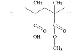
Chemical structure
Fig. 4: Structure of Eudragit® L 100
Product Form
Powder
Targeted Drug Release Area
jejunum
Dissolution
Dissolution between pH 6.0 and 7.0.
Characteristics
• Effective and stable enteric coatings with a fast dissolution in the upper Bowel
• Granulation of drug substances in powder form for controlled release
• Site specific drug delivery in intestine by combination with EUDRAGIT® S
grades
• Variable release profiles.
Chemical/IUPAC name
Poly (methacylic acid-co-methyl methacrylate) 1:1
INCI name
Acrylates Copolymer
Monographs Ph. Eur
Methacrylic Acid - Methyl Methacrylate Copolymer (1:1)
USP/NF
Methacrylic Acid Copolymer, Type A - NF
JPE
Methacrylic Acid Copolymer L
Weight average molar mass
approx. 125,000 g/mol
Acid Value
315 mg KOH/ g polymer
Glass Transition Temperature (Tg)
>130°C (+/- 5°C)
Viscosity / Apparent viscosity
60 - 120 mPa. s
Refractive index
1.390 - 1.395
Relative density
The suitability of Eudragit L 100 microspheresasoral carrier forpeptide drugs like insulin was evaluated. Insulin loaded EudragitL100 microsphereswere prepared using water-in-oil-in water (w/o/w) emulsion-solvent evaporation with polysorbate 20 as dispersing agent in internal aqueous phase and PVP/PVA as stabilizer in the external aqueous phase. In PBS pH 7.4, microspheres showed an initial burst release of 21% in 1 hr. and additional 35% release in next 5 hr. Thus, EudragitL100 microspheres have the potential to serve as an oral carrier forpeptide drugs like insulin.34
EUDRAGIT®S 100
It is an anionic copolymer based on methacrylic acic and methyl methacrylate.It is available only as an organic solution (Isopropanol) and solid.
Physical properties
It is a solid substance in form of a white powder with a faint characteristic odour.
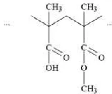
Chemical structure
Fig. 5: Structure of Eudragit® S 100
Form of Product
Powder
Targeted Drug Release Area
Colon delivery
Dissolution
Above pH 7.0
Characteristics
• Granulation of drug substances in powder form for controlled release
• Effective and stable enteric coatings with a fast dissolution in the upper Bowel
• Site specific drug delivery in intestine by combination with EUDRAGIT® S
grades
• Variable release profiles
Chemical/IUPAC name
Poly(methacylic acid-co-methyl methacrylate) 1:2
INCI name
Acrylates Copolymer
Monographs Ph. Eur.
Methacrylic Acid - Methyl Methacrylate Copolymer (1:2)
USP/NF
Methacrylic Acid Copolymer, Type B - NF
JPE
Methacrylic Acid Copolymer S
Weight average molar mass
approx. 125,000 g/mol
Acid Value
190 mg KOH/ g polymer
Glass Transition Temperature (Tg)
>130°C (+/- 5°C)
Viscosity / Apparent viscosity
50 - 200 mPa. S
Refractive index
1.390 - 1.395
Relative density
0.831-0.852
EudragitS100 microspheres have the potential to serve as an oral carrier forpeptide drugs like insulin. Insulin loaded PVA stabilized EudragitS100 microspheres showed maximum drug encapsulation released 2.5% insulin at pH 1.0 in 2 hr. Oral administration of PVA stabilized microspheres in normal albino rabbits (equivalent to 6.6 IU insulin/kg of animal weight) demonstrated a 24% reduction in blood glucose level, with maximum plasma glucose reduction of 76 ± 3.0% in 2 hours and effect continued upto 6 hr.43
The hypoglycemic effect of Eudragit S100 enteric-coated capsules containing sodium salicylate as an absorption promoter formulated with insulin in various ways: as physical mixture, by wet granulation or in suppository bases (polyethylene glycol 4000 or Witepsol W35) was studied in hyperglycemic beagle dogs. 25-30% reduction in plasma glucose levels and relative hypoglycemia (RH) of about 12.5% relative to subcutaneous injection of regular soluble insulin can be achieved by formulating insulin in Witepsol W35 (1 g) with sodium salicylate (50 mg) as an absorption promoter, reducing the resulting mass into particle size 180-315 microm, packing into hard gelatin capsules and coating with Eudragit S100.33 EUDRAGIT®S 12,5
It is an anionic copolymer based on methacrylic acic and methyl methacrylate.
Physical properties
Chemical structure
Fig. 6: Structure of Eudragit® S 12.5
Product Form
Organic Solution 12.5%
Targeted Drug Release Area
Colon Delivery
Dissolution
Above pH 7.0
Characteristics
• Granulation of drug substances in powder form for controlled release
• Effective and stable enteric coatings with a fast dissolution in the upper Bowel
• Site specific drug delivery in intestine by combination with EUDRAGIT® S
grades
• Variable release profiles
Chemical/IUPAC name
Poly(methacylic acid-co-methyl methacrylate) 1:2
INCI name
Acrylates Copolymer
Monographs Ph. Eur
Methacrylic Acid - Methyl Methacrylate Copolymer (1:2)
USP/NF
Methacrylic Acid Copolymer, Type B – NF
JPE
n/a
Weight average molar mass
approx. 125,000 g/mol
Acid Value
190 mg KOH/ g polymer
Glass Transition Temperature (Tg)
>130°C (+/- 5°C)
EUDRAGIT® FS 30 D
It is an aqueous dispersion with 30 % dry substance. EUDRAGIT® FS 30 D is the aqueous dispersion of an anionic copolymer based on methyl acrylate, methyl methacrylate and methacrylic acid. It is insoluble in acidic media, but dissolves by salt formation above pH 7.0. Apart from its enteric properties, its dissolution
at a higher pH value allows targeted colon delivery.
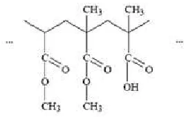
Chemical structure
Fig. 7: Structure of Eudragit® FS 30 D
Chemical/IUPAC name
Poly(methyl acrylate-co-methyl methacrylate-co-methacrylic acid) 7:3:1
INCI name: Acrylates Copolymer
Acid Value
70 mg KOH/ g polymer
Minimum Film Forming Temperature (MFT)
~14°C
Glass Transition Temperature (Tg)
43°C (+/- 5°C)
The ratio of the free carboxyl groups to the ester groups is approx. 1:10. It is milky-white liquid of low viscosity with a faint characteristic odour. The monomers are randomly distributed along the copolymer chain. The weight average molar mass (Mw) of EUDRAGIT® FS 30 D is approx. 280,000 g/mol.
The dispersion is miscible with water in any proportion, the milky-white appearance being retained. A clear or slightly cloudy, viscous solution is obtained by mixing 1 part EUDRAGIT® FS 30 D with 5 parts acetone. The same results are obtained by mixing with ethanol or isopropyl alcohol; initially, the polymer is precipitated, but then dissolves again in the excess organic solvent.
A clear or slightly cloudy liquid is obtained by mixing 1 part EUDRAGIT® FS 30 D with 2 parts 1 N sodium hydroxide.
Dissolution
Above pH 7.0
Viscosity / Apparent viscosity
Max. 20 mPa .s
pH: 2.0 - 3.5
Relative density
1.058 - 1.068
Monomers
Max. 100 ppm
Sample solution
dilute to 50.0 ml. Add 5.0 ml of the solution drop wise to 20 ml of a 70 % solution of methanol for chromatography in phosphoric acid pH 2 (adjust an appropriate volume of water with phosphoric acid 85 % to pH 2). Centrifuge until the supernatant is clear and use the supernatant solution as the sample solution.
Storage and handling
Store between 5 °C and 10 °C. Protect from freezing. Keep in well closed containers. Avoid contamination during sampling. Containers that have been opened for use should be closed again immediately and the content used up within the next few weeks. Matrix systems with EUDRAGIT® FS 30 D will
release 100% of the drug. Polymer amounts of 10 to 20 % are sufficient to get a pH-independent matrix.
EUDRAGIT® L 12,5
It is solution of EUDRAGIT® L 100 with 12.5% (w/w) dry substance in aqueous Isopropyl Alcohol Ph. Eur. / USP. The solution contains 3% (w/w) deionised water. The product contains 0.3 % Sodium Lauryl sulfate Ph. Eur. / NF on solid substance.
EUDRAGIT® L 100 is described as Copolymer (1:1), Type A or Copolymer L in the monographs.
It is colourless, clear to slightly cloudy liquids with the characteristic odour of isopropyl alcohol.
Chemical structure
Fig. 8: Structure of Eudragit® L 12,5
Dissolution
Dissolution between pH 6.0 and 7.0
Storage
Protect from warm temperatures (USP, General Notices). Store in tightly closed containers.
Monomers
Max. 70 ppm
Viscosity / Apparent viscosity
60 – 120 mPa .s
Refractive index
1.390 - 1.395
Relative density
0.831 - 0.852
It provides effective and stable enteric coatings with a fast dissolution in the upper Bowel and site specific drug delivery in intestine by combination with EUDRAGIT® S grades.
Table 1: Dissolution properties of enteric Eudrgit polymers
EUDRAGIT® polymer Product form Dissolution properties
Eudragit L 100 Powder Dissolution above pH 6.0 Eudragit S 100 Powder Dissolution above pH 7.0 Eudragit S 12,5 12.5 % organic solution Dissolution above pH 7.0 Eudragit FS 30 D 30 % aqueous dispersion Dissolution above pH 7.0 Eudragit L 12,5 12.5 % organic solution Dissolution above pH 6.0
Glass transition temperature (Tg)
The glass transition temperature is an important factor for describing the physical properties of polymers. On a macroscopic level it describes the solidification of an anisotropic polymer melt. The glass transition temperature has far-reaching consequences, e.g. for film formation, melt processing and storage of finished pharmaceutical dosage forms. Plasticizers, solvents or residual solvents (including water) that act as plasticizers usually cause a reduction in glass transition temperature, which is specifically exploited in application formulations. Most common plasticizer for EUDRAGIT polymers is triethyl citrate (TEC).
Effect of Plasticizers Compatibility
Polymers with high glass transition temperatures need plasticizer to obtain coatings which are not brittle. For example: Eudragit® L 100 in organic solution needs 10% Triethyl citrate (TEC). Dispersions from Polymers with high glass transition temperatures needs plasticizer to decrease the minimum Film-Forming Temperature and to optimize the film formation. For example: Redispersed Eudragit® L 100 needs 50% Triethyl citrate (TEC).
(Eudragit L and S) polymer films were formulated with a range of plasticizers for measuring Tg of polymer films in the wet state. This allows better prediction of polymer behavior in vivo conditions49.
A colon-specific drug delivery technology was designed to avoid the inherent problems associated with pH- or time-dependent systems. In this regard, Eudragithave severed to be a much better enteric coated polymer. There have been several studies where formulations have been enteric coated with different grades of Eudragits exploitingeither time, pH- dependent or microbial degradation mechanisms for targeting colonic release.
MICROPARTICLES
The microencapsulation process in which the removal of the hydrophobic polymer solvent, achieved by evaporation has been widely reported in recent years for the preparation of microspheres and microcapsules. The encapsulation of highly water soluble compounds including proteins and peptides presents formidable challenges to the researcher. The successful encapsulation of such entities requires high drug loading in the microspheres, prevention of protein degradation by the encapsulation method. To achieve these goals, solvent evaporation techniques and their innovative modifications have been attempted.
Different techniques of microencapsulation are 1.Single or multiple emulsion solvent evaporation (O/W, W/O, W/O/W).
2. Multiple emulsion system.
3. Double emulsion solvent evaporation. 4. Coacervation method.
a)
b)
Fig. 9: SEM micrographs of optimized insulin loaded a) Eudragit S 100 b) Eudragit L 100
ENCAPSULATION OF POORLY WATER SOLUBLE DRUGS
Basic Drugs
Poorly water soluble basic drugs are very sensitive to pH changes and their dissolution in the acidic stomach environment tends to precipitate them upon gastric emptying, which leads to compromised or erratic oral bioavailability. The oral bioavailability of such drugs can be
improved by encapsulation of drug within highly pH responsive Eudragit L microparticles using emulsion solvent evaporation method.50 Acidic Drugs
Sometimes, acidic drug encapsulated by emulsion solvent evaporation, are present in its crystalline form, which can affect drug release and produce negative impact on other characteristics of the final product. Henceforth, investigations were carried out to find factors that are responsible for the formation and inhibition of drug crystals in modified-release microparticles using Eudragit S or Eudragit L. It was concluded that the drug crystallization can be inhibited by optimizing the ratio of drug to polymer in the microparticles there by stabilizing this acidic drugs for drug delivery51. Water Soluble Drugs
Generally, highly water-soluble and poorly bioavailable drugs are unstable at gastric pH. Hence, to resolve this problem mucoadhesive microparticles were formulated using Eudragit S100 and EC using w/o/o double emulsion solvent diffusion method. Microparticles made with drug: Eudragit S100 (ratio 1:3) exhibited maximum entrapment efficiency and followed fickian diffusion with delayed release.52The
medium, while its acid solubility and microsphere's size had minor influences.53 ENHANCEMENT OF PROTEIN STABILITY
Purpose of such an approach was to formulate a stable formulation for proteins and peptides which are susceptible to denaturation, degradation, and conformational changes which render them inactive. Amongst the pioneering works in this regard, an oral colonic targeted heparin dosage form was fabricated, allowing the release of Low molecular weight heparins (LMWH) directly in the inflamed tissue using
pH-sensitive microspheres of Eudragit P 4135 F by double emulsion technique54, 55
COMBINATIONOF EUDRAGIT L 100 and EUDRAGIT S 100
Studies in human volunteers have confirmed that pH drops from 7.0 at terminal ileum to 6.0 at ascending colon, and Eudragit S based systems sometimes fail to release the drug. To overcome the shortcoming, combination of Eudragit S and Eudragit L which ensures drug release at pH < 7 has been advocated.56
a) b)
Fig. 10 SEM micrographs of optimized bovine insulin loaded a) chitosan and Eudragit L 100: Eudragit S100 (1:1) microparticles b) PLGA and
Eudragit L 100: Eudragit S100 (1:1) microparticles
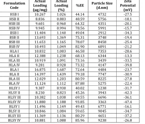
Table 2: Characterization of insulin loaded microparticles using Chitosan and different concentrations of Eudragit L 100, Eudragit S 100 and Eudragit L 100 : Eudragit S 100 (1:1)
Formulation Code
Actual Loading (μg/mg)
Actual Loading
(%)
%EE Particle Size (d.nm)
Zeta Potential
(mV)
Abbreviations
HSB I Human insulin loaded microparticles using Chitosan and 3% w/v Eudragit S 100 HSB II Human insulin loaded microparticles using Chitosan and 4% w/v Eudragit S 100 HSB III Human insulin loaded microparticles using Chitosan and 5% w/v Eudragit S 100 HSB IV Human insulin loaded microparticles using Chitosan and 6% w/v Eudragit S 100 BSB I Bovine insulin loaded microparticles using Chitosan and 3% w/v Eudragit S 100 BSB II Bovine insulin loaded microparticles using Chitosan and 4% w/v Eudragit S 100 BSB III Bovine insulin loaded microparticles using Chitosan and 5% w/v Eudragit S 100 BSB IV Bovine insulin loaded microparticles using Chitosan and 6% w/v Eudragit S 100 HLA I Human insulin loaded microparticles using Chitosan and 3% w/v Eudragit L 100 HLA II Human insulin loaded microparticles using Chitosan and 4% w/v Eudragit L 100 HLA III Human insulin loaded microparticles using Chitosan and 5% w/v Eudragit L 100 HLA IV Human insulin loaded microparticles using Chitosan and 6% w/v Eudragit L 100 BLA I Bovine insulin loaded microparticles using Chitosan and 3% w/v Eudragit L 100 BLA II Bovine insulin loaded microparticles using Chitosan and 4% w/v Eudragit L 100 BLA III Bovine insulin loaded microparticles using Chitosan and 5% w/v Eudragit L 100 BLA IV Bovine insulin loaded microparticles using Chitosan and 6% w/v Eudragit L 100
HLSY I Human insulin loaded microparticles using Chitosan and 3% w/v Eudragit L 100: Eudragit S 100 (1:1)
HLSY II Human insulin loaded microparticles using Chitosan and 4% w/v Eudragit L 100: Eudragit S 100 (1:1)
HLSY III Human insulin loaded microparticles using Chitosan and 5% w/v Eudragit L 100: Eudragit S 100 (1:1)
HLSY IV Human insulin loaded microparticles using Chitosan and 6% w/v Eudragit L 100: Eudragit S 100 (1:1)
BLSY I Bovine insulin loaded microparticles using Chitosan and 3% w/v Eudragit L 100: Eudragit S 100 (1:1)
BLSY II Bovine insulin loaded microparticles using Chitosan and 4% w/v Eudragit L 100: Eudragit S 100 (1:1)
BLSY III Bovine insulin loaded microparticles using Chitosan and 5% w/v Eudragit L 100: Eudragit S 100 (1:1)
BLSY IV Bovine insulin loaded microparticles using Chitosan and 6% w/v Eudragit L 100: Eudragit S 100 (1:1)
After observing these formulations, it was found that size of particles was in the range of 1-10 μm. which is ideal for cellular uptake. Also, zeta potential values indicate the good stability of particles. As the concentration of polymer increases in the formulation, % Entrapment Efficiency (EE) increases. This could be due to more coating of the Eudragit polymer and hence less leakage of the drug occurs due to less number of pores left.
CONCLUSION
The large variety of applications as well as the steadily increasing number of research workers engaged in studies of Eudragit polymers due to their unique properties, have made significant contributions to many types of formulations and suggest that the potential of Eudragit as novel and versatile polymer will be even more significant in future. Purpose of such an approach was to formulate a stable formulation for proteins and peptides like insulin which are susceptible to denaturation, degradation, and conformational changes.
REFERENCES
1. Keizer J and Magnus G. ATP-sensitive potassium channel and bursting in the pancreatic beta cell. A theoretical study.
Biophysical Journal. 1989;56(2):229– 242.
2. Moore C and Cooper G. Co-secretion of amylin and insulin from cultured islet beta-cells: modulation by nutrient secretagogues, islet hormones and hypoglycemic agents. Biochem Biophys Res Commun. 1991;17(1): 1–9.
3. Lang V and Light PE. The molecular mechanisms and pharmacotherapy of ATP-sensitive potassium channel gene mutations underlying neonatal diabetes. Pharmgenomics. Pers Med. 2010; 3:145–61.
4. Elayat AA, el-Naggar MM and Tahir M. An immunocytochemical and morphometric study of the rat pancreatic islets. Journal of Anatomy. 1995;186.(Pt 3):629–37.
5. Sleisenger, edited by Mark Feldman, Lawrence S. Friedman, Lawrence J. Brandt; consulting editor, Marvin H. Sleisenger & Fordtran's gastrointestinal and liver disease pathophysiology, diagnosis, management. 2009 (9th ed.). St. Louis, Mo.: MD Consult.
cellular composition and beta-cell viability in human islet preparations. American Journal of Transplantation. 2005;5 (7):1635–45.
7. Amisten S, Salehi A, Rorsman P, Jones PM and Persaud SJ. An atlas and functional analysis of G-protein coupled receptors in human islets of Langerhans. Pharmacol Ther. 2013. 8. Pérez-Armendariz M, Roy C, Spray DC
and Bennett MV. Biophysical properties of gap junctions between freshly dispersed pairs of mouse pancreatic beta cells. Biophysical Journal. 1991;59(1):76–92.
9. Hogan A, Pileggi A and Ricordi C. Transplantation: current developments and future directions; the future of clinical islet transplantation as a cure for diabetes. Frontiers of Bioscience. 2008;13(13):1192–205.
10. Kaushansky K. Lineage-specific hematopoietic growth factors. N Engl J Med. 2006;354(19):2034–45.
11. Kober L, Zehe C and Bode J. Optimized signal peptides for the development of high expressing CHO cell lines. Biotechnol Bioeng. 2013;110(4):1164– 73.
12. Soltys BJ, Falah MS and Gupta RS. Identification of endoplasmic reticulum in the primitive eukaryote Giardia lamblia using cryoelectron microscopy and antibody to Bip J Cell Science. 1996;109:1909-1917.
13. Endoplasmic reticulum. (n.d.). McGraw-Hill Encyclopedia of Science and Technology. Retrieved September 13, 2006, from Answers.com Web site: http://www.answers.com/topic/endop lasmic-reticulum
14. Levine T. Short-range intracellular trafficking of small molecules across endoplasmic reticulum junctions. Trends Cell Biol. 2004;14 (9):483–90. 15. Xu C. Endoplasmic Reticulum Stress:
Cell Life and Death Decisions. J Clin. Invest. 2005;115(10):2656–2664. 16. Steiner DF, Cunningham D, Spigelman L
and Aten B. Insulin Biosynthesis. Evidence for a Precursor Science. 1967;157(3789):697–700.
17. Hills CE and Brunskill NJ. Intracellular signalling by C-peptide. Exp Diabetes Res. 2008.
18. Mughal RS, Scragg JL and Lister P. Cellular mechanisms by which proinsulin C-peptide prevents insulin-induced neointima formation in human
saphenous vein. Diabetologia. 2010;53(8):1761–71.
19. Clark PM. Assays for insulin, proinsulin and C-peptide. Ann Clin Biochem. 1999;36:541-564.
20. Marques RG, Fontaine MJ and Rogers J. C-peptide: much more than a byproduct of insulin biosynthesis. Pancreas. 2004;29(3):231–8.
21. Melloul D, Marshak S and Cerasi E. Regulation of insulin gene transcription. Diabetologia. 2002;45(3):309–26. 22. Katsoyannis PG, Fukuda K, Tometsko A,
Suzuki K, Tilak M, Panayotis G Fukuda, Kouhei Tometsko, Andrew, Suzuki, Kenji, Tilak and Manohar. Insulin Peptides. X. The Synthesis of the B-Chain of Insulin and Its Combination with Natural or Synthetis A-Chin to Generate Insulin Activity. Journal of the American Chemical Society. 1964;86(5):930–932.
23. Kung YT, Du YC, Huang WT, Chen CC and Ke LT. Total synthesis of crystalline bovine insulin. Sci Sin. 1965;14(11):1710–6.
24. Steiner DF and Oyer PE. The biosynthesis of insulin and a probable precursor of insulin by a human islet cell adenoma. Proc Natl Acad Sci. U.S.A. 1967;57(2):473–480.
25. Menting JG, Whittaker J, Margetts MB, Whittaker LJ, Kong GK-W, Smith BJ, Watson CJ, Žáková L, Kletvíková E, JJ, Chan SJ, Steiner DF, Dodson GG, Brzozowski AM, Weiss MA, Ward CW and Lawrence MC. How insulin engages its primary binding site on the insulin receptor. Nature. 2013; 493(7431):241–245.
26. Dimitriadis G, Mitrou P, Lambadiari V, Maratou E and Raptis SA. Insulin effects in muscle and adipose tissue. Diabetes Res Clin Pract. 2011;93(Suppl 1):S52–9. 27. Drug Information Portal NLM –Insulin human [USAN:USP:INN:BAN] (http://druginfo.nlm.nih.gov/drugporta l/.
28. Bell GI, Pictet RL, Rutter WJ, Cordell B, Tischer E and Goodman HM. Sequence of the human insulin gene. Nature. 1980;284(5751):26–32.
30. Tanyolac S, Goldfine ID and Kroon L. Insulin Pharmacology, Type of Regimens and Adjustments Home PD, Alberti KG. The new insulins, their characteristics and clinical indications. Drugs. 1982;24(5):401–13.
31. Gray RS, Cowan P, di Mario U, Elton RA, Clarke BF and Duncan LJ. Influence of insulin antibodies on pharmacokinetics and bioavailability of recombinant human and highly purified beef insulins in insulin dependent diabetics. Br Med J (Clin Res Ed). 1985;290(6483):1687– 91.
32. Gowthamarajan K and Kulkarni, GT. Oral Insulin-Fact or Fiction. Possibilities of achieving oral delivery for insulin. Resonance. 2003;38-46.
33. Hosny EA, Al-Shora HI and Elmazar MA. Oral delivery of insulin from enteric coated capsules containing sodium salicylate: effect on relative hypoglycaemia of diabetic beagle dogs. Int J Pharm. 2002;237:71-76.
34. Jain D, Majumdar DK and Panda AK. Insulin loaded eudragit L100 microspheres for oral delivery preliminary in vitro studies. J Biomater Appl. 2006;21(2):195-211.
35. Hoffman A and Ziv E. Pharmacokinetic considerations of new insulin formulations and routes of administration. Drug Dispos. 1996;33:285–301.
36. Korytkowski M. When oral agents fail: practical barriers to starting insulin. Int J Obesity. 2002;26(3):S18-S24.
37. Lin YH, Mi FL and Chen CT. Preparation and characterization of nanoparticles shelled with chitosan for oral insulin delivery. Biomacromolecules. 2007;8:146-152.
38. Damge C and Socha M. Poly (Є-Caprolactone)/ Eudragit nanoparticles for oral delivery of aspart-insulin in treatment of diabetes. JPS. 2010;99(2):879-889.
39. Rick S. Oral protein and peptide drug delivery. In: Binghe W, Teruna S, Richard S, editors. Drug delivery: Principles and applications. New Jersey: Wiley Interscience. 2005;189.
40. Agarwal V and Khan MA. Current status of the oral insulin delivery. Pharmaceutical technology. 2001;76-90.
41. Nakamura K, Murray RJ, Joseph JI, Peppas NA, Morishita M and Lowman AM. Oral insulin delivery using
P(MAA-g-EG) hydrogels: effects of network morphology on insulin delivery characteristics. J Control Release. 2004;95:589-599.
42. Sajeesh S and Sharma CP. Cyclodextrin-insulin complex encapsulated polymethacrylic acid based nanoparticles for oral insulin delivery. Int J Pharm. 2006;325(1-2):147-154. 43. Jain D, Majumdar DK and Panda AK.
Eudragit S 100 entrapped insulin microspheres for oral delivery. AAPS Pharm Sci Tech. 2005;6(1):E100-E107. 44. Lee V.H. Oral route of peptide and
protein drug delivery, Chapter 16, Marcel Dekker Inc. 1991;691-738. 45. Kimura T and Sato K. Oral
administration of insulin as Poly (vinyl alcohol)-gel spheres in diabetic rats. Biol Pharm Bull. 1996;19(6):897-900. 46. Kapoor and Vinay Kumar (13 Jul 2011).
Large Intestine Anatomy. In Gest, Thomas R. Medscape. WebMD LLC. Retrieved 2013-08-20.
47. large intestine. Mosby's Medical Dictionary (8th ed.). Elsevier. 2009. ISBN9780323052900.
48. Colon Function And Health Information Retrieved on 2010-01-21.
49. Fadda HM, Khanna M, Santos JC, Osman D, Gaisford S and Basit AW. The use of dynamic mechanical analysis (DMA) to evaluate plasticization of acrylic polymer films under simulated
gastrointestinal conditions. Eur J Pharm Biopharm. 2010;76(3):493-7.
50. Alhnan MA, Murdan S and Basi. Encapsulation of poorly soluble basic drugs into enteric microparticles: A novel approach to enhancetheir oral bioavailability. Int J of Pharma. 2011; 416(1):55-60.
51. Alhnan MA and Basit AW. In-process crystallization of acidic drugs inacrylic microparticle systems: Influence of physical factors anddrug-polymer interactions. J Pharm Sci. 2011;100(8): 3284–93.
52. Garg Y and Pathak K. Design and in vitro performance evaluation of purified microparticles of pravastatin sodium for intestinal delivery. AAPS Pharm Sci Tech. 2011;12(2):673-82.
54. Meissner Y, Ubrich N, El Ghazouani F, Maincent P and Lamprecht A. Low molecular weight heparin loaded pH-sensitive microparticles. Int J Pharm. 2007;335(1-2):147-53.
55. Lamprecht A, Yamamoto H, Ubrich N, Takeuchi H, Maincent P and Kawashima Y. FK506 microparticles mitigate
experimental colitis with minor renal calcineurin suppression. Pharm Res. 2005;22(2):193-9.