Published online June 09, 2014 (http://www.sciencepublishinggroup.com/j/js) doi: 10.11648/j.js.s.2014020601.11
ISSN: 2330-0914 (Print); ISSN: 2330-0930 (Online)
Vascularized free fibula flap for reconstruction of
mandibular defects
Mohammad Akheel
1, Suryapratap Singh Tomar
2, Anuj Bhargava
31
Dept of Oral & maxillofacial surgery, NMCH, Nellore, India
2
Dept. of Neurosurgery, NMCH, Nellore, India
3
Dept of Oral & maxillofacial surgery, Index dental college, Indore, M.P., India
Email address:
drakheelomfs@gmail.com (M. Akheel), dr.suryapratap_singh_tomar@yahoo.com (S. S. Tomar)
To cite this article:
Mohammad Akheel, Suryapratap Singh Tomar, Anuj Bhargava. Vascularized Free Fibula Flap for Reconstruction of Mandibular Defects. Journal of Surgery. Special Issue: Craniofacial Surgery. Vol. 2, No. 6-1, 2014, pp. 1-5. doi: 10.11648/j.js.s.2014020601.11
Abstract:
Objective: To assess the versatility of vascularized free fibula flap in reconstruction of various defects of mandible. Study Design: Prospective study. Duration of Study: March 2009 to March 2012. Methodology: The study group consisted of 10 patients who underwent resection of mandible for various reasons and reconstruction of continuity defects using a vascularized free fibular flap. The mandible was resected for ameloblastoma in 4 cases, squamous cell carcinoma in 1 case, odontogenic keratocysts in 3 cases and ossifying fibroma in 2 cases. The type of reconstruction performed was primary in 9 patients in which osseous fibula flap was used and secondary in 1 patient in which osseocutaneous flap was used. Results: There were 10 patients which include 5 males and 5 females within age group of 20 to 50 years with mean age of 35 years. All flaps survived except in 1 patient who had donor site morbidity. Flap perfusion was seen immediately after anastomosis and was maintained throughout the follow-up period of minimum 6 months. All patients were kept innasogastric feeding for 5 days and then began oral feeding and walking with some aid in 2nd week and became completely
ambulant in 4th week postoperatively. Conclusion: In our study, we conclude that vascularized free fibula flap is a versatile option for reconstruction of large mandibular defects with its good quality and quantity of bone and ease of manipulation to restore the original anatomy of the mandible and permit implant based prosthetic rehabilitation.
Keywords:
Fibula Reconstruction, Ameloblastoma, Odontogenic Keratocysts1. Introduction
Pathologies predisposing to wide resection of mandible pose a great surgical challenge for head and neck surgeon for reconstruction and rehabilitation. Various techniques of flaps have been used since years. The goal of reconstructive surgery following ablation of soft and hard tissue loss is to reconstruct the defect at the time of surgery primarily to facilitate a good wound healing and cosmetic outcome1.
Reconstructive options for maxillofacial defects have improved tremendously beyond the primary closure, stainless steel reconstruction plates and skin grafts to a wide variety of pedicled flaps. More recently, due to advancements in surgical technique, improved knowledge in anatomy and various complications reported in literature, the surgeons have introduced the use of micro vascular free flaps to reconstruct the composite defects in order to match the resected tissues and provide a better quality of life2, 3.
The pedicled flaps which are anastomosed represent unique principle of transplantation, being suitable for reconstruction of a variety of complex and large defects. Among the various flaps used, vascularized free fibular flaps are considered to be a workhorse since hidalgo has used it for mandibular reconstruction4, 5, 6.
2. Materials & Methods
This prospective study was conducted in Department of Oral & Maxillofacial surgery in Sharad pawar dental college & hospital, India over a period of three years from march 2009 to march 2012 consisting of ten consecutive patients. Out of 10 patients, 5 patients were male and 5 patients were female within age group range from 20 to 50 years with mean age of 35 years. The mandible was resected for ameloblastoma in 4 cases, squamous cell carcinoma in 1 case, odontogenic keratocysts in 3 cases and ossifying fibroma in 2 cases. The type of reconstruction performed was primary in 9 patients in which osseous fibula flap was used and secondary in 1 patient in which osseocutaneous flap was used. (TABLE 1)
Table 1.
S.no. Case Type of defect Type of flap
1. Ameloblastoma L Osseous 2. Ameloblastoma Lcl Osseous 3. Squamous cell carcinoma Lc Osseocutaneous
4. Ossifying fibroma L Osseous 5. Odontogenic keratocyst Hl Osseous 6. Odontogenic keratocyst Hl Osseous 7. Ameloblastoma Hl Osseous 8. Odontogenic keratocyst Hl Osseous
9. Ameloblastoma L Osseous 10. Ossifying fibroma L Osseous
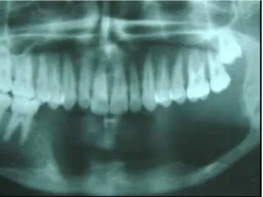
The patients included in this study were those needing mandibular resection and who were medically fit for surgery with no underlying systemic disorders. Pre operative CT angiography was done for all patients to rule out abnormalities of lower leg vascular anatomical variation like peroneus magnus and atherosclerotic blood vessels. The procedure was explained to all patients and their guardians with their postoperative complications and an informed consent was obtained preoperatively. Preoperative investigations like othopantomogram (Fig 1, 2), chest X-ray and a complete surgical profile were taken. All patients were evaluated by anesthetist and posted for surgical procedure under general anesthesia.
Fig 1. facial profile of a patient with odontogenic keratocyst on left side.
Fig 2. preoperative orthopantamogram showing multilocular odontogenic
keratocyst on left side involving the anterior mandible.
All mandibular defects were classified according to HCL classification. In our study 4 patients had defects in head region, 2 in central region and 4 patients in lateral region. After assessing the size of the defect, the free fibular flap was harvested from the donor site following standard protocols. 1 patient had osseocutaneous flap and 9 patients had osseous flap.
3. Procedure
Fig 3. dissection of vascularized fibula free flap.
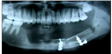
pedicle was isolate and divided from the donor site. The flap insetting was done and fixed with titanium reconstruction plate and miniplates. The anastamosis was done using 8-0 ethilon for peroneal and facial artery and vein with 9-0 ethilon. Vaccum drains were secured and wound closure was done in layers both in donor and recipient sites. Postoperative orthopantomogram was taken on 2st postoperative day (Fig4).
Fig 4. postoperative orthopantomogram showing fixation of vascularized
fibula free flap with miniplates.
4. Results
This study was carried out in 10 patients who underwent reconstruction of mandible with free vascularized fibula flap. Among these, 5(50%) patients were males and 5(50%) were females with a male to female ratio of 1:1.
The mandible was resected for ameloblastoma in 4 cases (40%), squamous cell carcinoma in 1 case (10%), odontogenic keratocysts in 3 cases (30%) and ossifying fibroma in 2(20%) case. The type of reconstruction performed was primary in 9(90%) patients in which osseous fibula flap was used and secondary in 1(10%) patients in which osseocutaneous flap was used.
Flap perfusion was assessed intraoperatively and postoperatively and throughout the follow up period for a minimum of 6 months. All patients were kept in nasogastric feeding for 5 days and then began oral feeding and walking with some aid in 2nd week and became completely ambulant in 4th week. Out of 10 patients 1(10%) patient had donor site morbidity with mild infection which was controlled by antibiotics and local antiseptic measures.
5. Discussion
Reconstruction of mandible represents a surgical challenge to the head and neck surgeon and has been revolutionized by
the modern microvascular techniques and complex anatomical landmarks. Ablative procedures of mandible
warrants mandibular reconstruction mandatory for
restoration of form and function. Other causes of mandibular defects include trauma, infection or inflammation, osteoradionecrosis, osteomyelitis and congenital deformities.
The anatomical location and size of the defect matters more in restoration of aesthetic and function of mandible reconstruction. The anterior mandibular reconstruction needs more skill and planning to mimic the original mandible to give function as well as aesthetics and is a prime concern when compared to the defect in posterior region of mandible. Restoration of functions like mastication, speech, deglutition and support for tongue and other tissues and muscles is lost. All these deformities warrants an appropriate procedure to address these problems. Hence mandibular reconstruction has become a mandatory procedure to address these problems9, 10, 11, 12.
Various materials and techniques were put forward to reconstruct the mandible. Free bone grafts taken from calvarium, iliac, tibia, and fibula were used to restore small defects of mandible. Over the past 20 years vascularized flaps have become popular to reconstruct large defects of mandible. In our study we have used vascularized free fibula bone flaps to reconstruct the mandible.
Due to advances in surgical skills, microvascular flaps are used for spontaneous and immediate repair of the recipient site. Hoffman et al in his study showed histological evidence of the healing of vascularized free flaps with bone continuity similar to that of a fracture13. This favors new bone formation and either immediate or delayed rehabilitation can be done with implant prosthesis. For anterior mandibular defects they have the ability of forming vascularized bone flaps in order to provide a solid arch to restore the form and function. Therefore, reconstruction with vascularized bone is the preferred method of mandibular reconstruction.
During the past few years, a variety of flaps have been discovered for reconstruction of mandible. Out of which fibula has been considered as workhorse for mandibular reconstruction as it has all the ideal features like adequate length, width, bone quantity and quality to connect the resected part of the defect. This fibula can be made as single or double barrel depending upon the height of mandible required to reconstruct14, 15,16,17,18.
According to the HCL mandibular defect classification19, the mandibular defects which were reconstructed in our study were classified as head region (n =4), central region (n = 2) and lateral region (n = 4). In 1 patient who had squamous cell carcinoma reconstruction was done after 6 months when there was no evidence of recurrence.
reconstruction of mandible. Reconstruction was planned only when no evidence of disease or recurrence was seen. The donor site complication is a very serious problem when planning a free tissue transfer. In our study the fibula free flap harvest had acceptable donor site morbidity with infection which was controlled by local antiseptic measures. And preservation of good foot and ankle function in most individuals. Garrett et al. in his study had increased talar tilt in one patient compared with the contralateral side but there was preservation of the ankle stability20. The results from our study regarding donor site morbidity are in accordance to the published studies, which also claim less donor site morbidity with vascularized free fibular graft21, 22.
The advantages of vascularized free fibula flap are achieved at the cost of the procedure that is longer than other conventional reconstructive procedures. Foster et al in his study concluded that additional operative time is required for a free flap reconstruction23. In our study average time for resection and reconstruction was 3 hours 25 minutes. Average hospital stay was 7 to 16 days with an average of 11.5 days. According to literature and recent advancements, vascularized fibula flap remains the first choice for all the large mandibular defects and for poor recipient bed which do not favor uptake of free bone grafts2,
15, 16, Nonvascularized bone grafts are effective for short
bony defects of mandible in non-irradiated tissue and in patients who are medically compromised to tolerate the additional operative time required for a free flap reconstruction.
6. Conclusion
Microvascular free flap recontruction is one of the modern means of restoring composite defects of the maxillofacial region. In our study the vascularized free fibula flap was a versatile and most appropriate option for microvascular reconstruction of large mandibular defects with low incidence of complication rates and good quality and quantity of bone stock to reconstruct defects in head and region. It also favors prosthetic rehabilitation and is always the first choice for the majority of mandibular reconstruction cases.
References
[1] Aydin a, emekli u, ere m, hafiz g. Fibula free flap for mandibular reconstruction. Kulak burun bogaz ihtis derg 2004; 13:62-6.
[2] Douglas ar, ariyan s, restifo r: use of the operating microscope and loupes for head and neck free microvascular tissue transfer: arch otolaryngol head neck surg 2003: 129. [3] Rosenthal e, carroll w, dobbs m, wax m, peters g.
Simplifying head and neck micro-vascular reconstruction. Head neck 2004; 26:930-6.
[4] Hidalgo da, rekow a. Review of 60 consecutive fibula free flaps for mandible reconstruction. Plast reconstr surg 1995;
96:585-96;discussion 597-62
[5] Hidalgo da. Fibula free flap: a new method for mandible reconstruction. Plast reconstr surg 1989; 84:71-9.
[6] Dean ad, casapi n, regev e, zeltser r: reconstruction of the mandible by fibula free flap: imaj 2002;4:600-602
[7] Blackwell ke: donor site evaluation for fibula free flap transfer: american journal of otolatyngology 1998;19: 89-95 [8] Daniel tr, thomas r, bell th, neligan pc. Functional outcome
of the foot and ankle after free fibula graft. Foot ankle int 2005; 26:597-601
[9] Stephen s, mark a , gregory p, michael j, gregory rd, geoffrey l : choice of flap and incidence of free flap success: american society of plastic surgeons 1996: 98 : 459-463. [10] Smolka k, kraehenbuehl m, eggensperger n: fibula free flap
reconstruction of the mandible in cancer patients: evaluation of a combined surgical and prosthodontic treatment concept: oral oncol 2007:10:10-16.
[11] Nicolic z, jeremic j, milosavjevic r. [use of free microvascular flaps in the management of the head and neck defects]. Vojnsanit pregl 2006; 63:713-20. Serbian
[12] Sieg p, zieron jo, bierwolf s, hakim sg : defect-related variations in mandibular reconstruction using fibula grafts: british journal of oral and maxillofacial surgery 2002: 40, 322–329
[13] Hoffmann j, ehrenfeld m, hwang s, schwenzer n: complications after microsurgical tissue transfer in the head and neck region: journal of cranio-maxillofacial surgery 1998: 26: 255-259.
[14] Hasse ps, zimmermann ce: versatility of vascularized fibula and soft tissue graft in the reconstruction of the mandibulofacial region: int j oral maxillofac surg 1999; 28:356-361.
[15] Joseph jd, richard mw, hidalgo da: long term evaluation of bone mass in free fibula flap mandible reconstruction: the ajos 1997: 174.
[16] Militsakh o, mohyuddin n, kriet dj: comparison of radial forearm with fibula and scapula osteocutaneous free flaps for oromandibular reconstruction: arch otolaryngol head neck surg. 2005;131:571-575.
[17] Wolff kd, ervens g, herzog k, hoffmeister b: experience with the osteocutaneous fibula flap: an analysis of 24 consecutive reconstructions of composite mandibular defects: j cranio-maxillofacial surgerv 1996 24, 330-338
[18] Wolff kd, holzle f, eufinger h: the radial forearm flap as a carrier for the osteocutaneous fibula graft in mandibular reconstruction: int j oral maxillofac surg 2003; 32: 614–618. [19] Neal df, wadsworth jt, villaret d, farwell dg : midface reconstruction with the fibula free flap: arch otolaryngol head neck surg. 2002;128:161-166
[20] Garrett a, ducic y, athre rs, motley t, carpenter b. Evaluation of fibula free flap donor site morbidity. Am j otolaryngol 2006; 27:29-32.
[22] Anthony jp, rawnsley jd, benhaim p, ritter ef, sadowsky sh, singer mi. Donor leg morbidity and function after fibula free flap mandible reconstruction. Plast reconstr surg 1995; 96: 146-52.