A Software Based Novel Approach:
Integrated Segmentation & Nuclei
Extraction of Overlapped Cervical Cell in
High Resolution MRI Images
1
Setu Garg, 2Shabana Urooj, 3RituVijay
1
Research Scholar, Deptt. Of Electronics, Banasthali University, Rajasthan, India
2
Asst.Prof, Deptt.of Electrical Engg.Gautam Buddha University, Greater Noida, UP, India,
3
HOD & Professor (AIM & ACT), Banasthali University, Rajasthan, India
Abstract - In this paper, we have proposed an algorithm to detect and locate the nuclei from cell masses and then segmentation ofoverlapped cervical in MRI images. Detection of cancerous area with fine boundaries in overlapped cell clusters is an important butchallenging task.In this proposed approach, multiple cell masses are detected from MRI images of overlapped cervical cells by generating superpixels and then multiple thresholding is performed. From cell masses, nuclei of multiple cells are detected and located by applying clustering thresholding. Finally, an integrated active contour algorithm is applied on cell nuclei for segmentation.
Index Terms: Cell Mass, Clustering Thresholding, Integrated Active Contour Method Multiple Thresholding, and Superpixel Generation.
I. Introduction
Cervical cancer has now become the most common cancer in women residing in developing countries. According to the report, in year 2000, approximately 400,000 cases of cervical cancerhave been recorded [5]. The very familiar, The Lancet Serieshas claimed that every year nearly 800000 women are dying because of cervical andbreast cancer. Althoughmany diagnostic methodologies and screenings are available to diagnose the cancer at early stage, but all are very costly and not affordable by everybody. Some cost effective and efficient screenings, which can prevent lots of deaths, are also there in medical science but not available everywhere in the world [1]. If the cancer of cervix is detected at early stage then it can be treated and fully cured in almost all the patients. But in poor resource countries and regions of the countries, it cannot be diagnosed and treated due to unawareness of high grade screening methodologies as well as unaffordability. The most commonly used screening method does the examination of visual color change of cervical cells when these cells are exposed in acetic acid. This methodology is cost effective too. The visual
screening of these exposed cervical cells can be improved by recording a film image of these exposed cells. This is the advanced technique of cervicography [8].
If the stage of the cancer is diagnosed then the treatment can be done. According to the staging of the cancer, treatments like radiotherapy, Chemotherapy, or combination of both are given to patients. A small surgery with minimum pain can cure the patient if cancer is diagnosed at early stage. To reduce postoperative sickness, there is a need to enhance surgical technologies based on nature of tumor. This enhancement will also preserve childbearing capability in women.
Magnetic resonance imaging (MRI) is more precise than computed tomography (CT) for the estimation of carcinoma volume and area, stage of the cancer, and parametrical invasion because the contrast resolution of tissues is remarkable [4,3,2].
Since last few years, software based methodologies are being used and a numerous number of algorithms have been resolute for the automatic segmentation and detection of cancer in MRI images. The segmentation of overlapped cervical cells is still a very challenging task due to inhomogeneous overlapping between cells.Few such challenges are well interpreted in ISBI 2014 and ISBI 2015 conferences. In both the conferences, images of overlapped cervical cells are obtained from Pap Smear Test and the data sets obtained in these conferences are used as references. A lot of experiments [13, 15, 16, 14, 17] have been done with the aim of detecting and segmenting overlapped cells and attained state-of-art level executions.
thresholding. Voronoi diagram has been applied on the located nuclei to segment the overlapped cells.Lu et al. [17] have been using shape based level set segmentation method [7] to segment overlapped cervical cells. The results obtained from this method are then refined using joint level set representation along with geodesic shape prior. Phoulady et al. [15] have been applying iterative thresholding to locate overlapped cell nuclei and then Gaussian mixture model (GMM) estimation approach with the fabrication of step-based distance map on the images with multiple focal has been using for the segmentation of these nuclei. Ramalho et al. [16] have detectedand polished the edges of overlapped cells by integrating the boundary information from extended depth-of-field (EDF) image and from images with multiple focal.
This research hasproposed a novel approach for the detection of fine boundaries of multiple overlapped cancerous cells inMRI images.First, to distinguish the area of interest; cell masses have beenidentified by using superpixel generation method. This method is thenfollowed by multiple thresholding. After extracting area of interest as cell masses, nuclei of these cell masses are identified and located by applying clustering thresholding. Finally, to outline the edges of the overlapped cells, a new integrated active contour method is applied in which, shape, edge, and region segmentation techniques are combined together.
II. Problem Formulation
We can categorize the normal cervical cells on the basis of its shape as well as its area, butcategorization of cytoplasm cervical cells is difficult. All the researchessay that accurate segmentation of overlapped cells is still a very challenging but importanttask. However,
segmentation itselfforms overlapped
clusterssometimes. Above all, many times upper layer cells can partlyabstrusein an overlapped image.
In this novel approach,the prime focus is on detecting the nuclei and cancer-affected area in overlapped cervical cells. The cytoplasmobjects, which are present in the MRI, image needs to be wellextracted with their nuclei so that the cancer-affected region can be marked accurately.
III. Proposed Approach and Workflow
The framework of proposedmethodology is well described in Fig.1. This approach is framed with three keystages:
Detecting cell masses by generating superpixels and then performing multilevel thresholding on the superpixels of the image,
From cell masses, all the cell nuclei are
located by applying clustering thresholding on the cell masses.
Now, to finally extract the cancer-affected area, an integrated active contour method is applied to perform segmentation.
Fig.1: Process Flow of the Algorithm
A. Cell mass Identification
Cell mass identification is required to extract the region of interest. In this, multiple cell masses that consist of nuclei as well as overlapped cells are identified.
The input MRI image acquired is firstly filtered using spatial filtering to reduce the noise occurred during the MRI process. Then from that filtered image, superpixels are generated using advanced Simple linear iterative clustering (SLIC) method [12].In SLIC superpixel segmentation method, firstly, doing sampling of image pixels at
Input MRI Image
Superpixel Clustering
Multilevel Thresholding
Cell Masses Superpixels
Clustering Thresholding
Cell Nuclei
Integrated Active Contour Method
fixed and regular grid steps initializes the cluster centers. These centers are then moved to lowest gradient point to ensure that not even a single superpixel is centered at the boundary. Then, for each pixel of every cluster, the distance from the center of cluster to the pixel is calculated to determine the threshold values. Now, to segment the image into multiple levels using multilevel thresholding, the mean and variance of the generated superpixel is calculated to determine the optimal threshold values. Using these multiple thresholding values, the image is segmented and the cell masses are extracted properly. Result of cell mass extraction is shown in Fig.2.
(a)
(b)
Fig.2: (a) Input Image, (b) Cell Masses
B.Locating Nuclei
Extraction of multiple distinct cell nuclei from an image with numerous cell masses is a necessary and important task. Since all the cell nuclei are coinciding so, the threshold value is adjusted in a way that a specified range of cell masses is amplified and remaining areais attenuated.Now, the value at which the combined range is minimized is calculated and marked as reference threshold.
Let us interpret the in-class variance as the weighted sum of the variance of each cell mass:
𝑣𝑖𝑛−𝑐𝑙𝑎𝑠𝑠2 𝑡 = 𝑥𝐵 𝑡 𝑣𝐵2 𝑡 + 𝑥𝑜(𝑡)𝑣𝑜2(𝑡)
Where,
𝑥𝐵 𝑡 = 𝑦(𝑖) 𝑡−1
𝑖=0
𝑥𝑜 𝑡 = 𝑦(𝑖) 𝑛−1
𝑖=𝑡
𝑣𝐵2 𝑡 is the variance of background pixels that are
below threshold, vo2 t is the variance of
foreground pixels that are above threshold,and intensity levels lie within the range of [0, n-1]. Now, subtract the in-class variance from overall variance of combined distributionand calculate mid–class variance:
𝑣𝑚𝑖𝑑2 𝑡 = 𝑣2− 𝑣𝑖𝑛−𝑐𝑙𝑎𝑠𝑠2 𝑡
= 𝑥𝐵 𝑡 𝛿𝐵 𝑡 − 𝛿 2 + 𝑥𝑜(𝑡)[𝛿𝑜 𝑡 − 𝛿]2
where, 𝑣2 is the overall variance of the pixels, and
𝛿 is the overall mean of the pixels.Now the mid-class variance (𝛿) is weighted variance of the mass means around the overall mean, so,
𝛿 = 𝑥𝐵 𝑡 𝛿𝐵 𝑡 + 𝑥𝑜(𝑡)𝛿𝑜(𝑡).
After substituting,
𝑣𝑚𝑖𝑑2 𝑡 = 𝑥𝐵 𝑡 𝑥𝑜 𝑡 𝛿𝐵 𝑡 − 𝛿𝑜 𝑡 2
Now, renovate the mid-class variance by using simple recurrence relations, as each threshold is tested consecutively:
𝑥𝐵 𝑡 + 1 = 𝑥𝐵 𝑡 + 𝑥𝑡 𝑥𝑜 𝑡 + 1 = 𝑥𝑜 𝑡 − 𝑥𝑡 𝛿𝐵 𝑡 + 1 =
𝛿𝐵 𝑡 𝑥𝐵 𝑡 + 𝑥𝑡(𝑡) 𝑥𝐵(𝑡 + 1) 𝛿𝑜 𝑡 + 1 =
𝛿𝑜 𝑡 𝑥𝑜 𝑡 − 𝑥𝑡𝑡 𝑥𝑜(𝑡 + 1)
Now, test all these quantities for the range of t = 0 to 256 and find out the threshold value that minimize the 𝑣𝑚𝑖𝑑2 𝑡 . This value is the reference
threshold value. Superpixels with lesser value of mean intensities than this threshold value are nuclei with outliers. These outliers are removed by neglecting both very small and with low circularity superpixels. The most of extracted nuclei are now cell nuclei with fine area.
C. Cytoplasm segmentation
As we have extracted nuclei, for the segmentation of multiple nuclei with overlapping, an Integrated Active Contour method is used. In this approach, three segmentation techniques (edge, shape, and region growing) are combined. To derive the segmentation function, the shape of nuclei is presumed as elliptical.
Now, level set method is used for shape prior function and is defined as,
𝐿𝑠𝑎𝑝𝑒 = (𝑓(𝑥
∝ ) − y(x))
2 ∇ 𝑓 𝛿(𝑓)𝑑𝑥
measurement of contour at 𝑓 = 0 This shape function is applied on the image, all the cell masses of interest similar to this function are extracted and the same time all the dissimilar masses are suppressed. This function can find maximum one and minimum zero cell masses.
To extract globally similar regions, a region growing function, with collaborating the shape function ψ, is defined as,
𝐿𝑟𝑒𝑔𝑖𝑜𝑛 y, B𝑖𝑛, B𝑜𝑢𝑡
= θ𝑖𝑛 𝛼
𝑆y𝑑𝑥 + θ𝑜𝑢𝑡 𝛼
𝑆−y𝑑𝑥
where, 𝑆y is the Heavisid function [6], θ𝑥= 𝐽 − 𝐵𝑥 2+ 𝜇 ∇ 𝐵𝑥2, and 𝑥 ∈ 𝑖𝑛, 𝑜𝑢𝑡 .B𝑖𝑛, B𝑜𝑢𝑡
are background and foreground respectively. Integrating all the functions,
𝐿 = 𝐿1+ 𝐿𝑟𝑒𝑔𝑖𝑜𝑛 y, B𝑖𝑛, B𝑜𝑢𝑡
𝐿1= 𝛽1𝐿𝑠𝑎𝑝𝑒 𝐶 + 𝛽2𝐿𝑒𝑑𝑔𝑒 𝑓, y
= 𝛽1((𝑓(x
∝ ) − y(x))
2)
+ 𝛽2(𝑒( ∇ 𝑞𝑒 ) ∇ 𝑓 𝛿(𝑓)𝑑x
where, eis a function of edge detection, 𝛽1 and 𝛽2
are some arbitrary positive constants to balance the edge, shape, and region terms.Putting all the values we get,
𝐿 = 𝛽1((𝑓(x
∝ ) − y(x))
2)
+ 𝛽2(𝑒 ∇ 𝑞𝑒 ∇ 𝑓 𝛿 𝑓 𝑑x
+ θ𝑖𝑛 𝛼
𝑆y𝑑x + θ𝑜𝑢𝑡 𝛼
𝑆−y𝑑x
Now, consider the image has numerous cell masses of almost same shape. As, we have assumed that all the nuclei are elliptical in shape; also assume that each pixel is correlated with multiple masses or the background. Assign one level set per cell mass and allow masses to overlap within the range of image. To segment these m-overlapped cells, integrated active contour function can be defined as,
𝐿 = 𝛽1((𝑓𝑙(x ∝
) − y(x))2) 𝑚
𝑙=1
+ 𝛽2(𝑒 ∇ 𝑞𝑒 ∇ 𝑓𝑙 𝛿 𝑓𝑙 𝑑x
+ 𝛽𝑟 (θ𝑖𝑛 𝛼
𝑆x1∨ x2)𝑑x
+ (θ𝑜𝑢𝑡
𝛼 − 𝑆x1∨ x2)𝑑x
+ 𝜔 𝑆x1∧ x2𝑑x + (𝑓𝑙− 𝑦𝑙)2
∝ 𝑚
𝑙=1 ∝
𝑙≠𝑘
where, 𝑆x1∨ x2 = 𝑆𝑦1+ 𝑆𝑦2− 𝑆𝑦1𝑆𝑦2 , 𝑆x1∧ x2 =
𝑆𝑦1𝑆𝑦2. Last two terms in the equation check for
the level set function and stops two functions to become identical. It also corrects the overlapped area between two regions that are to be segmented.
IV. Performance Analysis with Quantitative Measures
The proposed approach is being tested on more than 100 MRI images of overlapped cervical cells. To test the validity and performance, this proposed approach is also verified on the images provided in the ISBI 15.
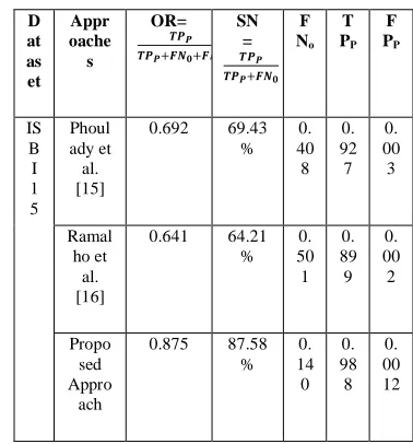
To validate this proposed approach, estimation of all the quantitative measurements like sensitivity (SN),overlap ratio (OR)nuclei based false negative rate (FNo) i.e. the number of nuclei overlooked by this method, nuclei based true positive rate (TPP) i.e. the number of correctly recognized nuclei from the cell masses, and false positive rates (FPP)i.e. the number of incorrectly recognized nuclei is being done. Table 1 is the summary of numerically calculated values of this proposed methodology and other state-of-the-art methodologies [15,16].
TABLE I
QUANTITATIVE MEASURES ON THE TRAINING SETS (ISBI 15)) AND COMPARATIVE ANALYSISWITH OTHER
APPROACHES
V. Results
The quality of the results of this proposed approach is shown in fig. 2. Very close to exact boundary is detected for multiple nuclei in these cervical MRI images. All the techniques proposed in this approach are implemented in MATLAB. The results show the successful detection of overlapped cervical cells.
D at as et Appr oache s OR= 𝑻𝑷𝑷 𝑻𝑷𝑷+𝑭𝑵𝟎+𝑭𝑷𝑷 SN = 𝑻𝑷𝑷 𝑻𝑷𝑷+𝑭𝑵𝟎 F No T PP F PP IS B I 1 5 Phoul ady et al. [15]
0.692 69.43
% 0. 40 8 0. 92 7 0. 00 3 Ramal ho et al. [16]
0.641 64.21
% 0. 50 1 0. 89 9 0. 00 2 Propo sed Appro ach
0.875 87.58
(a)
(b)
(c)
(d)
(e)
(f)
Fig.2: (a),(d) InputMRI Images, (b), (e)Extracted Cell masses, (c), (f) Segmentation of overlapped cells.
VI. Conclusion and Future Works
It can be summarized that this paper has proposed an advanced segmentation algorithmfor overlapped cervical carcinoma cells obtained fromMRI images. For detection of cell masses, superpixels aregenerated and superpixels undergo withmultilevel thresholding to extract the carcinoma cell masses. Cell nuclei are located by applying clustering thresholding on the cell masses. Now, for the segmentation of multiple overlapped cervical cells, an integrated active contour method is applied. Shape, edge, and the region of the single cell is segmented then all are integrated in one methodology to well segmentmultiple overlapped cells with nuclei. The sensitivity of proposed approach is nearly 90% that shows the accuracy of the approach in the comparison of other available state-of-the-art approaches. In qualitative results, boundaries of overlapped cervical carcinoma cells are detected with fine edges. This work can be extended as nuclei extraction in multi-focal images.
VII. References
[1] http://www.worldcancercongress.org/
[2] Sironi S, Zanello A, Rodighiero MG, Vanzulli A, Taccagni GL, Belloni C, Del Maschio A. Invasive carcinoma of the cervix uteri (Stage IB-IIB). Comparison of CT and MR for the assessment of the parametrium.
Radiol Med.81(5):671-7, May 1991.
Comput AssistTomogr, 17(4):633-40, Jul-Aug1993. [4] Subak LE, Hricak H, Powell CB, Azizi E, Stern JL.
Cervical Carcinoma: Computed tomography and Magnetic resonance imaging for preoperative staging. Obstetrics & Gynecology.86(1):43-50, July 1995.
[5] Parkin DM, Bray FI, &Devesa SS. Cancer burden in the year 2000. The global picture. European journal of cancer, 37(8):S4-66, Oct 2001.
[6] Chan TF, and Vese LA. Active contours without edges.
IEEE transactions on Image Processing. 10(2):266-277, Feb 2001.
[7] Rousson M, and Paragios N. Shape Priors for Level Set Representations. European Conference on Computer Vision. LNCS 2351: 78-92, May 2002.
[8] Jeronimo J, and Schiffman M. A Tool for Collection of Region Based Data from Uterine Cervix Images for Correlation of Visual and Clinical Variables Related to Cervical Neoplasia. Computer based medical systems. 17th IEEE Symposium. 558-562, June 2004.
[9] Matas J, Chum O, Urban M,Pajdla T. Robust wide-baseline stereo from maximally stable extremal regions.
Image and Vision Computing. 22(10): 761-767, 2004. [10] Arora S, Acharya J, Verma A, and PrasantaPanigrahi K.
Multilevel thresholding for image segmentation through a fast statistical recursive algorithm. Elsevier, Pattern Recognition Letters, 29: 119-125, 2008.
[11] Radau P, Lu Y, Connelly K, Paul G, Dick AJ, and Wright GA. Evaluation framework for algorithms segmenting short axis cardiac MRI. The MIDAS Journal – Cardiac MR Left Ventricle Segmentation Challenge. 2009. [12] Achanta R, Shaji A, Smith K, Lucchi A, Fua P, and
Susstrunk S. Slic superpixels compared to state-of-the-art superpixel methods. IEEE Transactions on Pattern analysis Machine Intelligence. 34(11): 2274-2282, Nov
2012.
[13] Nosrati MS, and Hamarneh G. A variational approach for overlapping cell segmentation. Overlapping Cervical Cytology Image Segmentation Challenge – ISBI 2014. 2014.
[14] Ushizima DM, Bianchi AGC, and Carneiro CM. Segmentation of subcellular compartments combining superpixel representation with voronoi diagrams.
Overlapping Cervical Cytology Image Segmentation Challenge – ISBI 2014, 2014.
[15] Phoulady HA, Goldgof DB, Hall LO, and Mouton PR. An approach for overlapping cell segmentationin multi-layer cervical cell volumes. Overlapping Cervical Cytology Image Segmentation Challenge – ISBI 2015, 2015. [16] Ramalho GLB, Ferreira DS, Bianchi AGC, Carneiro CM,
Medeiros FNS, and Ushizima DM. Cell reconstruction under voronoi and enclosing ellipses from 3d microscopy.
Overlapping Cervical Cytology Image Segmentation Challenge – ISBI 2015, 2015.
[17] Lu Z, carneiro G, and Bradley A. An improved joint optimization of multiple level set functions for the segmentation of overlapping cervical cells.IEEE Transactions on Image Processing. 24(4): 1261-1272, April 2015.
[18] PatilShweta R, and Patil VS. Similarity measurement using shape feature for image retrieval.
IJETT-ICGTETM-N5. Jan 2016.