University of Pennsylvania
ScholarlyCommons
Publicly Accessible Penn Dissertations
Fall 12-22-2010
The Pathological and Biochemical Characterization
of Leucine-Rich Repeat Kinase 2 in Parkinson’s
Disease
Jason P. CovyUniversity of Pennsylvania, [email protected]
Follow this and additional works at:http://repository.upenn.edu/edissertations Part of theNeurosciences Commons
This paper is posted at ScholarlyCommons.http://repository.upenn.edu/edissertations/1553
For more information, please [email protected]. Recommended Citation
Covy, Jason P., "The Pathological and Biochemical Characterization of Leucine-Rich Repeat Kinase 2 in Parkinson’s Disease" (2010). Publicly Accessible Penn Dissertations. 1553.
The Pathological and Biochemical Characterization of Leucine-Rich
Repeat Kinase 2 in Parkinson’s Disease
Abstract
Parkinson's disease (PD) is a debilitating and progressive neurodegenerative disorder that affects over 6 million people worldwide. Despite being the most common movement disorder in the U.S., there is still no effective treatment for halting the progression of disease. While generally considered a sporadic and idiopathic disorder, a number of mutations in genetic loci causal for PD have provided valuable insight into the etiology of disease. Mutations in the gene for leucine-rich repeat kinase 2 (LRRK2) are the single most common cause of both familial and sporadic forms of PD. LRRK2 is a large 2527‐amino acid protein with several distinct domains: leucine-rich repeats, Ras-like GTPase domain, C-terminal of ROC (COR) domain, serine/ threonine kinase domain, and WD40 repeats; however the understanding of LRRK2 function or how its aberration may lead to disease is still rudimentary. In 2006, we identified and characterized 3 patients with the G2019S LRRK2 mutation; however this search was limited to a few sequenced exons. An expanded screen identified 2 new patients with LRRK2 mutation, and the clinical and neuropathological findings for all these patients are provided herein. A novel system to express and purify the full-length protein with active in-vitro kinase activity revealed that the most common disease causing alteration (G2019S) markedly increases kinase activity. This highlighted overactive kinase activity as a possible intervention point for its aberrant effects. Screening for molecular inhibitors of kinase activity identified several compounds (Gö6976, K252a, and staurosporine) that share a basic indolocarbazole structure, which act as potent inhibitors of LRRK2 at low nanomolar concentrations. Increased kinase activity in the absence of outside factors is unlikely to account for the pathogenicity of the G2019S mutation. A more careful analysis of LRRK2 kinase activity revealed that this mutation, relative to the wildtype and other pathogenic mutations, may act in a novel pathway leading to disease by disrupting LRRK2 sensitivity to manganese kinase inhibition. Furthermore, based on kinetic data, we propose a novel hypothesis that LRRK2 may act as a cellular sensor of manganese levels, and disruption of this function may contribute to disease.
Degree Type
Dissertation
Degree Name
Doctor of Philosophy (PhD)
Graduate Group
Pharmacology
First Advisor
Dr. Benoit I. Giasson
Keywords
LRRK2, Parkinson's disease, neurodegeneration
Subject Categories
Neurosciences
THE PATHOLOGICAL AND BIOCHEMICAL CHARACTERIZATION OF
LEUCINE-RICH REPEAT KINASE 2 IN PARKINSON’S DISEASE
JASON P. COVY
A DISSERTATION
in
PHARMACOLOGY
Presented to the faculties of the University of Pennsylvania
in
Partial Fulfillment of the Requirements for the Degree of Doctor of Philosophy
2010
Supervisor of Dissertation
Signature ______________________________ Benoit I. Giasson, Ph.D.
Graduate Group Chairperson
Signature ______________________________ Vladimir R. Muzykantov, M.D., Ph.D.
Dissertation Committee:
Randall N. Pittman, Ph.D. (Committee Chair) Professor of Pharmacology Nancy Bonini, Ph.D., Professor of Biology and Neuroscience
ii
DEDICATION
To my family,
Your love and support has been the foundation for every day that has passed,
iii
ACKNOWLEDGMENTS
To reflect on everyone who has shaped my time here would require more room than the
thesis to follow, but I would like to highlight a few of these relationships:
To my friends back home, thank you for your constant encouragement. You’ve sought
higher education, gotten engaged or married, won softball leagues, began having kids,
and more. You’ve started so many new chapters in your life, yet you’ve always had time
to check in on mine. Thank you for your support and love. To my friends in
Philadelphia, thank you for your direction. You have provided insightful academic
conversations over lunch, opened new opportunities through Wharton’s Business Plan
Competition, broadened my views through TAL and others, and taught me discipline and
dedication at PMT/PJJ. You have contributed to my growth and development in ways
that I will always be grateful for.
I would like to specifically thank the members of the Giasson lab, past and present:
LaTasha Boeting, Irene Bukh, Kristel Emmer, Chenere Ramsey, and Elisa Waxman.
You have been a constant stream of support and entertainment in and outside of the lab.
With all sincerity, I know my time here has been enriched because of you.
I would especially like to thank the members of my committee. Virginia, I chose to come
to UPenn because of you, and after having the opportunity to work under your guidance,
I know that this was the best decision I could have made. Nancy, you provided some of
iv
you changed the way I approached my science. Harry, sitting down with you in your
office and forgoing my prepared presentation to talk about my work and the overall
literature was surprisingly one of the most enjoyable experiences of my graduate career.
Randy, there’s nothing nice I can say about you that hasn’t been said before. I’ve been
fortunate enough to have you down the hall from me, and I am thankful for the
encouragement and support you’ve always provided.
Most importantly I would like to thank my advisor, Benoit Giasson. I can think of no
better role model for the level of scientific excellence that I would like to operate on, than
you. Your tireless work ethic and vast knowledge is something I greatly admire, and I
am forever grateful that you have directed that energy towards me. The only thing I have
to be more appreciative than your guidance as a mentor is your camaraderie as a friend.
Whether it be on the bench, or at the squash court, I am glad that our relationship has
extended outside the academic realm. Thank you for everything.
v
ABSTRACT
THE PATHOLOGICAL AND BIOCHEMICAL CHARACTERIZATION OF
LEUCINE-RICH REPEAT KINASE 2 IN PARKINSON’S DISEASE
Jason P. Covy
Advisor:
Benoit I. Giasson Ph.D.
Parkinson’s disease (PD) is a debilitating and progressive neurodegenerative disorder that
affects over 6 million people worldwide. Despite being the most common movement
disorder in the U.S., there is still no effective treatment for halting the progression of
disease. While generally considered a sporadic and idiopathic disorder, a number of
mutations in genetic loci causal for PD have provided valuable insight into the etiology of
disease. Mutations in the gene for leucine-rich repeat kinase 2 (LRRK2) are the single
most common cause of both familial and sporadic forms of PD. LRRK2 is a large
2527‐amino acid protein with several distinct domains: leucine-rich repeats, Ras-like
GTPase domain, C-terminal of ROC (COR) domain, serine/threonine kinase domain, and
WD40 repeats; however the understanding of LRRK2 function or how its aberration may
lead to disease is still rudimentary.
In 2006, we identified and characterized 3 patients with the G2019S LRRK2 mutation;
vi
2 new patients with LRRK2 mutation, and the clinical and neuropathological findings for
all these patients are provided herein. A novel system to express and purify the
full-length protein with active in-vitro kinase activity revealed that the most common disease
causing alteration (G2019S) markedly increases kinase activity. This highlighted
overactive kinase activity as a possible intervention point for its aberrant effects.
Screening for molecular inhibitors of kinase activity identified several compounds
(Gö6976, K252a, and staurosporine) that share a basic indolocarbazole structure, which
act as potent inhibitors of LRRK2 at low nanomolar concentrations.
Increased kinase activity in the absence of outside factors is unlikely to account for the
pathogenicity of the G2019S mutation. A more careful analysis of LRRK2 kinase
activity revealed that this mutation, relative to the wildtype and other pathogenic
mutations, may act in a novel pathway leading to disease by disrupting LRRK2
sensitivity to manganese kinase inhibition. Furthermore, based on kinetic data, we
propose a novel hypothesis that LRRK2 may act as a cellular sensor of manganese levels,
vii
Table of Contents
DEDICATION ...ii
ACKNOWLEDGMENTS ... iii
ABSTRACT ... v
LIST OF FIGURES AND TABLES... x
CHAPTER ONE GENERAL INTRODUCTION ... 1
1. Parkinson’s Disease ... 2
1.1 Pharmacology and Anatomy of the Nigrostriatal System Relevant to PD ... 3
2. Pathogenesis of PD ... 5
2.1 Mitochondrial Damage ... 5
2.2 Environmental Risks and Toxins ... 7
2.3 Oxidative Stress ... 8
2.4 Ubiquitin Proteasome System ... 8
2.5 Alpha-Synuclein: The First Link to Protein Dysfunction and Disease ... 9
2.6 The Tau protein in PD ... 10
3. Genetic findings in Parkinson’s disease ... 11
3.1 PARK1/4 : Alpha-synuclein ... 13
3.2 PARK2: Parkin ... 14
3.3 PARK6: Pink1 ... 15
3.4 PARK9: ATP13A2 ... 16
3.5 PARK8: Leucine-Rich Repeat Kinase 2, Dardarin ... 16
4. The Role of LRRK2 ... 18
4.1 Expression and Localization ... 18
4.2 Kinase Domain Activity ... 20
4.3 ROC Activity: GTP Binding and GTPase Activity ... 23
4.4 Non-Enzymatic LRRK2 Domains ... 27
5. Mutations in LRRK2 and its Link to PD ... 30
5.1 The Pathology of Cases with LRRK2 Mutation ... 30
viii
5.3 The Common G2019S Pathological Mutation Increases Kinase Activity ... 35
5.4 LRRK2 Kinase Activity is Linked to Neurotoxicity ... 36
5.5 Mutations Outside of the Kinase Domain can Affect Kinase Activity ... 41
6.Scope of this Research ... 43
CHAPTER TWO CLINICAL AND PATHOLOGICAL CHARACTERISTICS OF PATIENTS WITH LEUCINE-RICH REPEAT KINASE-2 MUTATIONS ... 45
ABSTRACT ... 46
INTRODUCTION ... 47
MATERIALS AND METHODS ... 49
Antibodies ... 49
Molecular Genetic Analysis of LRRK2 ... 50
Immunohistochemistry and Immunofluorescence ... 51
RESULTS ... 52
Genetic Analysis of LRRK2: ... 52
Clinical and Pathological Findings in Cases Harboring the R793M and L1165P Mutations in LRRK2: ... 53
Phosphorylation of S129 in α-Synuclein in the Pathological Inclusions of Patients with LRRK2 Mutations ... 55
TDP-43 Cytoplasmic Inclusions in Patients with LRRK2 Mutations... 56
DISCUSSION ... 57
CHAPTER THREE IDENTIFICATION OF COMPOUNDS THAT INHIBIT THE KINASE ACTIVITY OF LEUCINE-RICH REPEAT KINASE 2 ... 62
ABSTRACT ... 63
INTRODUCTION ... 64
MATERIALS AND METHODS ... 65
Materials ... 65
Cell Culture ... 66
LRRK2 Expression Constructs ... 66
ix
In-Vitro LRRK2 Kinase assays ... 67
RESULTS AND DISCUSSION ... 69
Generation and Characterization of Active Full-Length GST-LRRK2. ... 69
Recombinant GST-LRRK2 was used to Screen a Range of Defined Kinase Inhibitors. ... 72
CHATPER FOUR THE PATHOGENIC G2019S MUTATION DISRUPTS SENSITIVITY OF LEUCINE-RICH REPEAT KINASE 2 TO MANGANESE KINASE INHIBITION ... 75
ABSTRACT ... 76
INTRODUCTION ... 77
MATERIALS AND METHODS ... 79
Materials ... 80
Cell Culture ... 80
LRRK2 Expression Constructs ... 80
Western Blotting Analysis ... 81
In-Vitro LRRK2 Kinase Assays ... 82
RESULTS ... 83
DISCUSSION ... 97
CHAPTER FIVE SUMMARY AND FUTURE DIRECTIONS ... 103
Patients With LRRK2 Mutations Present with Classical and Nonclassical Pathology, Regardless of Mutation Location ... 104
LRRK2 is an Active Kinase that can be Targeted for Possible Therapeutic Intervention ... 106
The G2019S Mutation May Alter LRRK2 Function, in Addition to Overall Activity 108 Future Directions for Studying LRRK2 in PD ... 111
x
LIST OF FIGURES AND TABLES
Chapter One
Figure 1-1. Neurochemical pathways of the basal ganglia involved in PD.
Table 1-1. Genetic loci implicated in Parkinson’s disease.
Figure 1-2. Human ROCO proteins.
Figure 1-3. Biochemical characterization of LRRK2 protein in human and mouse tissue.
Figure 1-4. Histological characterization of patients with LRRK2 mutations.
Figure 1-5. Characterization of LRRK2 antibodies.
Figure 1-6. LRRK2 aggregates in transfected COS-7 cells.
Chatper Two
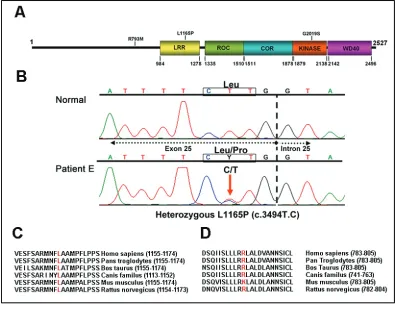
Figure 2-1. Identification of patients with LRRK2 mutations and sequence alignment of amino acids surrounding the mutations.
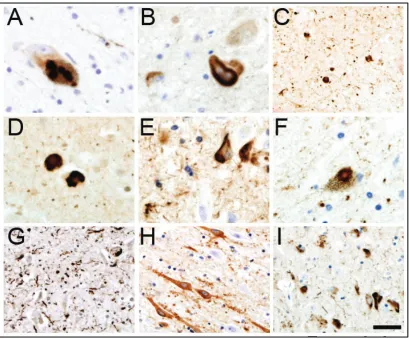
Figure 2-2. Histological characterization of patients with LRRK2 mutations.
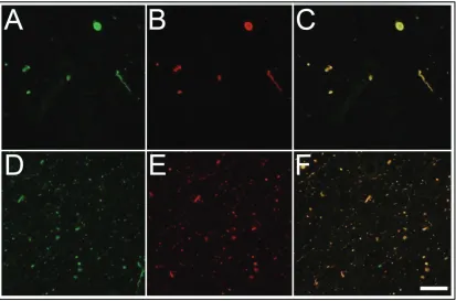
Figure 2-3. Double-labeling immunofluorescence analysis of α-synuclein phosphorylated
at S129 in pathological inclusions of patients with LRRK2 mutations
Figure 2-4. TDP-43 cytoplasmic inclusions in patients with LRRK2 mutations
Chapter Three
Figure 3-1. Characterization of recombinant GST-LRRK2 activity.
Figure 3-2. Analysis of GST-LRRK2 kinase activity in the presence of various inhibitors.
Chapter Four
Figure 4-1. Characterization of recombinant LRRK2 kinase activity
Figure 4-2. Analysis of wildtype and G2019S LRRK2 kinase activity in the presence of various inhibitors.
Figure 4-3. Substrate specificity of wildtype and G2019S LRRK2.
Figure 4-4. Concentration dependent affects of Mg2+ and Mn2+on the kinase activity of
xi
Figure 4-5. Concentration dependent affects of Mg2+ and Mn2+on the kinase activity of
wildtype, R1441C, G2019S, and I2020T LRRK2 on Nictide phosphorylation.
Figure 4-6. Concentration dependent affects of Mg2+ and Mn2+ on the
autophosphorylation activity of wildtype and G2019S LRRK2.
Figure 4-7. Kinetic characteristics of wildtype and LRRK2 mutations while varying the concentration of LRRKtide as a substrate.
Figure 4-8. Kinetic characteristics of wildtype and LRRK2 mutations while varying the concentration of ATP as a substrate.
Figure 4-9. Analysis of effects of sub-stoichiometric concentrations of Mn2+ on wildtype
1
CHAPTER ONE
2
1. Parkinson’s Disease
Parkinson’s disease (PD) is the most prevalent movement disorder in the United States
(190). It affects over 6 million people worldwide, and is the second most common
neurodegenerative disorder after Alzheimer’s disease (104, 292). PD primarily affects
those over the age of 55, and the incidence of disease sharply rises after the age of 65
with as many as 50,000 new cases identified each year in the United States (236).
The disease was first described in 1817 by James Parkinson in a paper entitled “An Essay
on the Shaking Palsy”, and his clinical observations still serve as the basis for disease
classification. PD presents as a broad clinical spectrum with the cardinal symptoms of
trembling at rest, rigidity, bradykinesia (slowness of movement), postural instability, and
a therapeutic responsiveness to L-dopa, the precursor of dopamine (292). A definite
diagnosis can only be confirmed by pathological post-mortem analysis (104), as a
number of other closely related neurological disorders (termed Parkinsonisms) present
with similar PD-like symptoms. Pathologically, PD is characterized by a loss of
(primarily dopaminergic) neurons in the substantia nigra pars compacta (SNpc) with
alpha-synuclein positive proteiniacous inclusions, known as Lewy bodies (LBs) and
Lewy neurites (LNs), present in some of the surviving neurons (58, 92, 93). During the
normal aging process, approximately 0.1-0.2% of the 400,000 dopaminergic neurons in
this area are lost per year, however, in the case of patients with PD, this rate is greatly
accelerated (40, 327). Symptoms of the disease manifest when ~70-80% of these
neurons have been lost (63, 252). Currently, the causes and mechanisms of the selective
3
1.1 Pharmacology and Anatomy of the Nigrostriatal System Relevant to PD
The progressive degeneration within the substantia nigra (SN) and subsequent loss of
dopaminergic output to the striatum is the major factor contributing to the disruption of
motor control in PD. The SN, caudate nucleus, putamen, globus pallidus (GP), and
subthalamic nucleus (STN) are the five major nuclei of the basal ganglia, which,
alongside the thalamus, receive and process input and provide feedback to the cerebral
cortex for the generation and initiation of voluntary movement.
The principal circuit associated with the basal ganglia is a loop projecting from the
cerebral cortex to the basal ganglia, to the thalamus, and back to the cortex (Figure 1-1).
These connections may be accomplished through two overlapping but distinct circuits:
the direct and indirect pathways. These pathways have competing effects on movement,
and the balance between them is involved in establishing and regulating tone. The direct
pathway facilitates movement and projects from the cortex to the striatum (the caudate
nucleus and the putamen), then to the internal segment of the GP (GPi), before
proceeding to the thalamus, and back to the cortex. The indirect pathway inhibits
movement and also projects from the cortex, but goes to the external segment of the GP
(GPe) followed by the STN before connecting to the GPi to complete the loop to the
thalamus and cortex. Axons leaving the striatum and GP use γ-aminobutyric acid
(GABA) as a neurotransmitter to make inhibitory synapses. Pallidal neurons are
tonically active, therefore inhibiting parts of the thalamus.
The SN projects to all areas of the striatum in a point-to-point fashion by way of very fine
4
to the dorsal putamen, are those predominately involved in motor coordination. This area
of the SNpc degenerates earlier and to a greater extent than other regions within the SN,
and destruction of this nigrostriatal pathway is the major factor causing the disruption of
motor control in PD. The SN pars reticulata (SNr) utilizes GABA while the SNpc uses
dopamine as its neurotransmitters. The SNpc projects to the striatum, exciting some
striatal neurons through D1 and D5 receptors, and inhibiting others through D2-D4
receptors. The striatum and STN both project to the SNr, which projects inhibitory
synapses to the thalamus. Loss of dopamine input to the basal ganglia from the SNpc
causes increased inhibition of the ventral anterior nucleus of the thalamus, which sends
excitatory glutamatergic projections to the motor cortex, leading to hypokinesia.
Supplementing the loss of dopamine with the dopamine precursor L-dopa remains the
5
Figure 1-1. Neurochemical pathways of the basal ganglia involved in PD. (Left) The neuroanatomy of the basal ganglia. Areas of degeneration are colored in yellow. (Right) The neurocircuitry of the basal ganglia’s direct and indirect pathways are both represented. Excitatory synapses are denoted by black arrows, while inhibitory synapses are denoted by red arrows. Figure adapted from Lozano et al (190).
2. Pathogenesis of PD
PD is primarily considered a sporadic and idiopathic disorder. An early study examining
the concordance rates of PD in 19,842 monozygotic and dizygotic male twins bolstered
this point of view when genetic factors played were found to play no major role in
causing typical PD (313). Generally, PD was believed to be caused by environmental
factors or toxins; a view that was greatly strengthened in 1982 through the discovery of a
small number of PD cases reported in a younger cohort of patients (191). Upon close
investigation of these patients, it was found that they all had contracted PD through use of
a tainted source of synthetic heroin. A faulty step in the purification process led to the
byproduct 1-methyl-4-phenyl-1,2,3,6-tetrahydropyridine (MPTP), which was found to be
a selective neurotoxin for dopaminergic neurons.
6
MPTP is metabolically converted by glia into the active metabolite MPP(+) (246, 272).
Attributed to its structural similarity to dopamine, MPP(+) is a selective substrate for the
dopaminergic transporter, and upon uptake by dopaminergic neurons, it can inhibit
complex I of the electron transport change, resulting in a modest decline of ATP and the
generation of reactive oxygen species (ROS) (80, 189, 301). As neurons are highly
metabolically active, mitochondrial impairment can be devastating to their function.
Furthermore, the mitochondria is a key area for regulation of apoptosis and cell survival
(188, 247).
Rotenone, an insecticide that also inhibitrs complex 1, has been shown to cause selective
loss of dopaminergic neurons in the SNpc, alpha-synuclein positive fibrillar inclusions,
and behavioral changes consistent to PD upon chronic intravenous administration in rats
(although, in this model, the depletion of striatal neurons and development of tau-positive
inclusions in cortical neurons have also been reported, inconsistent with PD) (22).
However, a more recent mouse model with a disruption in the gene for mitochondrial
transcription factor A (Tfam) in dopaminergic neurons more closely resembles the
etiological progression of PD (100). This conditional knockout has reduced
mitochondrial DNA expression and respiratory chain function in midbrain dopaminergic
neurons, intraneuronal inclusions and dopamine cell death, and adult onset impairment of
7
2.2 Environmental Risks and Toxins
The greatest risk factor for PD is age (226, 238, 330). As previously stated, nigrostriatal
loss of neurons is about 0.1-0.2% per year, however this number increases with age (40).
Outside of this common risk factor, there are a number of other not-so-common factors
that confer increased risk of Parkinsonism and PD. These range from rare insecticides
like paraquat, to more ubiquitous metals such as manganese (16, 181, 312).
The pesticide paraquat has some structural similarity to MPTP, and has been shown in
rats to cross the blood brain barrier and to lead to nigral dopaminergic cell demise (37).
The metabolism of paraquat leads to the production of reactive semiquinones and ROS,
which can induce oxidative stress and damage to the cell (227, 228) (discussed further
below). Manganese is an essential trace mineral necessary for normal development and
biological function (277). It is mainly taken in through the diet, and toxic levels are
usually not reached unless individuals experience dramatic reductions in excretion due to
liver failure (177); however, overexposure has been found in miners and welders (59,
268). Manganese causes preferential damage to the gloubus pallidus, which may be due
in part to the ability of manganese to activate this area’s glutamatergic machinery, which
may potentiate overall manganese cytotoxicity (14, 335). Manganese can initiate
apoptosis by disrupting the mitochondria, where it is rapidly taken up within the cell
(103). Here, it can promote disruption of complex I, calcium accumulation and
subsequent activation of the permeability transition pore, and activation of caspase 3
8
neurons as the dopamine transporter may also be involved in the accumulation of
manganese (8, 85).
2.3 Oxidative Stress
Post-mortem analysis of PD brains show increased lipid peroxidation, oxidative
modifications to proteins and DNA, depletion in levels of antioxidants such as
glutathione, and high levels of iron (71, 289); all markers of oxidative stress.
Dopaminergic neurons may already be subject to higher levels of oxidative stress from
the auto-oxidation and catabolism of dopamine, which produces electrophilic
semiquinones and quinones (90, 162). Monoamine oxidase, which is involved in the
breakdown of dopamine and other bicyclic amines, produces H202 as a normal by-product
of its metabolic process. Additionally, the auto-oxidation of dopamine into melanin can
produce reactive oxygen species (20, 128).
2.4 Ubiquitin Proteasome System
Protein deposits are common to many neurodegenerative disorders, and are usually the
result of decreased solubility, improper protein folding, and/or dysfunction in protein
clearance by the ubiquitin proteasome system (UPS) (237). The main function of the
UPS is to identify, ubiquitinate, and proteolytically degrade intracellular proteins (44,
333). Failure to do so causes protein accumulation which may lead to toxicity and
ultimately cell death (126). Within the context of PD, the proteinacious inclusions
known as LBs are rife with polyubiquitinated proteins, suggesting an inability of the
proteasome to degrade these proteins (319). Within the SN of PD patients, reports have
9
controls, as well as other brain regions, and there appears to be a selective loss of 26/20S
proteasome alpha-subunits (229, 230). ATP is required for the proper assembly and
function of the 26S proteasome complex (144), and both the 26S and 20S are selectively
vulnerable to oxidative stress (38). Altogether, mitochondrial deficits in complex I and
the generation of ROS both present problems for the UPS, making this a key area
affected in PD.
2.5 Alpha-Synuclein: The First Link to Protein Dysfunction and Disease
A number of protein candidates have been associated with PD, however the most well
known and prominent is the synuclein protein. Synuclein was originally identified in
1998 from Torpedo californica (221), and its name was derived at the time from its
apparent localization to the nuclear envelope of neurons as well as presynaptic nerve
terminals. There are three members of the synuclein family of proteins: alpha-, beta-, and
gamma-synuclein. They range in weight from 14-20 kDa, however the alpha and beta are
more closely related than the gamma-synuclein (46).
Alpha-synuclein is a 140 amino acid protein with three distinct regions: an amphiphatic
N-terminal region, a central hydrophobic region, and a highly acidic and proline-rich
region. The protein is natively unfolded and assumes a random-coil formation in the
cytosol, but will adopt a secondary structure upon binding to the membrane, or during the
process of aggregation (64, 83, 341). Shortly after PD associated mutations were
identified in alpha-synuclein (discussed below), an antibody developed against the
protein showed positive staining in LBs and LNs.(298, 299) It soon became clear that
10
polymerization of soluble alpha-synuclein into amyloid fibrils form the main structures of
these cellular inclusions (161, 325).
In an environment of oxidative stress, alpha-synuclein can undergo several
post-translational modifications that influence aggregation: tyrosine nitration, methionine
oxidation, and dopamine adduct formation (106, 184, 348). Oxidative cross-linking at
tyrosine residues can form alpha-synuclein dimers, which could be toxic in cellular
models (151, 185, 352). Little is known of the actual role alpha-synuclein plays within
the central nervous system, although it has been implicated in the function of the Golgi
apparatus, vesicle trafficking, and SNARE complexes as a molecular chaperone (2, 49,
317). Deletion of alpha-synuclein in mice does not lead to disease, thereby suggesting a
toxic gain of function for the protein itself, however even more controversial is the extent
that the alpha-synuclein proteinacious inclusions may play in PD; whether it is an active
toxin, a saving sponge, or just a passive artifact of another pathological pathway.
Regardless of the role, alpha-synuclein has a major presence in the pathology and
possibly pathogenesis of disease.
2.6 The Tau protein in PD
Tau is another dominant protein found in protein aggregates that are associated with
neurodegenerative disorders (196). Tau is part of the microtubule-associated protein
(MAP) family whose main function is to modulate the stability of microtubules. In
neuronal cells, tau expression is highest in the axons of neurons, however low levels can
also be detected in oligodendrocytes and astrocytes. Tau acts to assemble and stabilize
11
vesicles and organelles along microtubules, and morphogenesis (51, 281). Like
synuclein, tau can aggregate to form intracellular and extracellular protein inclusions, and
is a pathological hallmark in a variety of disorders including Alzheimer’s disease,
frontotemporal dementia, and Pick’s disease. In regards to PD, tau pathology has been
found in some LBs of cases with idiopathic PD (11), as well as other Parkinsonisms such
as progressive supranuclear palsy and corticobasal degeneration (300). In vitro,
alpha-synuclein and tau synergistically act to promote the fibrillization of each other (107), and
alpha-synuclein can bind to tau to stimulate tau phosphorylation by protein kinase A
(163). In vivo, mice expressing the aggregate enhancing A30P mutation of
alpha-synuclein accumulate hyperphosphorylated tau suggestive of an early or pre-aggregated
form, concomitant with alpha-synuclein aggregation (94). Together, these findings
suggest that alpha-synuclein may interact with tau to cause pathological changes in
disease.
3. Genetic findings in Parkinson’s disease
The discovery of various gene defects associated with PD has revolutionized the
mechanisms of disease pathogenesis. Since the previously mentioned twin study, a
number of specific genetic candidates have shed light on potential molecular pathways
leading to degeneration and malfunction of the nigrostriatal pathway. Linkage studies
have implicated components of protein overexpression leading to aggregation, alongside
dysfunction of the ubiquitin proteasome pathway exacerbating issues of protein
post-12
translational modification, and metal homeostasis. 5-10% of patients with PD are known
to have a monogenic form of the disease, and at least 15 genetic loci have been linked to
PD, of which 10 have been mapped to a specific gene (Table 1-1) (197, 344).
Locus
(gene) Map Position Protein Putative function Inheritance pattern
PARK1/ PARK4 SNCA
4q21 alpha-synuclein Presynaptic protein, neurotransmission, vesicle recycling
AD/EOPD with rapid progression and dementia, sporadic
PARK2
PARKIN 6q25–q27 parkin Ubiquitin E3 ligase, has neuroprotective function AR/juvenile and EOPD with slow progression, dystonia; sporadic
PARK3? 2p13 sepiapterin reductase Involved in biosynthesis of
tetrahydrobiopterin (BH4) AD/LOPD, dementia
PARK5
UCH-L1 4p14 ubiquitin hydrolase isozyme L1 (UCH-carboxyl-terminal L1)
Ubiquitin hydrolase AD/LOPD
PARK6
PINK1 1p35–p36 PTEN-induced putative kinase 1 (PINK1) Mitochondrial S/T-protein kinase, has neuroprotective function AR/EOPD with slow progression, tremor
PARK7
DJ-1 1p36 DJ-1 Chaperone, neuroprotective, RNA binding antioxidant, AR/EOPD, dystonia, psychiatric symptoms
PARK8
LRRK2 12q12 Leucine-rich repeat kinase 2 (LRR2) Protein kinase, protect cells from stress-induced mitochondrial dysfunction
AD/LOPD, tremor
PARK9 ATP13A 2
1p36 ATP13A2/probable
cation-transporting ATPase 13A2 Lysosomal ATPase Probable cation-transporting AR/juvenile Kufor-Rakeb syndrome, EOPD
PARK10
? 1p32 UPS24/ubiquitin carboxyl-terminal hydrolase 24 Involved in the ubiquitin-dependent proteolytic pathway Unclear/LOPD
PARK11
? 2q36–q37 GIGYF2/PERQ amino acid-rich with GYF domain-containing protein 2
Involved in regulation of tyrosine
kinase receptor signaling AD/LOPD
PARK12 Xq12–
q25 Unknown Unknown Unclear
PARK13 2p13 HTRA2/serine protease
HTRA2, mitochondrial Serine protease, may be involved in mitochondrial dysfunction Unclear
PARK14
? 22q13.1 PLA2G6/85 kDa calcium-independent phospholipase A2
Catalyzes the release of fatty
acids from phospholipids AR/juvenile,levodopa-responsive dystonia parkinsonism
PARK15
? 22q12–q13 FBXO7/F-box only protein 7 Involved in the ubiquitin-dependent proteolytic pathway AR/EO, parkinsonian-pyramidal syndrome
Not
assigned 2q22–q23 NR4A2/nuclear receptor subfamily 4 group A member 2
Probable nuclear receptor. May function as a general coactivator of gene transcription
AD?
Not
assigned 5q23.1–q23.3 SNCAIP/synphilin-1 Interact with alpha-synuclein, substrate of parkin, part of LBs Unknown
Not
13
Table 1-1. Genetic Loci implicated in Parkinson’s disease. Known and unknown genetic loci are listed alongside their chromosomal mapping, gene and protein product, putative protein function, and model of genetic inheritance. Question marks indicate candidates still under investigation. AD, autosomal-dominant; AR, autosomal-recessive; EO, early onset; LO, late onset; PD, Parkinson’s disease; PM, point mutations; HM, heterozygous mutations. Table adapted from Shadini et al 2010 (285).
3.1 PARK1/4 : Alpha-synuclein
SNCA (PARK1:alpha-synuclein) was the first gene identified to have a causal role in PD
(264). In 1990, Golbe et al reported a large family with an autosomal dominant form of
parkinsonism: greater than 60 family members over the course of five generations with a
diagnoses ranging from diffuse LB disease to typical PD (120). In 1996, Polymeropoulos
and colleagues identified linkage to the long arm of chromosome 4 (262), and the next
year a 209g>a (A53T) mutation was identified in alpha-synuclein for this kindred
alongside three unrelated Greek families (264). Soon after, two additional synuclein
mutations were found: an A30P mutation in a German family (186), and an E46K
mutation in a Spanish family (351).
In vitro, alpha-synuclein can form fibrils similar to those seen in LBs (47, 345). Fibril
formation is characterized by a slow lag phase that is nucleation dependent, followed by a
faster elongation phase (345). The A53T and the E46K mutations have both been shown
to increase the rate of polymerization; however, the effects of the A30P mutation on fibril
formation in vitro are not consistent between studies (47, 109, 129). Regardless, these
findings implicate a link between alpha-synuclein aggregation and disease. This theory
was strengthened by the discovery that the PARK4 loci also mapped to alpha-synuclein
through its triplication in an Iowa kindred and a Swedish-American family with
14
with typical disease onset (41). The dominant mode of inheritance from expansion of the
synuclein gene, paired with the greater severity and earlier onset for the triplication
versus the duplication, suggested a possible “SNCA gene dosage effect” leading to PD
(293). This theory is further bolstered by in vitro data which shows that the rate of
synuclein aggregation is concentration dependent (345). Consequently, this may explain
the pathogenicity behind the A30P mutation, as it has been proposed that this mutation’s
decreased ability to bind lipids may increase its intracellular pool, thereby facilitating
greater opportunities to aggregate (164).
3.2 PARK2: Parkin
The role of reduced protein degradation in PD was further emphasized with the
identification of mutations in genes affecting the ubiquitin proteasomal degradation
system: PARK2 (PARKIN:Parkin) and PARK5 (UCH-L1). Mutations in the PARKIN
gene were discovered in 1998, and are the most common form of recessive early-onset
Parkinsonism (225). Parkin is a 465 amino acid protein which is widely expressed in the
cell bodies of neurons in the midbrain, basal ganglia, cerebral cortex, and cerebellum,
where it functions as an E3 ubiquitin ligase (66). Traditionally, E3 ligases serve to
conjugate ubiquitin molecules to specific proteins, thus targeting them for degradation by
the proteasome (recent evidence suggests ubiquitin may also function in other various
signaling cascades) (146). In vitro, Parkin has been shown to interact with the E2
ubiquitin ligase UbcH7 and 8UbcH8 to promote ubquitination, and pathological
mutations decrease its ability to conjugate ubiquitin (287). In 2005, Wang et al found
15
solubility and increased the propensity of the protein to form aggresome-like aggregates,
suggesting a mechanism leading to its loss of function (336). In vivo, Parkin knock-out
mice show impaired mitochondrial function and increased markers of oxidative stress
linked to dysregulation of multiple proteins related to oxidative phosphorylation and
oxidative damage (253). In 2006, several studies linked Parkin to another genetic
candidate in PD, PARK6 (ARPD; PTEN-induced putative kinase 1, PINK1) (45, 256).
3.3 PARK6: Pink1
After DJ-1, PINK1 was the third gene to be associated with autosomal recessive PD
(328). Pink1 is a 581 amino acid protein ubiquitously expressed in all brain regions in
both neurons and glia (at lower levels), and localizes predominately to the mitochondria
(27, 290). Structurally, the protein contains a mitochondrial targeting motif and a highly
conserved serine/threonine kinase-like domain capable of autophosphorylation in vitro
(326, 328). Subsequent studies have confirmed Pink1’s ability to undergo in-vitro
autophosphorylation, and demonstrate that several pathological missense mutations
decrease kinase activity (18). While the function of this protein remains unclear, in vitro
models show that Pink1 overexpression protects against oxidative induced apoptosis
(259, 328). In one cellular model, Pink1 protected against oxidative stress by
phosphorylating TNF receptor-association protein 1 (TRAP1) at the mitochondria (265).
Pink1 has also been implicated in the phosphorylation and regulation of the
mitochondrial protease HtrA2 (261). Through interaction with p38 and HtrA2, Pink1
may increase HtrA2 protease activity in response to stress. In vivo, Pink1 knock-out
16
calcium levels, loss of mitochondrial membrane potential, reduced synaptic dopamine
release and plasticity in the striatum, and reduced viability of cortical neuron cultures
(114, 175). In Drosophila, Pink1 knockouts develop fragmented mitochondrial cristae,
hypersensitivity to oxidative stress, and muscle and neuronal degeneration which can be
rescued by Parkin overexpression; however, Pink1 cannot rescue Parkin deletion,
indicating that Parkin may function downstream of Pink1 in a disease-relevant pathway
(45, 256).
3.4 PARK9: ATP13A2
ATP13A2 was first associated with Kufor-Rakeb syndrome, a recessive atypical
Parkinsonism, in 2006 (270). Splicing and deletion mutations resulting in truncated
forms of the protein were found in the original family, as well as a family from Chile. In
2007, three additional missense mutations were found: G504R, T12M, and G533R (72).
ATP13A2 is a large 1180 amino acid protein belonging to the lysosomal type 5 P-type
ATPase family of transporters. Recently, ATP13A2 has been shown to be protective
against alpha-synuclein induced toxicity in yeast, C. elegans, and rat primary midbrain
neurons (115). In addition, ATP13A2 may play a role in sequestering heavy metal ions
possibly by acting as a lysosomal transporter, as it exhibits protective affects in yeast
against a number of metals including manganese, cadmium, nickel, and selenium (115,
283). Currently, little else is known of this protein’s function.
3.5 PARK8: Leucine-Rich Repeat Kinase 2, Dardarin
Of all the genetic loci mapped to PD, the PARK8 locus has generated the most attention
17
kindred with an autosomal dominant mode of PD inheritance (139), and five years later
they were able to determine linkage to the long arm of chromosome 12 (96). In 2004,
separate studies simultaneously identified the LRRK2 gene in a handful of other families
responsible for the PARK8 loci (250, 355). Since then, over 75 sequence variations in
the gene have been found, and it is regarded as the most common known cause of
familial and sporadic cases of PD (60).
The gene for LRRK2 spans ~7.5Mb and contains 51 exons. It encodes a large 2,527
amino acid protein with multiple complex domains, including N-terminal leucine-rich
repeats, a GTPase ROC (Ras of complex proteins) domain followed by COR (C-terminal
of Roc) domain, a mitogen-activated protein kinase kinase kinase (MAPKKK) catalytic
domain, and C-terminal WD40 repeats (Figure 2) (224). Early studies reported the
presence of ankyrin domains located in the N-terminal portion of the protein; however,
later modeling studies do not support their existence (219). Regardless, there are a
number of sequences unique to LRRK2 found throughout the N-terminal portion of the
protein that are conserved across species.
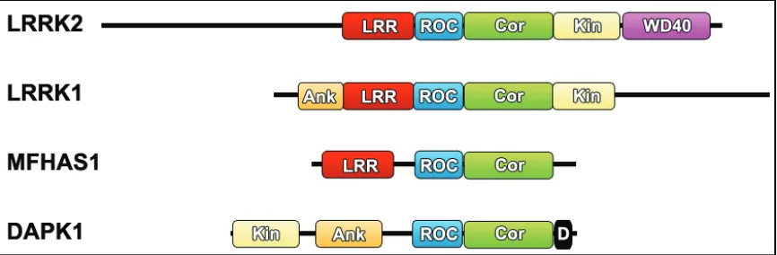
The characteristic ROC and COR domains of LRRK2 make it a member of the ROCO
protein family, alongside LRRK1, MFHAS1/MASL1, and DAPK1, which are found in
humans (Figure 1-2) (219). At least 40 other ROCO proteins have been identified in
eukaryotes and prokaryotes, and have been shown to play a role in cytokinesis, cell
18
Figure 1-2. Human ROCO proteins. Schematics to scale for each of the ROCO proteins expressed in humans with functional domains labeled. LRR, leucine-rich repeat; ROC, Ras of complex protein; Cor, C-terminal of ROC; Kin, kinase; WD40, WD40 repeat; D, death domain.
4. The Role of LRRK2
Currently, little is known of the biological function of LRRK2. Multiple studies have
attempted to elucidate its physiological binding partners, pathological kinase substrates,
nucleotide binding activity, and signaling pathways; and while a substantial amount of
information has been reported, a significant number of these findings seem to contradict
each other. However, despite these inconsistencies, a general role for LRRK2 is
beginning to emerge.
4.1 Expression and Localization
Expression of LRRK2 has been examined at the mRNA and protein level with some
minor differences. LRRK2 is found in numerous tissues including the lungs, heart, liver,
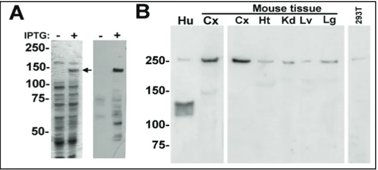
kidney, spleen, testes, and brain (Figure 1-3) (105, 205, 215). Within the CNS, LRRK2
mRNA expression is found in most regions including those affected in PD. A common
finding among in-situ hybridization studies utilizing rodent, primate, and human brain, is
19
pathogenesis of PD (101, 232). Particularly high expression is found in the cerebral
cortex (pyramidal projection neurons and various interneurons throughout most layers),
olfactory tubercle, and the striatum (output projection neurons, medium-sized spiny
neurons) (101, 291). Medium expression levels have also been identified in the
cerebellum and hippocampus, and similar results have been obtained at the protein level
(137, 148, 234).
Figure 1-3. Biochemical characterization of LRRK2 protein in human and mouse tissue. (A) (Left) Coomassie-stained gel showing the expression of recombinant His-tagged human LRRK21245-2527 protein
(~145kDa) as indicated by the arrow. (Right) Western blotting analysis showing that LRRK2 antibody AP7099b specifically detects LRRK2 protein and some breakdown products. (B) Immunoblot analysis with rabbit anti-LRRK2 antibody AP7099b detecting LRRK2 in postmortem human brain cortex (Hu) (~250kDa) and as multiple species between approximately 120 to 130kDa. In various freshly dissected mouse tissues (Cx = cortex; Ht = heart; Kd = kidney; Lv = liver; Lg = lung) and in HEK293T cells, LRRK2 is predominantly expressed as an ~250kDa protein. Figure adapted from Giasson et al (105).
Initial studies had failed to identify LRRK2 mRNA in dopaminergic neurons themselves,
specifically within the SN (101, 232). However, late in 2006, a number of new LRRK2
antibodies allowed for the detection of moderate levels of LRRK2 immunoreactivity
35S-20
labeled oligionucleotides, in lieu of the 33P-labeled methods used previously, and were
subsequently able to detect low levels of mRNA expression within the SN (148), and this
finding has since been confirmed in several other studies (137, 291, 316). Besides
differences in methodologies, it has been suggested that discrepancies in detecting
LRRK2 in the SN may result from the instability of LRRK2 mRNA, its short half-life, or
its transportation to distal sites within nigral dopaminergic pathways (149). In addition,
at the protein level, LRRK2 may have a long half-life in this population of neurons thus
requiring low levels of mRNA.
4.2 Kinase Domain Activity
Aberrant protein phosphorylation is a common finding in a variety of neurodegenerative
disorders. Alongside Pink1, LRRK2 is one of two kinases implicated in monogenetic
forms of PD. Within the family of human ROCO genes, kinase domains can be found in
LRRK1, LRRK2, and DAPK1. The DAPK1 kinase domain bears closest resemblance to
the calcium/calmodulin-dependent kinase (CAMK) family,while both LRRK genes were
classified within the tyrosine kinase-like (TKL) sub-family, with LRRK2 bearing closest
homology to the mitogen-activated protein kinase kinase kinase (MAPKKK) family (32,
219) and the receptor-interacting protein kinase (RIPK) family (130).
The study of LRRK2 kinase activity has been complicated by the varied complex
domains and overall size of the protein. As such, most studies to assess biochemical
activity have relied on smaller truncated versions, while only a few have successfully
utilized the full-length protein for qualitative analysis. The first obstacle in assessing the
21
has the ability to autophosphorylate (117, 342). Sites of autophosphoyrlation within the
kinase domain have been suggested to take place on residues T1967, T1969, T2031,
S2032, and T2035 (residues in bold have been confirmed in at least 2 or more studies)
(116, 134, 167, 203, 213) and can occur in a cis or trans manner (134, 213).
Autophosphorylation appears to be critical for subsequent kinase activity and proper
dimerization of LRRK2 (213, 284), and conversely, some kinase inhibitors have the
ability to disrupt this dimerization (284).
In addition to autophosphorylation, myelin basic protein (MBP) has proven to be a well
tolerated generic substrate, illustrating LRRK2’s ability to phosphorylate targets beside
itself. To extend the identification of kinase substrates of LRRK2 towards physiological
targets, Jaleel and colleagues utilized a kinase substrate tracking elucidation (KESTREL)
screen of brain tissue from rodents. In doing so, they were able to identify moesin (for
membrane organizing extension spike protein), a member of the ezrin, radixin, moesin
(ERM) family of proteins (160). ERM proteins crosslink actin filaments with membranes,
and contain the following domains: an N-terminal globular FERM domain, and extended
alpha-helical domain, and a charged C-terminal domain (36). This protein family has
been shown to be involved in cell shape, growth, and motility (89). Since its initial
discovery as a LRRK2 substrate in 2007, only one study from 2009 has been able to
show physiological evidence linking the ERM protein family to LRRK2 (255). Therein,
primary neurons cultured from G2019S LRRK2 transgenic mice showed increased
numbers of pERM-positive and F-actin enriched filopodia, which coincided with
22
substrate, the conserved sequence surrounding the phosphorylation motif in the ERM
family was synthesized into a peptide and termed LRRKtide (RLGRDKYKTLRQIRQ).
This peptide is specifically and robustly phosphorylated by LRRK2 in vitro (53, 160),
and has become the standard substrate for biochemical testing.
Because LRRK2 bears sequence homology to the MAPKKK family, attempts have been
made to identify potential substrates within the mitogen-activated protein kinase (MAPK)
cascade. The MAPK cascade is a three-tiered signaling pathway beginning with
extracellular stimulation of MAPKKKs. Upon stimulation, they will phosphorylate a
MAP kinase kinase (MAPKK), which will then go on to phosphorylate a MAPK: signal
transducing enzymes that regulate diverse cellular responses (173, 307). MKK3/6 and
MKK4/7 were found to be mildly phosphorylated by LRRK2 in vitro at residues
necessary for their activation of c-JUN and p38 respectively (118). This finding was
subsequently replicated for MKK3, 6, and 7, albeit weakly, but not for MKK4 (153).
Regardless, in both events, there has been no discernable difference in MKK
phosphorylation states in vivo associated with LRRK2 mutation.
Recent studies in Drosophila initially indicated that 4E-BP (eukaryotic initiation factor
4E (eIF4E)-binding protein) was a potential substrate for LRRK2 (157, 308). eIF4E
binds to capped mRNA species and promotes their translation, while binding of 4E-BP
inhibits eIF4E and therefore represses translation (334). In this vein, LRRK2 would
phosphorylate 4E-BP (at T37/T46), which would then recruit other kinases to
phosphorylate the protein at secondary sites (S65/S70) thereby activating 4E-BP and
23
phosphorylation of 4E-BP by LRRK2, and found that levels of 4E-BP phosphorylation
were extremely weak, even lower than autophosphorylation of LRRK2 (187).
Furthermore, they showed that the 4E-BP phosphorylation state in HEK cells stably or
transiently expressing LRRK2 is unaffected. Therefore, the authenticity of 4E-BP as a
physiological kinase substrate remains in question.
Qing and colleagues recently proposed that alpha-synuclein is phosphorylated by LRRK2
at S129 (267). As mentioned previously, alpha-synuclein is known to be phosphorylated
in LBs, and some studies suggest that phosphorylation at S129 is the dominant
modification in LBs (9, 95, 125). Unfortunately, we and many other groups have been
unable to replicate this finding. The original study relied on crude cellular extracts which
may contain a number of contaminating kinases known to phosphorylate synuclein at
S129 (casein kinase-2, G-protein coupled receptor kinase-2,-5 and polo-like kinases), and
they failed to provide a kinase-dead version of LRRK2 to bolster confirmation of direct
phosphorylation of alpha-synuclein. Like 4E-BP, the validity of alpha-synuclein as a
direct substrate for LRRK2 is questionable at best.
4.3 ROC Activity: GTP Binding and GTPase Activity
The ROC domain of LRRK2 shares closest sequence homology to the Ras/Rab-related
small GTPase family (219). This subfamily of GTPases is known to be involved in cell
growth, differentiation, vesicle and membrane trafficking, vesicle formation, and
membrane fusion (122, 303, 304). These GTPases are generally activated by the binding
of GTP and deactivated by its subsequent hydrolysis to GDP, a process facilitated by
24
(17, 30). Due to the requirement of these GEFs and GAPs, measuring GTP/GDP binding
and GTPase activity in vitro can be difficult. The first study to examine LRRK2 guanine
nucleotide binding and enzymatic activity metabolically labeled transiently transfected
non-neuronal (HEK 293) and neuronal (Neuro-2A) cells (159) and found that LRRK2
was capable of binding both GTP and GDP; however, LRRK2 recovered from cells
persisted only in the GTP bound state. While the majority of GTPases usually exist in
the GDP-bound state, there are a few that remain preferentially bound to GTP (e.g.
Di-Ras) (182). Deficiencies in GTPase activity usually account for preferential binding to
GTP, and here, it was concluded that LRRK2 lacked the ability to hydrolyze GTP.
Subsequent studies of the GTPase domain found that this enzymatic activity is present,
however it was very weak (201). The rate limiting step was shown to be the release of
GDP, and the kcat for the hydrolysis of was reported in two studies as ~0.025min-1 (136,
205) and a third study at ~0.23sec-1 (210).
Given that both the GTPase and kinase domain are active in LRRK2, it is not surprising
that a functional link exists between the two domains. Mutating the K1347 to A in the
guanine nucleotide phosphate-binding loop (P-loop) prevents GTP/GDP from binding
(201, 296, 342). As a result of this mutation, levels of autophosphorylation and
phosphorylation of MBP are drastically decreased, suggesting that an active GTPase
domain (or one bound to GTP/GDP) is required for functional activity. Conversely,
binding of GTP or non-hydrolyzable GTP within the ROC domain stimulates LRRK2
25
Despite the ROC domain’s ability to function in the absence of an active kinase domain,
the ROC domain itself contains several residues proposed to be constitutively
phosphorylated and/or autophosphorylated that may affect GTPase activity:
T1343/S1345, T1348/T1349, T1368, S1403/T1404, T1410, T1452, T1491, and T1503
(residues in bold have been confirmed in at least two separate studies) (116, 133, 167).
The majority of these residues cluster around the P-loop, which may indicate a possible
role for modulation of GTPase activity by phosphorylation. T1343 alongside R1398
occupy structurally equivalent positions in Ras (G13 and Q61) which are known to
interact with the gamma phosphate of GTP (70). Similarly to inactivating mutations
found at these residues in Ras, mutating these residues in LRRK2 (T1343G and R1398Q)
disrupts GTP binding and results in lowered kinase activity. Adjacent to the
aforementioned K1347 residue in the P-loop, mutating the T1348 to N (mimicking the
Ras S17N inactivating mutation found in a conserved S/T residue of GTPases) also
results in the ablation of kinase activity (159). Currently, how these phosphorylation
sites may affect endogenous kinase activity is unknown, but this data strengthens the role
for the GTPase domain’s regulation of LRRK2 kinase activity.
Aside from enzymatic activity, it has also been shown that the ROC domain is sufficient
for dimerization of truncated and full-length forms of LRRK2 (70, 134, 176, 206).
Co-immunopreciptation of differentially tagged LRRK2, yeast two hybrid assays, native
PAGE, and gel filtration analyses support the ability for LRRK2 to interact with itself
and form dimeric as well as oligiomeric structures in vitro and in cell culture systems.
26
interaction between the two monomers (70). Each monomer contains 5 alpha-helices and
6 beta-strands with loops in-between distributed through the head, neck and body
domains. The head domain and first half of the neck domain from one monomer interacts
with the body domain of the other monomer. Berger recently demonstrated that
compared to the monomer, the LRRK2 dimer binds GTP more efficiently, has increased
kinase activity, and is enriched in the membrane (21). This study also found that the
membrane-bound pool of LRRK2 had a 30% decrease in phosphorylation levels
compared to the cytostolic pool.
In addition to self-interaction, the Roc domain is sufficient for interaction with a variety
of binding partners. The carboxy terminus of HSP70-interacting protein (CHIP), which
acts to ubiquitinate and direct LRRK2 for degradation by the proteasome (76, 179),
interacts with LRRK2 through the ROC domain. In addition, the ROC domain can pull
down ribosomal binding proteins S8 and L3, as well as alpha/beta tubulin heterodimers
(102). In primary hippocampal neurons, endogenous LRRK2 has been shown to
colocalize with alpha/beta-tubulin colocalize in the cell body and along neuronal
processes (102), and this finding has also been seen in HEK293 cells for beta-tubulin
(117). A functional interaction has been shown in-vitro through the phosphorylation of
beta tubulin by LRRK2 (112), although this has not yet been replicated. However, this
same study found that LRRK2 knockout mice display a ~33% reduction in
phosphorylated beta-tublin compared to wild-type controls. Multiple lines of other
evidence suggest a role for LRRK2’s interaction with the cytoskeleton. For example, in
27
significant increase in process length, while disruption of the ROC domain leads to
shortened processes (214).
4.4 Non-Enzymatic LRRK2 Domains
There are three defined non-enzymatic domains that have consequential effects on
LRRK2 functionality. Starting from the C-terminus, the first of these is the WD40
domain. First described in 1986, WD40 domains have been implicated in signal
transduction, pre-mRNA processing, and cytoskeleton assembly (91, 295, 305). The
LRRK2 WD40 domain is a notable point of divergence between the LRRK1 homologue,
and therefore may be critical for its unique function. Recently, T2483 in the WD40
domain was identified as a site of autophosphorylation (116), and deletion of this domain
results in a loss of autophosphorylation as well as decreased phosphorylation of MBP
(134, 156, 160, 165). In addition, the WD40 deletion disrupts the ability of LRRK2 to
dimerize (134, 165), while its addition to the ROC-Cor-Kinase fragment strengthens
dimerization (134). Deletion of the WD40 domain in the zebrafish homologue for
LRRK2 (zLRRK2) results in loss of dopaminergic neurons in the diencephalon alongside
defects in locomotion (286). Aside from dimerization, the WD40 domain may also play
a critical role in functionally linking LRRK2 to other proteins.
LRRK2 associates with a number of membrane-bound organelles and vesicular
membranous structures, including lipid rafts and the outer membrane of the mitochondria
(24, 140, 342). In yeast, another similar complex of proteins known to associate with the
mitochondrial membrane is the Fis1.Mdv1.Dnm1 complex (240). Dnm1
28
stably retained through interaction with the WD repeat containing adaptor protein Mdv1,
where further interaction with Fis1 triggers mitochondrial fission. Given that LRRK2 has
both a GTPase and WD40 repeat region, the WD40 domain may play a similar role,
linking LRRK2 and other protein interactors at the level of the membrane.
While a number of studies implicate the ROC domain’s ability to regulate kinase activity,
it is unclear how this signal is transduced to the kinase domain. When the structure of the
ROC domain was published in 2008, there was note of a strong intrinsic interaction
between the ROC and Cor domains, stronger than that of the ROC domain and full-length
LRRK2 (70). It has therefore been hypothesized that upon activation of the ROC
domain, the Cor domain may act as a molecular hinge to facilitate the dimerization and
subsequent autophosphorylation of the kinase domain (70, 331). The structure of the
highly conserved Roc-Cor domain of the C. tepidum prokaryotic homologue of LRRK2
was published in 2008 and showed that this region contained two subdomains connected
by a long, slightly flexible single polypeptide chain (127). This study proposed that
instead of the ROC domain, dimerization is facilitated through the COR domains’
interaction at its C-terminal subdomains. In this formation, the ROC domains partake in
a more freely mobile state.
In addition to dimerization, the Cor domain has been implicated in an interaction between
LRRK2 and Parkin (297). Co-expression studies of these two proteins in HEK293T
cells show that both proteins CO-IP with each other, and this interaction is facilitated
through the Cor domain of LRRK2 and the RING2 domain of parkin. Co-expression also
29
ubiquitination; however, unlike CHIP, there is no direct evidence that parkin is involved
in the ubiquitination of LRRK2. It is unclear why the amount of ubiquitinated aggregates
increased, however the coexpression of LRRK2 increased the autoubiquitination activity
of parkin 25-fold, which may lead to the overall stimulation of the ubiquitin proteasome
pathway.
While few functional findings have been reported for the leucine-rich repeat (LRR)
region of LRRK2, Shin and colleagues discovered that this portion of the protein interacts
with Rab5b (288). Rab5b is a small GTPase involved in synaptic function by modulating
endocytosis of synaptic vesicles (202). Its interaction with LRRK2 is isoform specific as
neither Rab5a nor Rab5c interact with the protein. Both proteins were detected in the
soluble synaptosome fraction (although LRRK2 was also found in the cytosol and other
membrane fractions) in rat cortex, and both proteins co-localized in the cell body and
neurites as well as with presynaptic vesicle markers (synaptobrevin-2, synaptophysin) in
rat hippocampal neurons. Functionally, LRRK2 overexpression causes a decrease in the
rate of synaptic vesicle endocytosis, and this effect can be rescued by the expression of
both wildtype and a constitutively active Rab5b, but not a dominant negative form.
Deletion of the LRR region does not appear to affect localization of transiently expressed
LRRK2 in SH-SY5Y cells, however it does prevent toxicity by disrupting its ability to
induce caspase 3 activation and nuclei condensation (156). In vitro kinase activity is not
affected by the loss of this domain, and so presumably this effect is mediated by the LRR
30
interactions may be regulated by a narrow stretch of serine residues directly preceding the
LRR region, which is a site of high constitutive phosphorylation (116).
5. Mutations in LRRK2 and its Link to PD
Autosomal dominant missense mutations in the gene for LRRK2 are the most common
known cause of PD (110, 249, 355). Over 75 sequence variations have been identified in
LRRK2, of which at least 5 missense mutations are considered definitely pathogenic
(R1441C/G, Y1699C, G2019S, and I2020T), and 2 others are considered increased risk
factors for disease (R1628P, G2385R) (60). Understanding how these mutations lead to
the dysfunction of LRRK2 may shed new light on potential pathways in the progression
of PD.
5.1 The Pathology of Cases with LRRK2 Mutation
To date, a relatively low number of cases have come to autopsy with mutations in
LRRK2. A wide range of pathological phenotypes have been reported in these patients,
ranging from classic degeneration of the SN with traditional Lewy pathology similar to
presentation in idiopathic cases of PD, to pure nigral degeneration with a lack of Lewy
pathology and the presence of tau neurofibrillary tangles (NFTs) (56, 62, 96, 98, 99, 105,
111, 113, 172, 222, 269, 276, 346, 347, 355). These varying pathologies are even present
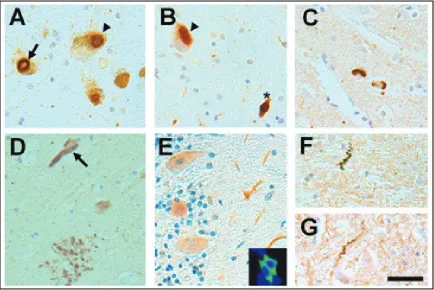
with patients who carry the same mutation (Figure 1-4): in three patients with the
G2019S mutation, two (Patient A and B) demonstrated classic PD with LBs, while the
third (Patient C) had a paucity of Lewy pathology. Patient A also displayed LBs in the
limbic cortex, while patient B had concurrent neocortical senile plaques and occasional
31
Figure 1-4. Histological characterization of patients with LRRK2 mutations. (A, B) Staining for alpha-synuclein in the SNpc of Patient A using antibody Syn 505 demonstrating the presence of classic LBs (arrow) and cytoplasmic alpha-synuclein inclusions (arrowheads) in dopaminerginergic neurons containing neuromelamine. An alpha-synuclein spheroid is indicated by an asterisk in B. (C) Cortical LBs stained with anti–alpha-synuclein antibody Syn 505 in the cingulated cortex of Patient A. (D) A neurofibrillary tangle (NFT) (arrow) and tau-positive dystrophic neurites within a senile plague (SP) in the hippocampus of Patient B stained with anti–tau antibody 17026. (E) The staining pattern of LRRK2 depicted with rabbit anti–LRRK2 antibody AP7099b. Note the intense staining in Purkinje neurons and their processes. (inset) Immunofluorescence analysis showing the cytoplasmic pattern of HA-tagged LRRK2(1245-2527) expressed in HEK293T (green) and DAPI staining of the nuclei (blue). (F, G) Dystrophic neurites in the SN of Patient C displaying accumulation of LRRK2 as stained with the rabbit anti–LRRK2 antibody. Scale bar = 40μm. Figure from Giasson et al (105).
A paucity of Lewy pathology has been reported in 1 out of 17 additional G2019S cases
(99, 269), 6 of the 8 autopsied patients from the Hasegawa and Kowa Japanense kindred
32
the 4 R1441C cases from the Canadian family D (74, 142, 346, 355), and 2 of the 4
Y1699C cases (172, 355). The additional neuropathological findings for these atypical
cases have been mixed. For the 2 Y1699C cases, amyotrophy in addition to
parkinsonism was observed, and there was a presence of “ubiquitin-positive” cytoplasmic
and nuclear inclusions (355). For the R1441C mutations carried within the Canadian
family D, one of the documented cases presented tau pathology reminiscent of
progressive supranuclear palsy while another displayed nonspecific loss of dopaminergic
neurons with ubiquitin-positive inclusions in the absence of Lewy pathology (346, 355).
Despite the pleomorphy of pathological inclusions observed throughout the literature, all
cases report degeneration of the SN.
Given studies showing that LRRK2 forms aggregates in some cellular models (131,
297), several laboratories began examining LRRK2’s presence in LBs (4, 131, 148, 234,
235, 269, 353). In our initial 2006 study of the 3 G2019S patient described above, no
LRRK2 staining of LBs was observed, nor was LRRK2 found in brain Lewy inclusions
from an additional 80 patients with either classic PD (46 cases) or DLB (34 cases).
Shortly thereafter, Zhu and colleagues published a letter indicating that LRRK2 could be
found in LBs when using two newly designed antibodies by Novus Biologicals (354).
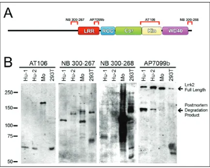
We tested these antibodies and found that their specificity was lacking (Figure 1-5), and
concluded the staining of LBs could be non-specific (55); other laboratories have
corroborated these results (280). Because we cannot account for batch differences among
commercial antibodies, we designed and developed our own. We engineered three
33
protein. Using these antibodies, again, a paucity of staining was observed for LRRK2 in
LBs (340). Regardless, a handful of studies have used the nonspecific antibodies
mentioned above to ascribe the inclusion of LRRK2 in LBs (4, 131, 148, 234, 235, 269,
353); however, staining is inconsistent and therefore inconclusive. It remains to be seen
whether LRRK2 is truly a component of Lewy pathology, but our studies indicate that it
is not.
Figure 1-5. Characterization of LRRK2 antibodies. A. Schematic of LRRK2 and its
associated domains with antibodies AT106 (Alexis Biochemicals, San Diego, CA), NB