ARTICLE
Neurophysiologic Assessment of Neonatal Sleep
Organization: Preliminary Results of a Randomized,
Controlled Trial of Skin Contact With
Preterm Infants
Susan M. Ludington-Hoe, PhDa, Mark W. Johnson, PhDb, Kathy Morgan, BSNb, Tina Lewis, BSNc, Judy Gutman, BAb, P. David Wilson, PhDc, Mark S. Scher, MDa,b
aSchool of Nursing, Case Western Reserve University, Cleveland, Ohio; Departments ofbPediatric Neurology andcNursing, University Hospitals of Cleveland, Cleveland, Ohio;cDepartment of Epidemiology, University of Maryland, College Park, Maryland
The authors have indicated they have no financial relationships relevant to this article to disclose.
ABSTRACT
BACKGROUND.Sleep is important to brain organization, but few strategies to promote
sleep among premature infants have been tested. Behaviorally based measures of sleep have shown increased quiet sleep (QS) and decreased active sleep (AS) during skin-to-skin contact (SSC) with the mother, but these results have not been confirmed with objective electroencephalographic/polysomnographic measures of sleep organization. Important differences exist between behavioral and electroen-cephalographic/polysomnographic definitions of sleep state.
METHODS.Data for the first 28 relatively healthy, preterm subjects of an ongoing
randomized trial of one 2- to 3-hour session of SSC or incubator care between feedings are reported here. Infants were positioned prone, inclined, and nested in an incubator during the 2- to 3-hour pretest period, were fed, and then went into the test period of SSC or incubator care. Infants were left largely undisturbed throughout testing. A mixed-model regression analysis compared the test-pretest differences in outcome measures within and between groups.
RESULTS.Results showed that arousals were significantly lower in the SSC group,
compared with the control group, for the entire study period and for test-pretest matched segments of QS and AS. Rapid eye movement was significantly lower for the SSC group for the study period and AS segments. Indeterminate sleep was significantly lower for the SSC group when confounding environmental variables were included in the regression analysis. When 4 subjects who experienced excessive ambient light levels during SSC were removed from analysis, QS in-creased during SSC.
CONCLUSIONS.The patterns demonstrated by the SSC group are analogous to
more-mature sleep organization. SSC may be used as an intervention to improve sleep organization in this population of preterm infants.
www.pediatrics.org/cgi/doi/10.1542/ peds.2004-1422
doi:10.1542/peds.2004-1422
Key Words
electroencephalography, brain maturation, sleep, preterm infants, skin-to-skin contact
Abbreviations SSC—skin-to-skin contact PMA—postmenstrual age IS—indeterminate sleep AS—active sleep QS— quiet sleep EOG— electrooculographic EEG— electroencephalographic REM—rapid eye movement ABSS—Anderson Behavioral State Scale
Accepted for publication Oct 26, 2005 Address correspondence to Susan M. Ludington-Hoe, PhD, Case Western Reserve University, Bolton School, 10900 Euclid Ave, Cleveland, OH 44106-4904. E-mail: susan. ludington@case.edu
A
LTHOUGH CONSIDERABLE ATTENTIONhas been given to preterm infant sleep patterns and the influence of the NICU environment on newborn sleep,1,2 littleattention has been directed to the study of strategies designed to improve sleep organization. One strategy is skin-to-skin contact (SSC). SSC is the upright prone position of the diaper-clad preterm infant skin-to-skin between the mother’s breasts. The SSC technique has been shown to alter sleep organization, as measured with behavioral state indices.3–8 SSC increases the
amount of time spent in behaviorally determined quiet sleep (QS)3,9,10 and decreases the time spent in active
sleep (AS)6,10and awake states,6,10compared with
incu-bator time, regardless of who (mother, father, grandpar-ent, or surrogate parent) provides the SSC.11,12The need
to verify behavioral sleep findings with more-rigorous neurophysiologic assessments exists because behavioral states among preterm infants are immature,13behavioral
state assessments are subject to observer bias, and blind-ing of the observers is not possible. Examinblind-ing sleep organization with blinded observers scoring from elec-troencephalographic (EEG)/polysomnographic records would serve this purpose; however, no reported studies that used this method could be found, although neonatal states can be detected with EEG recording as early as 26 to 28 weeks.14,15
Important differences in terminology and definitions of neonatal states exist among authors. The definitions used in behavioral observation scales can result in con-fusion when they are compared with the definitions used for polysomnography. Table 1 compares the defi-nitions in the Anderson Behavioral State Scale (ABSS)16,17and the relationship between sleep state and
physiologic signals recorded commonly in neonatal polysomnography. It is important to note that AS ac-cording to the polysomnographic definition used herein is most similar to the ABSS irregular sleep state and the ABSS active and very active sleep states are most similar to indeterminate sleep (IS) or AS and QS disrupted by microarousals. Finally, the polysomnographic definition of QS is based primarily on the EEG trace discontinu pattern and is associated with, but not defined by, reg-ular respiration, lack of rapid eye movement (REM), and lack of or slight body movements. In contrast, the ABSS regular quiet sleep state is much stricter in excluding any respiratory irregularities and all but slight movement. Furthermore, the ABSS scale uses the highest-numbered state seen within a 30-second epoch, whereas polysom-nographic definitions typically use the dominant state within an epoch. On the basis of these differences in definition, reported increases in the ABSS regular quiet sleep state with SSC are analogous to decreases in mi-croarousals during polysomnographic QS. Similarly, re-ported decreases in ABSS active and very active sleep states are analogous to decreases in microarousals in polysomnographic AS or QS.
SSC provides the infant with physical boundaries (containment), maternal heartbeat sounds, rhythmic movement with maternal breathing, increased body warmth, and prone positioning,18ie, simultaneous
gen-tle stimulation across the proprioceptive, auditory, ves-tibular, thermal, and tactile sensory systems, which con-tributes to comfort and stabilization of state.6 Because
poor neonatal sleep organization is associated with later developmental disabilities,19SSC has been suggested as a
simple inexpensive intervention to improve sleep orga-nization.13
The purpose of this randomized, controlled trial was to test the effect of SSC on 5 neonatal sleep organization features assessed with EEG/polysomnographic measures at postmenstrual age (PMA) of 32 weeks. The EEG/sleep measures represent different neural networks through-out the neural axis that contribute to sleep organization. Complex interconnections among multiple neuronal networks that subserve sleep allow phenotypic expres-sion of defined states of sleep, arousal, and wakefulness, punctuated by phasic activities such as motor activities (including REM). The effects of SSC on these measures have not been tested previously. We hypothesized that SSC would alter EEG/sleep organization.
METHODS
Design
An institutional review board-approved, pretest-test, randomized, controlled trial was conducted. Seventy-one premature infants were tested between October 2002 and June 2004; data for 28 have been analyzed to date, with 14 in the SSC group and 14 in the control group. Infants were assigned randomly to the SSC or control group with a computerized minimization tech-nique20that matched subjects with respect to 5 variables,
ie, gender, gestational age, severity of illness (assessed with the Neurobiologic Risk Scale21,22), age (in days) at
the time of recruitment, and body weight at the time of recruitment. This randomization technique was chosen to maintain equivalence between groups with the inclu-sion of each new subject.
Subjects
Subjects were recruited before PMA of 32 weeks,23after
Setting
Infants were tested in 1 of the 7 nursery rooms of the NICU or in the step-down unit at Rainbow Infants’ and Children’s Hospital. Each room accommodates 1 to 6 infants. The step-down unit is composed of private or semiprivate rooms that contain an incubator or crib and sleeping accommodations for the mother. Some rooms have large windows.
Conditions
Recordings were conducted in 2 consecutive interfeed-ing periods, beginninterfeed-ing at approximately 9:00AM. Infants
were left undisturbed between feedings. For the pretest period, all infants wore only a diaper if in an incubator. If the infant was in an open-air crib, then he or she wore a diaper and shirt and was covered with a blanket. In the pretest period, infants were positioned prone at a 30% incline and nested with blanket rolls around the sides and head within a commercially hooded (IsoCover model 92042A-DS; Child Medical Ventures, Boston, MA) OHIO CarePlus incubator (Air-Shields, Philadel-phia, PA), or within an open-air crib that was inclined similarly, until the next feeding, which was conducted by a staff nurse. Mothers were absent during the test period if the infant was in the control group. All control group feedings were conducted in the incubator. Control infants continued in the pretest incubator or open-air crib conditions for the test period, whereas SSC infants were positioned with SSC as the mother reclined in a lounger at a 40% incline by the side of the incubator, behind privacy screens. Each mother wore a standard hospital gown and held the infant in a flexed position beneath a receiving blanket folded in fourths. Mothers were asked not to disturb the infant if he or she appeared to be sleeping. Maternal movement was recorded through direct observation and videotape review, to dis-tinguish mother-induced from spontaneous neonatal arousals. Data collection ended when the next scheduled feeding began.
Equipment
A Nihon Koden 9100-PSG EEG system (Nihon Koden, Foothill Ranch, CA) was used to record EEG and poly-somnographic data. Data were collected with the Nihon Koden Neurofax software program. Ten-millimeter, gold, EEG electrodes (Grass, Waterford, CT) were placed at standard locations (C3, C4, T3, T4, Cz, O1, O2, and ground). Standard disposable electrodes (Nicolet Bio-medical, Madison, WI) were used for polygraphic mon-itoring of 2 electromyographic electrodes on the chin, 1 electrooculographic (EOG) electrode at the outer can-thus of each eye, and 2 electrocardiographic electrodes. Polygraphy also included 2 inductive respiratory bands (Respiband; SensorMedics, Yorba Linda, CA), placed on the chest and abdomen, and 1 pulse oximeter sensor (Masimo SET; Masimo Corp, Irvine, CA), placed over the
ball of the infant’s foot. Neurophysiologic data were sampled at 1000 samples per second. Ten-20 conductive paste (Weaver, Aurora, CO) was used to affix electrodes to the scalp, with a subset of the standard 10 –20 inter-national protocol for electrode placement. EEG, EOG, and electromyographic electrodes with 1.0-m lengths were wrapped together in a stockinette, and the infant’s head was covered with a mesh head net (NeuroSupplies, Waterford, CT). Digital EEG data were reviewed and scored with Insight (Persyst, Prescott, AZ), with a sensi-tivity of 7 V at 20 seconds per page. Synchronized digital video (model CVXV18NSSEC; Sony, Tokyo, Ja-pan) was also recorded during the study. The study was conducted by a board-certified EEG technician assisted by a skilled neonatal nurse, who annotated the record online for incidental events such as movements, proce-dures, and environmental occurrences. Ambient light was measured with an EVTECH Instruments lux meter (model L565969; EVTECH Instruments, Taiwan), and sound was measured with a decibelometer (model 33– 2055; Tandy Corp, Fort Worth, TX). The light and sound meters were placed near the infant’s head in the incu-bator and on the mother’s shoulder during SSC. Light and sound recordings were performed before each study and then every 5 minutes. Infant abdominal skin tem-perature was recorded with the incubator’s thermistor attached 1 cm below the right costal margin on the infant’s abdomen, beneath a Mylar patch (Kentex Corp, Irvine, CA). All instruments were autocalibrated.
Recording Procedure
After parental signatures on the institutional review board-approved consent form were obtained, the day for study was scheduled within 2 weeks of the infant having a PMA of 32 weeks. When the 9AMfeeding was over, an
event marker was activated, signaling the beginning of data collection. A second event marker signaled the end of the pretest and test periods. SSC mothers arrived 30 minutes before the feeding that concluded the pretest period, so that they could change into the hospital gown and pump breast milk, as needed. SSC mothers were then seated in the recliner and given their infants before the feeding. Infants were fed in the SSC position. When either 120 minutes (for feedings every 2 hours) or 180 minutes (for feedings every 3 hours) of SSC were com-pleted, data collection ceased and the infant was re-turned to the incubator, after which the electrodes were removed.
Visually Scored EEG Sleep Measures
Measurement
QS
Electrographically, quiescence or discontinuity (trace discontinu) is the primary measure defining rudimen-tary QS among preterm infants of⬍36 weeks’ PMA.24It
is characterized by periods of low-amplitude EEG activ-ity (⬍20 V, excluding artifacts) across all channels, typically having a duration of 2 to 10 seconds and re-peating 3 to 8 times per minute. A trained neonatal neurologist marked the beginning and end of all discon-tinuity segments throughout the record.
AS
Continuous EEG sleep background activity characterizes AS. REM is usually present during AS and was used as an outcome measure but was not used to define AS. Periods of continuous EEG activity with no discontinuity forⱖ60 seconds and⬍30 seconds of microarousal were defined as rudimentary AS.
Arousals
Arousals punctuate the underlying EEG continuity-dis-continuity architecture. EEG arousal is characterized by a desynchronization or change in the EEG pattern (loss of sleep background activity), which usually is associated with body movements, muscle activity, alterations in the respiratory pattern, and/or eye opening.25–28In this
anal-ysis, a microarousal (⬍30 seconds) is a brief disruption of the ongoing state and is not scored as a change in state. In polysomnographic tracings, there is often little dis-tinction, other than duration, between microarousals, more-extended arousals, and IS. This is significantly dif-ferent from a behavioral state scale that assigns a change in state to a brief microarousal.
IS
Epochs that did not show normal continuous or discon-tinuous sleep background activity or contained⬎30 sec-onds per minute of arousal were defined as IS.29 In
polysomnographic tracings, there is often little distinc-tion, other than duradistinc-tion, between microarousals, ex-tended arousals, and IS. This is significantly different from a behavioral state scale that assigns a state change to even a very brief microarousal.
Cycling Architecture
A macroscopic sleep cycling architecture encompasses the microstructure of preterm neonatal sleep features. Typically, neonatal sleep states cycle between QS (for
⬃20 minutes) and AS (for⬃40 minutes), with varying degrees of arousal and IS scattered throughout both QS and AS. The scoring of EEG sleep measures was per-formed on a continuous time basis. The raw scoring was aggregated into minute-by-minute epoch state scores with computerized analysis. Commonly, investigators use smoothing or filtering techniques to aggregate states over several minutes.24,30In this analysis, the onset of QS
was defined as the beginning of a segment in which 3 consecutive minutes or 3 of 4 consecutive minutes were scored as QS. Similarly, the onset of AS was defined as the beginning of a segment in which 3 consecutive min-utes or 3 of 4 consecutive minmin-utes were scored as AS. In general, the onset of a state was not allowed at the first epoch of a recording. Cycle duration was defined as the time from the onset of QS through a required period of AS (and IS if present) to the onset of the next QS segment. QS duration was the time from the onset of QS to the onset of AS, excluding any IS epochs at the transition. AS duration was the time from the onset of AS to the onset of QS, excluding any IS epochs at the transition. Typically, 1 or 2 complete sleep cycles were recorded per test or pretest period, with additional par-tial cycles occurring at the beginning and end of each period. Understanding this macrostructure is important to understanding how and why QS, for example, can contain a finite percentage of AS, percentage of IS, and seconds of arousal.
Outcome Measures
Twenty-one outcome variables were analyzed. The mea-sures were selected to encompass a broad range of phys-iologic sleep parameters. Some measures were based on visual scoring, and others were based on computerized analysis. Each measure was summarized for both the test and pretest periods. All outcome measures were ana-lyzed as test-pretest changes. Most measures were sum-marized across study periods (the whole test period, compared with the whole pretest period), but several measures were summarized across comparable test and pretest segments of rudimentary QS or rudimentary AS, where appropriate. The measures were as follows.
Changes in discontinuity were measured across the study period and within QS. The outcome measures were defined as the test-pretest change in the mean percentage of time occupied by discontinuous segments. Changes in REM counts were measured across the study period and within AS. Rudimentary AS among preterm infants of⬍36 weeks’ PMA is defined by periods of continuous EEG sleep background activity (no discon-tinuity)24and is usually associated with eye movements.
con-tainedⱖ1 polysomnographic REM or visually observed eye movement.
Changes in arousals were measured across the study period and within QS and AS. EEG arousal is defined as a desynchronization of the EEG activity (loss of sleep background activity), which is usually associated with body movements, muscle activity, alterations in the re-spiratory pattern, and/or eye opening.16,28 The arousal
outcome measures were defined as the test-pretest change in the percentage of time of microarousal and extended arousal within the respective time periods.
Changes in the mean duration of the cycle, QS, and AS were measured. Rudimentary QS, AS, and IS (as defined above) were derived from visual scoring of EEG discontinuity and arousals. The mean duration outcome measures were defined as the test-pretest change in cycle or segment duration.
Changes in percentages of QS, AS, and IS were mea-sured. States were scored on a continuous basis, not epoch by epoch, although many analyses were summa-rized on a minute-by-minute basis. The percentage of each state was the total percentage of the study period (test or pretest) that was occupied by that state, with QS being discontinuous EEG activity excluding any mi-croarousals, AS being continuous EEG sleep background activity excluding any microarousals, and IS encompass-ing any arousals, IS, and rare wakefulness. The outcome measures were defined as the test-pretest change in percentage for each state.
Changes in the respiratory ratio and respiratory rate were measured. The respiratory ratio is a computer-calculated measure of the regularity of respiration. It is a measure of the spread of energy in the frequency do-main. A sinusoidal signal has all of its energy focused at a single frequency, resulting in a respiratory ratio of 0. The energy of a chaotic signal is spread very widely across the frequency spectrum, with a respiratory ratio approaching 1. In general, the regular respirations of QS have a low respiratory ratio, the irregular respirations of AS have higher values, and the chaotic respirations of IS have the highest values. The respiratory rate was taken from a measure of the center frequency in the respira-tory ratio calculation. These 2 outcome measures were the test-pretest changes calculated from the minute-by-minute averages for each subject.
Changes in the EEG /␣ ratio and EEG left/right hemisphere correlation were assessed. These 2 measures were derived from computer calculations of the EEG signals. Historically, neurologists have separated the EEG frequencies into several bands, including ␣ (8 –13 Hz) and(13–22 Hz). The EEG/␣ratio is a unitless mea-sure of the energy in the-band versus the energy in the
␣-band, which shows fairly robust changes between QS and AS; it is a modification of measures described by Scher et al.31–33The measure was calculated for a number
of electrode pairs for each minute, expressed in
logarith-mic units. The median value across the electrode pairs was used because it limits the effects of artifacts if they are present in a limited number of channels. The EEG left/right hemisphere correlation was calculated as the cross-covariance between the C3-T3 (left) and C4-T4 (right) homologous electrode pairs. The measure was selected because it changes with age and development. The EEG outcome measures were the test-pretest change in the minute-by-minute values averaged over the study period.
Changes in heart rate mean and SD and blood oxygen saturation mean and SD were measured. The oximeter averaging time was set to 2 seconds. The means and SDs of the heart rate and blood oxygen saturation values measured with the Masimo pulse oximeter were calcu-lated for each 1-minute epoch. Each outcome measure was the test-pretest change in the minute-by-minute values averaged over the study period.
EEG/Sleep Record Analysis
A single neonatal neurophysiologist (M.S.S.), who was blinded with respect to both study group and pretest-test periods, visually analyzed all records. Digital annotations were made on each record, marking the beginning and end of each interburst interval (measure of discontinu-ity), the beginning and end of each arousal, and each REM (identified as an out-of-phase signal on the 2 EOG channels). Each record was reviewed multiple times by the same reader, to determine whether notations had consistent entries (eg, beginning and end of interbursts and arousals and REM occurrences). The raw annota-tions made by the technician and neurophysiologist were transferred into a database, where they were checked again for consistency and then used in analyses of the sleep architecture.
Statistical Plan
differences between infants. The regression equation for an example outcome variable, X, is YXi ⫽ o_X ⫹
SSC_X ⫻ (SSC group)i ⫹ pretest_X ⫻ (Xi_pretest ⫺ Xpretest_mean) ⫹ ⑀Xi, where YXi is fit to the test minus
pretest differenceXfor theith subject,o_Xis the
aver-age X for the control group,SSC_X is the change inX
(the effect) attributable to SSC, pretest_X is the
adjust-ment for baseline differences among the infants, and⑀Xi
is the residual error not explained by the regression model. All statistical results are reported ascoefficients (effect size) and theirPvalues, which indicate whether the coefficients are statistically different from 0. Sig-nificance was set atPⱕ.05.
The effect of potential confounding variables was also analyzed by adding the effects of confounding variables to the regression equation given above, ie, Y*Xi ⫽ YXi ⫹⌺j [Cj_X ⫻ (Cji ⫺ Cj_mean)], where the effects are
summed for each confounding variable, Cj. Cj_Xis the
coefficient that describes the reduction in the residual error attributable to inclusion of Cjin the model. With
subtraction of the mean valueCj_meanfrom the
individ-ual values, Cj_Xcauses no net change in the expected
value of o_X. The potential confounding variables
in-cluded in the analysis were (1) study location (NICU versus step-down unit), (2) length of feeding interval (2 hours versus 3 hours), (3) type of bed unit (incubator versus open-air crib), (4) gender, (5) gestational age, (6) PMA at the time of study, (7) age (in days) on the day of study, (8) birth weight, and (9) study weight.
RESULTS
Demographic Features
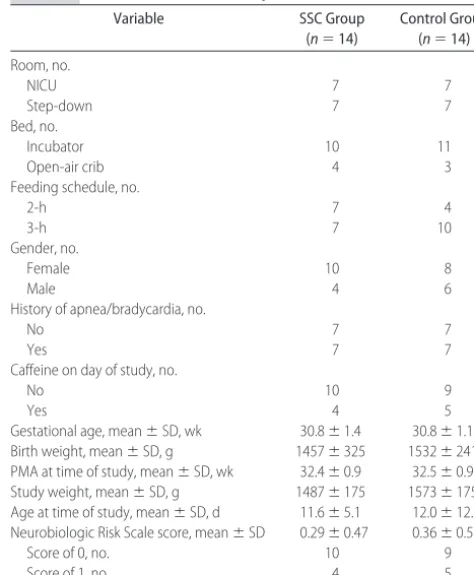
Table 2 presents the characteristics of the subjects. No differences between the groups were present, which confirms the balance established by the randomization procedure.
Sleep Organization Variables
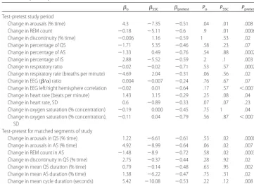
Regression analysis results for 21 outcome variables without confounding variable effects are reported in Ta-ble 3. Significant SSC effects were found in 3 change-in-arousal variables and 2 change-in-REM count variables. With the addition of confounding variable effects, the change in percentage of IS also showed significant SSC effects. The study period was defined as the full test period, compared with the full pretest period. The per-centage of time of arousals was significantly lower for the SSC group, compared with the control group, across the study period (SSC ⫽ ⫺7.35;P ⫽ .015), as well as
during QS (SSC ⫽ ⫺6.61; P ⫽ .02) and AS (SSC ⫽ ⫺8.99;P⫽.02) when analyzed separately. The control group showed a smaller but significant increase in the percentage of time of arousal in the test period, com-pared with the pretest period (o⫽ ⫹4.3;P⫽.037). The
increase in control group arousals was not significant
when QS and AS were analyzed separately. Controlling for pretest values reduced significantly the residual re-gression deviance for most of the outcome variables analyzed (18 of 21 variables; P ⱕ .05). The arousal pretest regression coefficients werepretest⫽ ⫺0.61 (P⫽
.00002) for QS,pretest⫽ ⫺0.64 (P⫽.0066) for AS, and
pretest⫽ ⫺0.51 (P ⫽.0075) for the study period. The
sign of the pretest coefficients was negative for all 21
output variables, which indicates that, if a pretest value was higher or lower than the group mean, then the test-pretest difference,⌬X, tended to be in the direction more toward the group mean. In the case of the arousals, if the infant had a particularly high pretest arousal level, then the test period arousal level tended to be a little lower. Conversely, if the pretest arousal level was par-ticularly low, then the test period arousal level tended to be a little higher. Most importantly, with inclusion of the pretest levels in the regression analysis, the baseline scatter was accounted for and the regression analysis was improved significantly.
Two change-in-REM count variables were found to have significantly lower values for the SSC group, com-pared with the control group. The change-in-REM count values analyzed were the percentage of 10-second ep-ochs that containedⱖ1 REM, as recorded either through direct observation during the study or through polysom-nographic scoring. REM counts during AS (SSC⫽ ⫺8.9; P⫽.029) and during the study period (SSC⫽ ⫺5.11;P ⫽.013) were significantly lower in the SSC group,
com-TABLE 2 Characteristics of the Subjects (Nⴝ28)
Variable SSC Group
(n⫽14)
Control Group (n⫽14)
Room, no.
NICU 7 7
Step-down 7 7
Bed, no.
Incubator 10 11
Open-air crib 4 3
Feeding schedule, no.
2-h 7 4
3-h 7 10
Gender, no.
Female 10 8
Male 4 6
History of apnea/bradycardia, no.
No 7 7
Yes 7 7
Caffeine on day of study, no.
No 10 9
Yes 4 5
Gestational age, mean⫾SD, wk 30.8⫾1.4 30.8⫾1.1 Birth weight, mean⫾SD, g 1457⫾325 1532⫾241 PMA at time of study, mean⫾SD, wk 32.4⫾0.9 32.5⫾0.9 Study weight, mean⫾SD, g 1487⫾175 1573⫾175 Age at time of study, mean⫾SD, d 11.6⫾5.1 12.0⫾12.0 Neurobiologic Risk Scale score, mean⫾SD 0.29⫾0.47 0.36⫾0.50
Score of 0, no. 10 9
pared with the control group. The control group showed no significant difference from 0 (o). However, the
pre-test values contributed significantly to the regression analysis, with coefficients ofpretest⫽ ⫺0.72 (P⫽.0003)
for AS and pretest ⫽ ⫺0.60 (P ⫽ .0006) for the study
period. Again, the negative sign indicates that outliers in the pretest period tended back toward the mean in the test period.
Regression Analysis With Confounding Variables
As a broad assessment tool, all 21 outcome variables were analyzed by including all confounding variables (except high light exposure) in the regression model, with attention to several important possible concerns, as follows. (1) Were any of the confounders themselves significant? (2) Did they “importantly” change the SSC regression coefficients? (3) Did they change the signifi-cance of any regression coefficients? Firstly, with a few exceptions, almost no confounding variables had statis-tically significantcoefficients. Secondly, the addition of the confounding variables had only minor effects on the SSC coefficients and did not change the impact or inter-pretation of the results. Thirdly, the confounding vari-ables often improved the fit (reduced thePvalues) of the SSC coefficients; in other words, they better explained the scatter in the data, although they were not them-selves significant, givenN⫽28. Five outcome variables (changes in arousals, percentage of IS, percentage of QS, REM counts, and heart rate means across the study period) were analyzed in more depth after the initial
confounding variable analysis. In the analysis, the least-significant confounding variables were removed from the analysis in a stepwise manner, until only the most important confounding variables remained in the regres-sion model. Table 4 reports the regresregres-sion results for these 5 outcome variables, controlling for confounding variables. The analysis was performed for all variables, but these variables were chosen because the SSC effect was significant when controlling for confounding vari-ables. The change-in-arousal and change-in-REM count variables (described above) during QS and AS are not shown but reflect similar changes.
Table 4 shows the  coefficient, SE, t value, and P
value for each input variable included in the model, as well as the degrees of freedom and the overall (null) deviance and residual deviance not explained by the regression model. For each outcome variable, 2 or 3 regression results are shown, ie, (1) without ing variables, (2) with the 3 most-significant confound-ing variables, and (when significant) (3) with the same variables plus the variable high light. High light exposure is not a strictly confounding variable because it is not a pretest variable. Rather, it is derived from measurements of light levels during the pretest and test periods. Light levels could be controlled fairly consistently for record-ing sessions conducted in the private or semiprivate rooms of the step-down unit but not in the NICU. The light could be controlled to some degree in the incubator portion of NICU studies. In 4 of the SSC studies reported here, the light was substantially higher in the test period,
TABLE 3 Regression Analysis Results for Outcome Variables Without Confounding Variable Effects (Nⴝ28)
o SSC pretest Po PSSC Ppretest Test-pretest study period
Change in arousals (% time) 4.3 ⫺7.35 ⫺0.51 .04 .01 .008
Change in REM count ⫺0.18 ⫺5.11 ⫺0.6 .9 .01 .0006
Change in discontinuity (% time) ⫺0.006 1.16 ⫺0.59 1 .53 .02
Change in percentage of QS ⫺1.71 5.35 ⫺0.46 .58 .23 .07
Change in percentage of AS ⫺1.33 0.49 ⫺0.76 .54 .88 .0002
Change in percentage of IS 2.88 ⫺5.52 ⫺0.59 .2 .1 .003
Change in respiratory ratio ⫺0.02 ⫺0.02 ⫺0.71 .53 .57 .0002
Change in respiratory rate (breaths per minute) ⫺4.69 2.04 ⫺0.31 .06 .56 .02 Change in EEG (/␣) ratio 0.004 ⫺0.007 ⫺0.24 .76 .67 .07 Change in EEG left/right hemisphere correlation ⫺0.02 0.01 ⫺0.64 .17 .57 ⬍.0001 Change in heart rate (beats per minute) 1.43 3.15 ⫺0.29 .25 .08 .04
Change in heart rate, SD 0.6 ⫺0.89 ⫺0.33 .07 .07 .23
Change in oxygen saturation (% concentration) ⫺0.19 0.000 ⫺0.45 .75 1 .04 Change in oxygen saturation (% concentration),
SD
⫺0.11 0.04 ⫺0.79 .56 .87 ⬍.0001
Test-pretest for matched segments of study
Change in arousals in QS (% time) 1.22 ⫺6.61 ⫺0.61 .53 .02 .00003 Change in arousals in AS (% time) 4.92 ⫺8.99 ⫺0.64 .06 .02 .007
Change in REM count in AS ⫺1.48 ⫺8.9 ⫺0.72 .58 .02 .0003
TABLE 4 Regression Analysis Results for Selected Outcome Variables With Most-Significant Confounding Variable Effects (Nⴝ28)
Test-Pretest Differences Across Study Period  SE tValue PValue Residual
Deviance
Degrees of Freedom
Arousals (% time) 2002.9a 27
Without confounding variables
Intercept 3.7 2 1.9 .07 1320.4 25
SSC group ⫺7.3 2.8 ⫺2.6 .01
Pretest ⫺0.5 0.2 ⫺2.9 .008
With confounding variables but not high light
Intercept 4.7 1.8 2.6 .02 910.5 22
SSC group ⫺9.3 2.6 ⫺3.6 .002
Pretest ⫺0.5 0.2 ⫺3.2 .004
2-h versus 3-h feeding ⫺6.1 3.1 ⫺2 .06
Incubator versus crib 6.2 3.5 1.8 .09
PMA ⫺2.8 1.7 ⫺1.7 .10
With confounding variables including high light
Intercept 4.4 1.7 2.6 .02 755.9 21
SSC group ⫺11.2 2.6 ⫺4.3 .0003
Pretest ⫺0.5 0.1 ⫺3.1 .005
High light 8.4 4.1 2.1 .051
Incubator versus crib 6.9 3.3 2.1 .048
PMA ⫺2.9 1.6 ⫺1.9 .08
2-h versus 3-h feeding ⫺3.7 3.1 ⫺1.2 .25
Percentage of IS 2290.6a 27
Without confounding variables
Intercept 2.9 2.2 1.3 .2 1591.4 25
SSC group ⫺5.5 3.2 ⫺1.7 .097
Pretest ⫺0.59 0.18 ⫺3.2 .003
With confounding variables but not high light
Intercept 3.9 2.1 1.9 .08 1219.5 22
SSC group ⫺7.6 3.1 ⫺2.4 .024
Pretest ⫺0.61 0.17 ⫺3.5 .002
Incubator versus crib 7.6 4 1.9 .07
PMA ⫺2.9 1.9 ⫺1.5 .15
2-h versus 3-h feeding ⫺5.4 3.6 ⫺1.5 .15
With confounding variables including high light
Intercept 3.5 2.1 1.7 .1 1099.4 21
SSC group ⫺9.0 3.2 ⫺2.8 .01
Pretest ⫺0.55 0.17 ⫺3.2 .004
High light 7.5 5 1.5 .14
Incubator versus crib 8.3 4 2.1 .049
PMA ⫺2.9 1.9 ⫺1.6 .13
2-h versus 3-h feeding ⫺3.0 3.8 ⫺0.79 .44
Percentage of QS with high light-exposed infants removed 3017.7a 23
Without confounding variables
Intercept ⫺1.6 3.0 ⫺0.5 .61 2665.3 21
SSC group 5.9 4.7 1.2 .23
Pretest ⫺0.4 0.3 ⫺1.3 .21
With confounding variables
Intercept ⫺3.7 3.2 ⫺1.2 .26 2058.8 18
SSC group 10.3 5.0 2.1 .05
Pretest ⫺0.4 0.3 ⫺1.6 .13
Study weight 10.9 8.5 1.3 .21
Incubator versus crib ⫺14.8 6.8 ⫺2.2 .04
Gestational age ⫺9.9 7.1 ⫺1.4 .18
REM count 1192.1a 27
Without confounding variables
Intercept 2.6 1.4 1.9 .07 638.2 25
SSC group ⫺5.1 1.9 ⫺2.7 .01
Pretest ⫺0.6 0.2 ⫺3.9 .0006
With confounding variables
Intercept 2.6 1.3 2 .06 488.4 22
SSC group ⫺5.1 1.8 ⫺2.8 .01
Pretest ⫺0.7 0.2 ⫺4 .0006
Incubator versus crib ⫺4.8 2.5 ⫺1.9 .07
Gender ⫺3.9 2.1 ⫺1.9 .07
compared with the pretest period (more than sevenfold increase in mean lux value) (Table 5). These 4 studies were designated with the high light input variable. The overall (null) deviance is a measure of scatter in an outcome variable without accounting for any regression effects. The residual deviance is a measure of the re-maining scatter in the outcome variables after account-ing for any regression effects.
For the first outcome variable (change in arousal across the study period), the 3 most-significant con-founding variables were 2-hour versus 3-hour feeding interval, incubator versus crib studies, and PMA of the infant. The overall deviance of the change in arousals was 2002.9. The primary regression analysis presented in Table 4 reduced the deviance to 1320.4, which was further reduced to 910.5 with the addition of 3 con-founding variables and to 755.9 with the further addi-tion of high light. Although none of the confounding variables or high light exposure showed remarkably strong effects, the variables served to improve the regres-sion analysis, with SSC showing more strongly lower
values and greatly increased significance levels (P⫽.002 and P ⫽ .0003, compared with P ⫽ .01 without con-founders).
When confounding variables were added to the re-gression analysis for change in percentage of IS across the study period, the results paralleled those for change in arousals. The overall deviance of change in percentage of IS decreased from 2290.6 to 1591.4 with the primary regression analysis, was further reduced to 1219.5 with
the addition of confounding variables, and was reduced to 1099.4 with the further addition of high light. Again, none of the confounding variables or high light showed remarkably strong effects, but the variables served to improve the regression analysis, with SSC showing
more strongly lower values and increased significance levels (P⫽.024 andP⫽.01, compared withP⫽.097 without confounding variables). Change in percentage of QS reached significance only when the 4 neonates with high light exposure were eliminated from the re-gression analysis.
When confounding variables were added to the re-gression analysis for change in REM counts across the study period, the regression analysis showed a modest reduction in residual deviance, but the variables had virtually no effect on the primary effects. The variable high light also had no significant effect on the regression analysis and so was not included in Table 4.
When confounding variables were added to the re-gression analysis for heart rate mean across the study period, the result was quite different from the results for the variables reported above. Four confounding vari-ables were found to have significant effects; in order of significance, they were birth weight, study weight, age (in days), and gender. Two additional variables (PMA of the infant and study location) were included in the regression analysis but were not shown to be statistically significant. With inclusion of these confounding vari-ables, SSC was shown to have a significant effect on mean heart rate (SSC ⫽ 4.8; P ⫽ .008). Because the
outcome variable was the test-pretest difference, the significance of the confounding variable coefficients in-dicates that there is an afternoon versus morning rela-tionship between heart rate and the combination of these 6 confounding variables. The confounding vari-ables taken individually did not show any significance.
TABLE 4 Continued
Test-Pretest Differences Across Study Period  SE tValue PValue Residual
Deviance
Degrees of Freedom
Heart rate mean 703.3a 27
Without confounding variables
Intercept ⫺1.6 1.2 ⫺1.3 .21 511.0 25
SSC group 3.2 1.7 1.8 .08
Pretest ⫺0.3 0.1 ⫺2.2 .04
With confounding variables
Intercept 0.7 1.06 0.66 .51 268.9 20
SSC group 4.6 1.56 2.95 .008
Pretest ⫺0.14 0.13 ⫺1.07 .3
Birth weight 0.039 0.01 3.47 .0026
Study weight ⫺0.03 0.01 ⫺2.4 .027
Age 0.6 0.22 2.67 .015
Gender 3.56 1.67 2.13 .046
PMA ⫺2.09 1.11 ⫺1.89 .075
NICU 4.2 2.29 1.83 .082
aNull deviance.
TABLE 5 Summary of Light Levels During Studies
Light Level, Mean⫾SD, lux
Pretest Period Test Period
DISCUSSION
The results show benefits of SSC for neurophysiologic organization of sleep among preterm infants with PMA of 32 weeks; all benefits were independent of any sleep position-related changes in respiratory rate, heart rate, and oxygen saturation. The benefits included reductions in arousals during both QS and AS in the SSC group and throughout the SSC period. Similarly, the percentage of epochs withⱖ1 REM decreased significantly during the SSC period and during AS in the SSC group, compared with the control group. These changes were indepen-dent of the confounders, including sleep in the home-like step-down unit, rather than in the NICU. The lack of differences attributable to multiple confounders supports the robust effect of SSC without contaminants.
The mean percentage of time of arousals decreased during the SSC period in all measures and increased (albeit insignificantly) during incubator care and in the control group in all measures. These data suggest that the decreases in arousals seen in EEG sleep recordings might be equivalent to the reported increases in behav-iorally based QS that have been seen in the multiple behavioral state studies of SSC, especially if linearity is assumed. At 32 weeks’ PMA, relatively healthy preterm infants experience fewer and shorter arousals during QS than during AS.28,32
A decrease in arousals is a positive change for preterm infants. A pattern of decreased arousal is consistent with more-mature sleep organization and maturation of spe-cific neuronal processes of the central nervous system.34
A study of breastfeeding mothers found greater central nervous system maturity, as measured by increased QS and decreased arousals, among infants of breastfeeding mothers who had high levels of docosahexaenoic acid, a long-chain polyunsaturated fatty acid that correlates positively with enhanced central nervous system integ-rity.35 A decrease in arousals also suggests better sleep
organization, with the infant sustaining sleep states for longer time periods. Fewer arousals may have physio-logic benefits as well, in part because arousals have been linked to apnea among spontaneously breathing in-fants.13
A possible explanation for decreased arousals from QS and AS during SSC may be intensification of the prone position when the infant is on the mother’s chest. Prone position itself decreases arousals. At 36 to 38 weeks’ PMA, arousal from AS and QS is less likely to occur when the infant sleeps prone.36Because the SSC infants
were in the prone position during the pretest period and because the control infants were positioned prone in the incubator throughout the pretest and test periods, ma-ternal influences intensifying the prone position experi-ence during SSC might have had a role. For example, the infant might hear maternal heartbeat sounds during SSC; heartbeat sounds induce sleep.37 When heartbeat
sounds are coupled with gentle rocking (somewhat
sim-ilar to the rhythmic rise and fall of the mother’s chest as she breathes), earlier development of distinct sleep pat-terns occurs38and the amount of QS increases.39,40SSC
includes an element of containment, in that the infant is placed prone between the mother’s breasts, beneath a blanket, with the mother’s arms holding the infant in place. Containment evokes quiescence and the onset of QS,41as well as a decrease in arousals from QS.42
There-fore, the SSC prone position plus containment may be more potent than the prone position alone in changing arousals. Another possible explanation for the decrease in arousals during SSC may be that SSC in some way increases the arousal threshold in QS and AS, although increases in arousal threshold during QS among preterm infants are not usually seen until 44 to 45 weeks’ PMA.43
The reductions in arousals and REM counts were clearly the most robust findings of this investigation and were independent of environmental conditions. How-ever, other indicators of sleep organization may be re-sponsive to environmental influences. To look at this possibility, we examined the influence of other con-founders. We found that the presence of high light levels in the NICU outside the incubators influenced several sleep indices. When the light level was much higher during the SSC test period, compared with the pretest period, the amount of QS did not increase and instead decreased slightly. The decrease failed to reach statistical significance (n⫽4) but showed an influence of ambient lighting on QS. A mixed-model regression analysis was performed after removal of the 4 subjects who were tested under the high lighting condition (n⫽ 24). Sig-nificant changes from the pretest period to the study period in the SSC group were seen in percentage of QS (increase;  ⫽ ⫹10.3; P ⫽ .05), percentage of IS (de-crease;  ⫽ ⫺9.9; P ⫽ .008), arousals (decrease;  ⫽
⫺10.3; P ⬍ .0005), and REM counts (decrease;  ⫽
⫺5.1;P⫽.02). Percentage of QS increased significantly when ambient light was⬍240 lux. These findings are in contrast to those of Hellstrom-Westas et al,44who found
no differences in duration of and percentage of time spent in QS, with amplitude-integrated EEG recording, when 32- to 34-week preterm infants were in a hooded incubator that provided low ambient light levels or in an uncovered incubator that provided high illumination levels. The conflict may be explained by the difference in being in an uncovered incubator, for which light levels are still lower than outside the incubator, although over-head lighting is similar.45Also the studies by
Hellstrom-Westas et al44 were single-channel recordings, which
differ-ences in medical outcomes.47 Future studies that
evaluate polysomnographic sleep under low and high lighting conditions are needed.
The increase in QS in the SSC group is an encouraging finding, because QS is needed for brain maturation. Hip-pocampal processing of waking experiences occurs dur-ing QS,48and preterm infants need a dominance of QS
before being able to express AS.49The succession of REM
sleep after QS also plays an important role in memory processing, because certain types of memory are laid down in QS50,51and other types of memory are processed
in AS.52,53As the premature infant matures, the
percent-age of time in QS increases. Increased duration of QS requires the greatest amount of neural control, making QS the most organized sleep state.54
No changes in respiratory rate and oxygen saturation between the periods or for the SSC group were seen. These data confirm cardiorespiratory stability during the pretest and test periods for both groups and are similar to previous findings with SSC.7,55–57Heart rate did increase
during SSC when the influence of confounding variables was controlled. Heart rate increased by ⬃10 beats per minute during SSC, similar to previous SSC findings7,58
and within the range expected because of increased body temperature,7,59 upright position of SSC57,58 or upright
position alone,60,61or being in the prone position during
QS episodes.62
Our findings showed no difference in changes in sleep parameters when infants were observed in the NICU or in a home-like step-down unit. This is not surprising, because the pretest-test design of the study was intended to control for these factors. However, additional analysis is warranted to assess whether any of these confounding variables were correlated with baseline sleep parameters (not changes in parameters). It was suggested that we may wish to analyze the effects of other confounding variables, such as history of apnea of prematurity and caffeine administration, as well as analyzing the effect of SSC on apnea and bradycardia and their correlations with the well-documented temperature effects of SSC.
Firstly, a contribution of this study is that it is the first report of a neurophysiologic method for accurate state assessment while an infant is being held by his or her mother. Secondly, the randomized, controlled design removed the effects of multiple confounders that cannot be controlled for when clinical trials are conducted with preterm infants. Thirdly, the minimization technique is a useful method of randomization when numerous con-founders are anticipated.
Because promotion of sleep organization is so impor-tant for normal neurodevelopment, one would expect that the infant’s sleep status could guide clinical care. However, this is not yet a well-defined goal for physician or nursing personnel. In general, developmental care instructs that care should be based on the infant’s indi-vidual ability to handle the stimulation provided by the
caregiver, by the physical environment, and by parental contact, without promoting autonomic and behavioral disorganization. The goal of developmental care is to promote stable, well-organized infants who can con-serve energy for growth and development.63,64
Physio-logic stability can be promoted through the use of SSC, as reflected by behavioral state stability.13Although state
organization is also a laudable goal for developmental care,65state organization is not a primary focus of
devel-opmental care among NICU staff members. Nurses do not assess systematically the levels of sleep or wakeful-ness among infants,66 although responses to
interven-tions are mediated by state transiinterven-tions. Caregiving itself influences state expression; QS increases if infants are undisturbed, whereas waking states increase when care-givers interact with children.1,67In fact, 50% of
prema-ture infants in one study did not experience any waking before nursing interventions,68 and subsequent nursing
interventions disrupted sleep predominantly. The more intrusive the care, the more disruption to sleep there is, as measured with transitions from sleep to wakefulness. Nurses have been taught that developmental care means reduction of stimulation to the infant, especially tactile stimulation.69,70 Minimal handling, including minimal
handling and holding by parents, is considered desir-able.69 As minimal handling protocols have become
common, noxious handling has not been differentiated from beneficial handling. Gentle human touch has mul-tiple benefits for preterm infants, which are seldom ac-knowledged.71Parental involvement is an inherent
com-ponent of SSC, and clearly SSC is a form of beneficial handling, assuming that stimulation is mediated through the limbic system.72Parental involvement in NICU care
is recommended but has advanced slowly.73,74SSC is not
practiced routinely in the United States,75 although its
implementation is increasing76and it is recommended as
a strategy to increase parental involvement and to pro-vide infants with pleasing experiences.77,78Because the
beneficial effects of improved sleep organization on neu-rodevelopment are gradual and subtle, SSC should be practiced more widely for extended periods during post-natal life.
Previous research showed that the physiologic and behavioral state effects of SSC are not sustained once SSC is discontinued,9,58,59,79–83which suggests a limited
impact from just 1 session of SSC. Repeated use of SSC to improve sleep organization may result in improved sleep organization by 40 weeks’ PMA, as a reflection of enhanced neuroplasticity leading to improved neu-robehavioral development. Better sleep organization at 40 weeks’ PMA might result in sleep among pre-term infants that is more similar to that of pre-term in-fants.31Therefore, future studies should measure sleep
CONCLUSIONS
Previous reports advocated environmental adjustments that promote better sleep organization while neonates are in the NICU.13 SSC can improve the integrity of
sleep.13 Changes in sleep organization reported in this
study, namely, decreased arousals, REM counts, and IS, with increased QS percentages, are alterations in sleep organization that may promote improved brain matura-tion. These data suggest that SSC is a nonpharmacologic interventional treatment that affects sleep organization, and ultimately neurodevelopment, positively.
ACKNOWLEDGMENTS
This study was supported in part by National Institutes of Health grants 5R01 NR04926 and 1RO3 NR08587 (to S.M.L.-H.).
REFERENCES
1. Brandon DH, Holditch-Davis D, Beylea M. Nursing care and the development of sleeping and waking behaviors in preterm infants.Res Nurs Health.1999;22:217–229
2. Rivkees SA, Mayes L, Jacobs H, Gross I. Rest-activity patterns of premature infants are regulated by cycled lighting.Pediatrics.
2004;113:833– 839
3. Bauer K, Pyper A, Sperling P, Uhrig C, Versmold H. Effects of gestational and postnatal age on body temperature, oxygen consumption, and activity during early skin-to-skin contact between preterm infants of 25–30-week gestation and their mothers.Pediatr Res.1998;44:247–251
4. Eichel P. Kangaroo care: expanding our practice to critically ill neonates.Newborn Infant Nurs Rev.2001;1:224 –228
5. Feldman R, Weller A, Sirota L, Eidelman AI. Skin-to-skin contact (kangaroo care) promotes self-regulation in premature infants: sleep-wake cyclicity, arousal modulation, and sus-tained exploration.Dev Psychol.2002;38:194 –207
6. Feldman R, Eidelman AI. Skin-to-skin contact (kangaroo care) accelerates autonomic and neurobehavioural maturation in preterm infants.Dev Med Child Neurol.2003;45:274 –281 7. Ludington-Hoe SM, Anderson GC, Swinth JY, Thompson C,
Hadeed AJ. Randomized controlled trial of kangaroo care: car-diorespiratory and thermal effects on healthy preterm infants.
Neonatal Netw.2004;23:39 – 48
8. Messmer PR, Rodriguez S, Adams J, et al. Effect of kangaroo care on sleep time for neonates.Pediatr Nurs.1997;23:408 – 414 9. de Leeuw R, Colin EM, Dunnebier EA, Mirmiran M. Physio-logical effects of kangaroo care in very small preterm infants.
Biol Neonate.1991;59:149 –155
10. Ludington SM. Energy conservation during skin-to-skin con-tact between preterm infants and their mothers.Heart Lung.
1990;19:445– 451
11. Anderson GC, Dombrowski MA, Swinth JY. Kangaroo care: not just for stable preemies anymore. Reflect Nurs Leadersh.
2001;27:32–34, 45
12. Ludington-Hoe SM, Hashemi MS, Argote LA, Medellin G, Rey H. Selected physiologic measures and behavior during paternal skin contact with Colombian preterm infants.J Dev Physiol.
1992;18:223–232
13. Lehtonen L, Martin RJ. Ontogeny of sleep and awake states in relation to breathing in preterm infants.Semin Neonatol.2004; 9:229 –238
14. Lehtonen L, Johnson MW, Bakdash T, Martin RJ, Miller MJ, Scher MS. Relation of sleep state to hypoxemic episodes in
ventilated extremely-low-birth-weight infants.J Pediatr.2002; 141:363–368
15. Selton D, Andre M, Hascoet JM. Normal EEG in very prema-ture infants: reference criteria. Clin Neurophysiol. 2000;111: 2116 –2124
16. Gill NE, Behnke M, Conlon M, McNeely JB, Anderson GC. Effect of nonnutritive sucking on behavioral state in preterm infants before feeding.Nurs Res.1988;37:347–350
17. Gill NE, Behnke M, Conlon M, Anderson GC. Nonnutritive sucking modulates behavioral state for preterm infants before feeding.Scand J Caring Sci.1992;6:3–7
18. Ludington-Hoe SM, Swinth JY. Developmental aspects of kan-garoo care.J Obstet Gynecol Neonatal Nurs.1996;25:691–703 19. Whitney MP, Thoman EB. Early sleep patterns of premature
infants are differentially related to later developmental disabil-ities.J Dev Behav Pediatr.1993;14:71– 80
20. Conlon M, Anderson GC. Three methods of random assignment: comparison of balance achieved on potentially confounding variables.Nurs Res.1990;39:376 –379
21. Brazy JE, Eckerman CO, Oehler JM, Goldstein RF, O’Rand AM. Nursery Neurobiologic Risk Score: important factor in predicting outcome in very low birth weight infants.J Pediatr.
1991;118:783–792
22. Brazy JE, Goldstein RF, Oehler JM, Gustafson KE, Thompson RJ Jr. Nursery Neurobiologic Risk Score: levels of risk and relationships with nonmedical factors. J Dev Behav Pediatr.
1993;14:375–380
23. Engle WA. Age terminology during the perinatal period. Pedi-atrics.2004;114:1362–1364
24. Curzi-Dascalova L, Mirmiran M.Manual of Methods for Recording and Analyzing Sleep-Wakefulness States in Preterm and Full-Term Infants. Paris, France: Les E´ditions INSERM; 1996
25. American Sleep Disorders Association, Sleep Disorders Atlas Task Force. EEG arousals: scoring rules and examples: a pre-liminary report from the Sleep Disorders Atlas Task Force of the American Sleep Disorders Association. Sleep. 1992;15: 173–184
26. Crowell DH, Brooks LJ, Corwin M, et al. Ontogeny of arousal.
J Clin Neurophysiol.2004;21:290 –300
27. Crowell DH, Kulp TD, Kapuniai LE, et al. Infant polysomnography: reliability and validity of infant arousal as-sessment.J Clin Neurophysiol.2002;19:469 – 483
28. Scher MS, Richardson GA, Salerno DG, Day NL, Guthrie RD. Sleep architecture and continuity measures of neonates with chronic lung disease.Sleep.1992;15:195–201
29. Anders T, Emde R, Parmelee AH. A Manual of Standardized Terminology, Techniques, and Criteria for Scoring of State of Sleep and Wakefulness in Newborn Infants. Los Angeles, CA: UCLA Brain Information; 1971
30. Kulp TD, Corwin MJ, Brooks LJ, et al. The effect of epoch length and smoothing on infant sleep and waking state archi-tecture for term infants at 42 to 46 weeks postconceptional age.
Sleep.2000;23:893– 899
31. Scher MS, Jones BL, Steppe DA, Cork DL, Seltman HJ, Banks DL. Functional brain maturation in neonates as measured by EEG-sleep analyses.Clin Neurophysiol.2003;114:875– 882 32. Scher MS, Dokianakis SG, Steppe DA, Banks DL, Sclabassi
RJ. Computer classification of state in healthy preterm neo-nates.Sleep.1997;20:132–141
33. Scher MS, Steppe DA, Sclabassi RJ, Banks DL. Regional differ-ences in spectral EEG measures between healthy term and preterm infants.Pediatr Neurol.1997;17:218 –223
34. Scher MS, Sun M, Steppe DA, Banks DL, Guthrie RD, Sclabassi RJ. Comparisons of EEG sleep state-specific spectral values between healthy full-term and preterm infants at comparable postconceptional ages.Sleep.1994;17:47–51
Lammi-Keefe CJ. Higher maternal plasma docosahexaenoic acid during pregnancy is associated with more mature neonatal sleep-state patterning.Am J Clin Nutr.2002;76:608 – 613 36. Horne RS, Bandopadhayay P, Vitkovic J, Cranage SM,
Adam-son TM. Effects of age and sleeping position on arousal from sleep in preterm infants.Sleep.2002;25:746 –750
37. Salk L. Mothers’ heartbeat as an imprinting stimulus.Trans N Y Acad Sci.1962;24:753–763
38. White-Traut RC, Pate CM. Modulating infant state in prema-ture infants.J Pediatr Nurs.1987;2:96 –101
39. Barnard KE, Bee HL. The impact of temporally patterned stim-ulation on the development of preterm infants. Child Dev.
1983;54:1156 –1167
40. Lacy JB, Ohlsson A. Behavioral outcomes of environmental or care-giving hospital-based interventions for preterm infants: a critical overview.Acta Paediatr.1993;82:408 – 415
41. Lipton EL, Steinschneider A, Richmond JB. Swaddling, a child care practice: historical, cultural and experimental observa-tions.Pediatrics.1965;35(suppl):519 –567
42. Gerard CM, Harris KA, Thach BT. Spontaneous arousals in supine infants while swaddled and unswaddled during rapid eye movement and quiet sleep.Pediatrics.2002;110(6). Avail-able at: www.pediatrics.org/cgi/content/full/110/6/e70 43. Horne RS, Sly DJ, Cranage SM, Chau B, Adamson TM. Effects
of prematurity on arousal from sleep in the newborn infant.
Pediatr Res.2000;47:468 – 474
44. Hellstrom-Westas L, Inghammar M, Isaksson K, Rosen I, Stjernqvist K. Short-term effects of incubator covers on quiet sleep in stable premature infants. Acta Paediatr. 2001;90: 1004 –1008
45. Lee YH, Malakooti N, Lotas M. A comparison of the light-reduction capacity of commonly used incubator covers. Neona-tal Netw.2005;24:37– 44
46. Graven SN. Sound and the developing infant in the NICU: conclusions and recommendations for care.J Perinatol.2000; 20:S88 –S93
47. Kennedy KA, Fielder AR, Hardy RJ, Tung B, Gordon DC, Reynolds JD. Reduced lighting does not improve medical out-comes in very low birth weight infants.J Pediatr.2001;139: 527–531
48. Buzsaki G, Carpi D, Csicsvari J. Maintenance and modification of firing rates and sequences in the hippocampus: does sleep play a role? In: Maquet P, Smith C, Stickgold R, eds.Sleep and Brain Plasticity. Oxford, England: Oxford University Press; 2003:247–270
49. Benington JH, Heller HC. Does the function of REM sleep concern non-REM sleep or waking?Prog Neurobiol.1994;44: 433– 449
50. Giuditta A, Mandile P, Montagnese P, Piscopo S, Vescia S. The role of sleep in memory processing: the sequential hypothesis. In: Maquet P, Smith C, Stickgold R, eds.Sleep and Brain Plas-ticity. Oxford, England: Oxford University Press; 2003:157–180 51. Smith C. The REM sleep window and memory processing. In: Maquet P, Smith C, Stickgold R, eds.Sleep and Brain Plasticity. Oxford, England: Oxford University Press; 2003:117–134 52. Stickgold R. Human studies of sleep and off-line memory
re-processing. In: Maquet P, Smith C, Stickgold R, eds.Sleep and Brain Plasticity. Oxford, England: Oxford University Press; 2003:41– 64
53. Prechtl HF. The organization of behavioral states and their dysfunction.Semin Perinatol.1992;16:258 –263
54. Nader R, Smith C. A role for stage 2 sleep in memory process-ing. In: Maquet P, Smith C, Stickgold R, eds.Sleep and Brain Plasticity. Oxford, England: Oxford University Press; 2003: 87–100
55. Fohe K, Kropf S, Avenarius S. Skin-to-skin contact improves
gas exchange in premature infants. J Perinatol. 2000;20: 311–315
56. Schrod L, Walter J. Effect of head-up body tilt position on autonomic function and cerebral oxygenation in preterm in-fants.Biol Neonate.2002;81:255–259
57. Tornhage CJ, Stuge E, Lindberg T, Serenius F. First week kangaroo care in sick very preterm infants.Acta Paediatr.1999; 88:1402–1404
58. Bohnhorst B, Heyne T, Peter CS, Poets CF. Skin-to-skin (kan-garoo) care, respiratory control, and thermoregulation.J Pedi-atr.2001;138:193–197
59. Ludington-Hoe SM, Nguyen N, Swinth JY, Satyshur RD. Kan-garoo care compared to incubators in maintaining body warmth in preterm infants.Biol Res Nurs.2000;2:60 –73 60. Fifer WP, Greene M, Hurtado A, Myers MM. Cardiorespiratory
responses to bidirectional tilts in infants.Early Hum Dev.1999; 55:265–279
61. Sahni R, Schulze KF, Kashyap S, Ohira-Kist K, Myers MM, Fifer WP. Body position, sleep states, and cardiorespiratory activity in developing low birth weight infants.Early Hum Dev.
1999;54:197–206
62. Tuladhar R, Harding R, Cranage SM, Adamson TM, Horne RS. Effects of sleep position, sleep state and age on heart rate responses following provoked arousal in term infants.Early Hum Dev.2003;71:157–169
63. Byers JF. Components of developmental care and the evidence for their use in the NICU.MCN Am J Matern Child Nurs.2003; 28:174 –180
64. Symington A, Pinelli J. Developmental care for promoting development and preventing morbidity in preterm infants.
Cochrane Database Syst Rev.2001;(4):CD001814
65. Als H, Gilkerson L. The role of relationship-based developmen-tally supportive newborn intensive care in strengthening out-come of preterm infants.Semin Perinatol.1997;21:178 –189 66. Liaw JJ. Use of a training program to enhance NICU nurses’
cognitive abilities for assessing preterm infant behaviors and offering supportive interventions.J Nurs Res.2003;11:82–92 67. Ingersoll EW, Thoman EB. Sleep/wake states of preterm
infants: stability, developmental change, diurnal variation, and relation with caregiving activity.Child Dev.1999;70:1–10 68. Symanski ME, Hayes MJ, Akilesh MK. Patterns of premature
newborns’ sleep-wake states before and after nursing interven-tions on the night shift.J Obstet Gynecol Neonatal Nurs.2002; 31:305–313
69. Aita M, Goulet C. Assessment of neonatal nurses’ behaviors that prevent overstimulation in preterm infants.Intensive Crit Care Nurs.2003;19:109 –118
70. Aita M, Snider L. The art of developmental care in the NICU: a concept analysis.J Adv Nurs.2003;41:223–232
71. Modrcin-Talbott MA, Harrison LL, Groer MW, Younger MS. The biobehavioral effects of gentle human touch on preterm infants.Nurs Sci Q.2003;16:60 – 67
72. Olausson H, Lamarre Y, Backlund H, et al. Unmyelinated tac-tile afferents signal touch and project to insular cortex. Nat Neurosci.2002;5:900 –904
73. Davis L, Mohay H, Edwards H. Mothers’ involvement in caring for their premature infants: an historical overview.J Adv Nurs.
2003;42:578 –586
74. Heermann JA, Wilson ME. Nurses’ experiences working with families in an NICU during implementation of family-focused developmental care.Neonatal Netw.2000;19:23–29
75. Engler AJ, Ludington-Hoe SM, Cusson RM, et al. Kangaroo care: national survey of practice, knowledge, barriers, and perceptions.MCN Am J Matern Child Nurs.2002;27:146 –153 76. Field T, Hernandez-Reif M, Feijo M, Freedman L. Prenatal,
77. Aucott S, Donohue PK, Atkins E, Allen MC. Neurodevelop-mental care in the NICU.Ment Retard Dev Disabil Res Rev.2002; 8:298 –308
78. Verklan M, Walden M, eds.Core Curriculum for Neonatal Inten-sive Care Nursing. St Louis, MO: Elsevier Saunders; 2004 79. Bosque EM, Brady JP, Affonso DD, Wahlberg V. Physiologic
measures of kangaroo versus incubator care in a tertiary-level nursery.J Obstet Gynecol Neonatal Nurs.1995;24:219 –226 80. Constantinou JC, Adamson-Macedo EN, Stevenson DK,
Mirmiran M, Fleisher BE. Effects of skin-to-skin holding on
general movements of preterm infants. Clin Pediatr (Phila).
1999;38:467– 471
81. Gray L, Watt L, Blass EM. Skin-to-skin contact is analgesic in healthy newborns. Pediatrics. 2000;105(1). Available at: www.pediatrics.org/cgi/content/full/105/1/e14
82. Ludington-Hoe SM, Ferreira CN, Goldstein MR. Kangaroo care with a ventilated preterm infant. Acta Paediatr. 1998;87: 711–713
DOI: 10.1542/peds.2004-1422
2006;117;e909
Pediatrics
Gutman, P. David Wilson and Mark S. Scher
Susan M. Ludington-Hoe, Mark W. Johnson, Kathy Morgan, Tina Lewis, Judy
Infants
Results of a Randomized, Controlled Trial of Skin Contact With Preterm
Neurophysiologic Assessment of Neonatal Sleep Organization: Preliminary
Services
Updated Information &
http://pediatrics.aappublications.org/content/117/5/e909
including high resolution figures, can be found at:
References
http://pediatrics.aappublications.org/content/117/5/e909#BIBL
This article cites 70 articles, 5 of which you can access for free at:
Subspecialty Collections
http://www.aappublications.org/cgi/collection/neurology_sub Neurology
sub
http://www.aappublications.org/cgi/collection/fetus:newborn_infant_ Fetus/Newborn Infant
following collection(s):
This article, along with others on similar topics, appears in the
Permissions & Licensing
http://www.aappublications.org/site/misc/Permissions.xhtml
in its entirety can be found online at:
Information about reproducing this article in parts (figures, tables) or
Reprints
http://www.aappublications.org/site/misc/reprints.xhtml
DOI: 10.1542/peds.2004-1422
2006;117;e909
Pediatrics
Gutman, P. David Wilson and Mark S. Scher
Susan M. Ludington-Hoe, Mark W. Johnson, Kathy Morgan, Tina Lewis, Judy
Infants
Results of a Randomized, Controlled Trial of Skin Contact With Preterm
Neurophysiologic Assessment of Neonatal Sleep Organization: Preliminary
http://pediatrics.aappublications.org/content/117/5/e909
located on the World Wide Web at:
The online version of this article, along with updated information and services, is
by the American Academy of Pediatrics. All rights reserved. Print ISSN: 1073-0397.