Successful
Direct
Extubation
of Very
Low Birth
Weight
Infants
From Low Intermittent
Mandatory
Ventilation
Rate
Eun H. Kim, MD, and Walter
C. Boutwell,
MD
From the Newborn Division, Department of Pediatrics, Santa Clara Valley Medical Center, San Jose, California
ABSTRACT. It is common practice to use endotracheal continuous positive airway pressure for various time
pe-nods up to 24 hours before attempting extubation in infants who are mechanically ventilated. A few studies in newborns have indicated that airway resistance is in-creased through small endotracheal tubes. This increases the work of breathing and the likelihood of subsequent ventilatory failure. In this study, 27 very low birth weight infants who were ‘/2 to 28 days old at the time of extu-bation were randomly divided into two groups. One group of 13 study infants were extubated directly from inter-mittent mandatory ventilation rates of six to ten per minute, and the other 14 control infants were placed on continuous positive airway pressure through endotra-cheal tubes for six hours prior to an attempt to extubate. There was no difference between the two groups in ges-tational age, postnatal age, weight, or severity of lung disease at the time of extubation. All 13 study infants were successfully extubated without significant apnea or respiratory acidosis. Of the 14 control infants, only seven were successfully extubated; six infants had significant apnea and in one infant respiratory acidosis with pH 7.13 and Pco2 65 developed while receiving continuous posi-tive airway pressure (13/13 v 7/14, P < .005). The seven infants who failed the preextubation trial of continuous positive airway pressure were later extubated from low intermittent mandatory ventilation rates without signif-icant apnea or respiratory acidosis. Furthermore, slight
CO2
retention developed in the very low birth weight infants after six hours of continuous positive airway pressure but not after direct extubation (mean changePco2 ± SD: 4.43 ± 3.87 v -0.23 ± 3.79 mm Hg, P < .01). This study demonstrates that the recommended preex-tubation trial of continuous positive airway pressure
Received for publication Aug 4, 1986; accepted Dec 11, 1986. Presented, in part, at the Society of Pediatric Research, May 1986, Washington, DC.
Dr. Bothwell is currently with the Division of Neonatology, Temple University School of Medicine, 3401 N Broad St, Phil-adelphia, PA 19140.
Reprint requests to (E.H.K.) Newborn Division, Department of Pediatrics, Santa Clara Valley Medical Center, 751 S Bascom Aye, San Jose, Ca 95128.
PEDIATRICS (ISSN 0031 4005). Copyright © 1987 by the American Academy of Pediatrics.
through an endotracheal tube is not only unnecessary but detrimental to very low birth weight infants, in whom
more apnea and slight CO2 retention develop probably
because of increased airway resistance through small
endotracheal tubes. Pediatrics 1987;80:409-414; extuba-tion, intermittent mandatory ventilation, very low birth weight infant, mechanical ventilation, continuous positive airway pressure, apnea, airway resistance.
ABBREVIATIONS. CPAP, continuous positive airway pressure; VLBW, very low birth weight; IMV, intermittent mandatory
ventilation
It is common practice to use continuous positive
airway pressure (CPAP) of 2 to 3 cm H20 for
periods of up to 24 hours before attempting extu-bation in infants after mechanical ventilation.’8 Despite the lack of evidence to support its uses, this practice is recommended to ensure that the patient will not become exhausted without mechanical yen-tilation.7 We observed several very low birth weight
(VLBW) infants who were stable while receiving
intermittent mandatory ventilation (IMV) rates of
less than ten per minute, and who did not tolerate preextubation trials of CPAP, but who tolerated self-extubation from low IMV rates. It has been
shown that airway resistance is significantly
in-creased through small endotracheal tubes compared with infants’ natural airways911 and that
ventila-tory muscle fatigue can be caused by increased work of breathing resulting in ventilatory failure.’1’3
We hypothesize that the use of CPAP via small endotracheal tubes for preextubation trials may
increase airway resistance and critically increase
work of breathing. This may cause ventilatory
mus-cle fatigue and subsequent failure to extubate
TABLE 1. Patient Characteristics
Patient Characteristic Control Study Infants Infants
14 990 ± 118
963 ± 99
28.5
± 2.213
1,085 ± 101
1,042 ± 108 28.9 ± 1.7
7.1 ± 7.2 9.2 ± 8.5
35.4 ± 7.8 38.1 ± 9.8
2.4 ± 0.95 2.85 ± 1.16
12 11
rates of extubation with and without preextubation trials of CPAP.
MATERIALS
AND
METHODS
All infants with birth weights less than 1,250 g who were ventilated for more than 12 hours but
less than 28 days were eligible for this prospective
study. They were ventilated through endotracheal
tubes of 2.5 mm by Bird Mark VII (Bird Corpora-tion, Palm Springs, CA) or Sechrist (Sechrist In-dustries, mc, Anaheim, CA) pressure-limited yen-tilators in a conventional manner with solid state
electronic monitors to confirm ventilatory settings. Any infant who had a neuromuscular disorder or who was receiving CNS depressants was excluded
from the study.
Infants were weaned from mechanical ventilators
using usual clinical criteria until the following
set-tings were obtained: flow rate s8 L/min, peak
inspiratory pressure <16 cm H20, inspiratory time O.5 seconds, positive end-expiratory pressure 3 cm H2O (1 kg trial weight) or 2 cm H2O (<1 kg trial
weight), and IMV rate six to ten cycles per minute.
Only at the time when the attending neonatologist determined that the infant was ready for extubation
was the patient randomly assigned to either the
study or control group.
Thirteen infants were directly extubated from the low ventilatory settings (study) and 14 infants received preextubation trials of CPAP for six hours before extubation (control). The amount of CPAP
used in each control infant was the same as that of
the pretrial positive end-expiratory pressure. All control infants and ten study infants were
random-ized and placed on the test when they were
deter-mined to be stable at an IMV rate of six per minute
and three other study infants at an IMV rate of
eight or ten per minute. Patient characteristics are summarized in Table 1. During six hours following
discontinuation of IMV, nursing staffwere assigned
Patients (No.)
Birth wt (mean g ± SD) Trial wt (mean g ± SD) Gestational age (mean wk
± SD)
Postnatal age (mean d ± SD)
Pretrial Pco2 (mean mm Hg ± SD)
Pretrial P02/Fi02 (mean mm Hg/% ± SD)
Patients theophylline used (n)
at all times to the infants for observation of apnea, bradycardia, and alteration in oxygen requirement
determined by transcutaneous Po2 monitoring to maintain the Pao2 within the range of 50 to 70 mm Hg. Blood gases were analyzed either from blood obtained by heel stick or indwelling arterial
cathe-ter but not both for any infant at #{189},one, and six
hours and at least one more between one and six
hours during the test period. A chest x-ray film was
taken two hours after extubation.
L\PCO2 was defined as the difference in Pco2 compared with preextubation Pco2, ie, Pco2 =
trial Pco2 - pretrial Pco2. Likewise, %P#{248}2/Fi02
was defined as (trial P02/Fi02 #{247}pretrial P#{248}2/Fi02)
x
100.Apnea was defined as a cessation of spontaneous
breathing for 20 seconds by chest wall impedance. Bradycardia was defined as a heart rate of <100 beats per minute as measured by R-R interval. All
bradycardia was considered to be associated with apnea.
The following criteria were used to initiate
re-sumption of IMV:
(
1)
two or more episodes of severeapneafbradycardia requiring vigorous stimulation within 30 minutes, (2) any profound apnea/brady-cardia that would respond only to intermittent
pos-itive pressure ventilation, or (3) respiratory acidosis with pH <7.3.
Infants who failed extubation in one arm of the study were reventilated at levels of support that
provided normal blood gas values. They were again weaned to the study criteria. Patients were then
assigned to the alternative treatment arm
(cross-over).
This study protocol was approved by The
Re-search and Human Subjects Review Committee of
Santa Clara Valley Medical Center. Informed con-sent was obtained from the parents of the infants.
The numbers of successfully extubated infants were
compared between the two groups using x2 analysis. The apnea density (episodes per hour) was also observed and compared between the two groups
using t test and
x2
analysis. The changes in respi-ratory rate, Pco2, and the P02/Fi02 during the six hours of the study, ie, during CPAP and after direct extubation, were analyzed using Student’s t test.RESULTS
Gestational age, postnatal age, weight, Pco2, and
alveolar-arteriolar 02 gradient as measured by the
P02/Fi02 at the time of trial were similar between
the two groups (Table 1). Birth weight but not trial weight of the study group infants was greater than
that of the control group. All 13 infants directly extubated from IMV tolerated extubation according
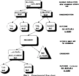
Fig 1. Experimental flow chart.
Ratio Changes Between Pretrial and Trial
Measure-Hours
n
Control Infants Study Infants
Pco2 (mm Hg)
P02/Fi02 (mm Hg/%)
n Pco2
(mm Hg)
Pao2/Fio2 (mm Hg/%)
-Pretrial
/2
1 6
14 10 10 6
34.7 ± 8.6 40.2 ± 9.0 41.2 ± 11.7 36.7 ± 8.4
2.45 ± 0.94 1.86 ± 0.48 2.03 ± 0.58 2.48 ± 1.24
13 10 11 10
38.1 ± 9.8 36.8 ± 11.2 37.2 ± 8.2 37.4 ± 9.4
2.86 ± 1.16 2.29 ± 0.84 2.44 ± 0.97 2.70 ± 1.39
* Results are
means ± SD.
first received CPAP via endotracheal tubes prior to
extubation, seven tolerated the six-hour trial prior to extubation and subsequently tolerated extuba-tion; significant apnea developed in six infants and significant respiratory acidosis with capillary pH
7.13 and Pco2 65 mm Hg developed after two hours of CPAP in one infant (a change from pretrial
capillary pH 7.34 and
Pco2
41 mm Hg). Thus, directextubation was associated with a significantly higher rate of successful extubation (13/13 v 7/14,
x2 8.7, P < .005). All seven infants who could not
be extubated were reventilated at the time failure
criteria were met; of these seven infants, all
even-tually were successfully extubated directly from
IMV. Three of the seven infants who could not be
extubated from CPAP were able to be weaned to extubation criteria within 24 hours and successfully extubated directly from IMV. Their results when
combined with above data also showed the same findings (16/16 v 7/14, x2 = 10.43, P < .002).
No
significant changes were noted in postextu-bation chest roentgenograms, and there were nosignificant changes in respiratory rates in any group during the trial. Changes in Pco2 and the P02/Fi02 ratio in both groups of infants between pretrial and trial measurements are shown in Table 2. No sig-nificant differences were noted in the absolute Pco2
and P02/Fi02 values between control and study infants. However, control infants who could not
eventually be extubated from CPAP had higher
Pco2
values at #{189}and one hour compared with infants who tolerated CPAP (45.8 ± 7.4 v 27.3 ±0.6 mm Hg, P < .05; 45.4 ± 4.1 v 30.7 ± 6.4 mm
Hg, P < .03, respectively) and with study infants
(45.8 ± 7.4 v 36.8 ± 11.2 mm Hg, P < .05; 45.4 ± 4.1 v 37.2 ± 8.2 mm Hg, P < .01, respectively). The
PATFNT PCPt.LAT1ON
SItI1Ofl aiteia
RNDOP4ZATION
WTCOPE
X2=tim
B CO.OO
asAn t
r
extiionRVER
TABLE 2. Pco2 and Po2 to Fi02
ments*
OUTCOPd -Combined X2- .43 S-i
0 U
0
5
4
3
2
0
-1
-2
PRE 0.5 (hoirs) 6
S 110
0
I
z
7 0 SUCCEID
. FAIL
6
5
4
3,
2
1 0
0
00
0 ‘ 000000000
.
S
S S
S
0 000000
STUDY CONTROL
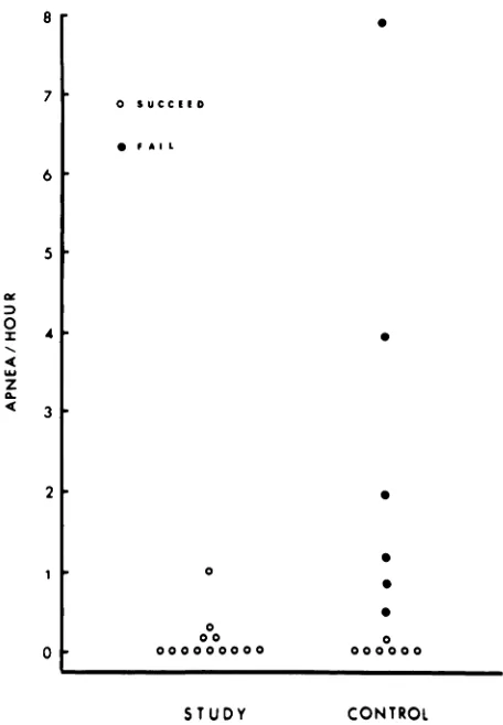
Fig 3. Apnea density, control v study infants. Number of moderate to severe apnea
(x2
= 5.84, df = 1, P < .025).Apnea density > 0.4 episodes per hour
(x2
= 4.89, df = 1,P= .03). same trend could not be statistically demonstrated
thereafter because only two control infants who
could not be extubated from CPAP remained on
CPAP beyond one hour of trial.
Because blood gas values were measured from
either capillary or arterial specimens from each
infant, relative differences in Pco2 (Pco2) values
and P02/Fi02 (%P02/Fi02) ratios were also
evalu-ated (Fig 2). As shown in Fig 2, the Pco2 values
of the entire control group was significant at all
intervals compared with pretrial Pco2 values.
Ar-terial 2Pco2 value in the control group was also significant at ‘/2 and six hours after CPAP in spite
of a limited number of arterial blood gas
measure-PRE 05 1 6
(hours)
Fig 2. Mean Pco2 and %P02/Fi02 values, study v con-trol infants. Top, Pco2 = trial Pco2 - pretrial Pco2.
Bottom, %Po2/Fi02 = (Po2/Fio2 #{247}baseline P02/Fi02) X
100. 0, continuous positive airway pressure via endotra-cheal tube prior to extubation; , direct extubation from
low intermittent mandatory ventilatory rates; numerals,
numbers of determinations at each time; circles con-nected with lines, values from arterial blood gas measure-ments; circles pointed by arrows, values from both arte-rial and capillary blood gas measurements. *P .05
compared with baseline value; ** < .01 compared with baseline value; *** < .001 compared with baseline value. Pco2 of control group was significantly higher than that of study group during test period.
ments. The differences were more pronounced
when compared with the study group (at #{189}hour P
< .001, at one hour P < .01, and at six hours P <
.01).
ArterialPco2
was also significantly larger inthe control than in the study group at six hours (P
<
.05).
The study group Pco2 values were neverelevated compared with pretrial values.
Although large alterations in alveolar-arterial 02 gradient as measured by the %P02/Fi#{248}2 ratio were
not seen, they were statistically different. The %P02/Fi02 decreased somewhat at #{189}and one hour after extubation in study infants compared with
pretrial values (Fig 2). This was confirmed by
ar-terial blood gas measurement, which remained sig-nificantly low at six hours after extubation although
less significantly. The control group %P02/Fi02 was decreased but not significantly at all times after initiation of CPAP via endotracheal tube, but a
small number of arterial blood gas measurements showed significant decreases in %P02/Fi02 ratios
at #{189}hour after initiation of CPAP.
Apnea was the most common cause of respiratory failure during the CPAP trials (Fig 3). The mci-dence of apnea overall was not altered, but apnea
requiring any stimulation was more common in
control infants. Apnea density greater than 0.4
episodes per hour was also more common in control infants.
Of 27 infants, 23 received therapeutic doses of theophylline prior to reaching study entry criteria.
Of the four infants not receiving theophylline, two were assigned to the control group and the other
two to the study group. Neither control infant could
be extubated from CPAP, and both study infants
tolerated direct extubation. If all infants who did not receive theophylline prior to the trial were
excluded, the success rate would be still
signifi-cantly different (11/11 v 7/12,
x2
=5.86,
P < .02). Control infants who could not be extubated from CPAP had a greater birth weight than those control infants who could (mean birth weight 1,031 v 950 g; t = 2.93, P < .001) but were similar in weight tostudy infants.
DISCUSSION
The use of CPAP of two to three cm H2O via
endotracheal tube for a period of up to 24 hours
before attempting extubation has been
recom-mended for infants after mechanical ventilation.’8 This practice has been suggested to ensure that the
patient will not become exhausted without mechan-ical ventilation.7 The current study demonstrated
that the recommended use of CPAP for a
preextu-bation trial is not only unnecessary but detrimental to VLBW infants prior to extubation. CPAP via an
endotracheal tube in VLBW infants was associated with mild CO2 retention after six hours of CPAP
even in those infants who tolerated the treatment.
In infants who could not eventually be extubated after CPAP, the increase in Pco2 value was
dra-matic during one hour of CPAP, but this could not be demonstrated thereafter because of limited
num-bers of blood gas measurements in control infants
who remained on CPAP for more than one hour.
These findings are consistent with previous studies by Fox et al3 in spontaneously breathing infants before and after extubation. In one study, those
infants who could not be extubated from CPAP
tended to have higher Pco2 values associated with a significantly higher airway resistance. In that
study, those infants who failed extubation were
considerably smaller than those who succeeded.
The findings of our study duplicated the increase
in PC02 seen in control infants who could not be
extubated; even control infants who could be extu-bated had slightly elevated PC02 values. Elevated
Pco2 values during CPAP would suggest that CPAP increases airway resistance and the work of breath-ing.9’1#{176}We have not measured work of breathing in
VLBW
infants, but LeSouef et al1’ demonstratedthat airway resistance is significantly increased
through endotracheal tubes compared with
new-born infants’ natural airways which increased dia-phragmatic work.11
In our study, all VLBW infants were either placed on CPAP or directly extubated from IMV
rates of six to ten per minute. This sudden removal of IMV was initially suggested by Gregory,5 which
may be more aggressive treatment than that used
by others who use IMV rates of less than six per minute before preextubation trials of CPAP.4’14 We do not believe that our method of weaning of
VLBW infants from IMV of six to ten breaths per minute significantly altered the success rate of
ex-tubation between the two groups, because we used
the same weaning method from IMV in both groups of infants after they were determined to be stable
in similar ventilatory settings. In addition, they were randomly assigned to either group only when
the primary physician determined that they were ready to be extubated.
Absolute values of P02/Fi02 during the trials did
not differ significantly from pretrial values in this study. However, in our VLBW infants low %Po2/
Fi02 values were found during the entire six hours after direct extubation but not during CPAP via an
endotracheal tube except at 30 minutes after
initi-ation of CPAP when arterial %P02/Fi02 values
were low. This is contrary to the results of previous
studies on larger newborn infants whose P02 values
were unchanged during small amounts of CPAP compared with that after extubation.”2 The results
of this study suggest that, in VLBW infants, even CPAP of 2 to 3 cm H2O may prevent subsegmental ventilation/perfusion abnormalities which may be associated with the lowered functional residual
Ca-pacity and lowered P02/Fi02 ratios. Further study
is warranted to determine “physiologic” CPAP of VLBW infants by comparing functional residual
capacity during IMV with low positive
end-expira-tory pressure and after extubation.
Failure of VLBW infants to tolerate CPAP was
caused most frequently by apnea rather than res-piratory acidosis. It is not possible to compare this finding with previously published studies because
the criteria for extubation failure were not
men-tioned.’4 Because the elevation of Pco2 was modest even in most control infants who were not to be
extubated from CPAP, we speculate that the
prox-imate cause of apnea in infants failing low amounts
of CPAP via endotracheal tubes is an increased
work of breathing due to increased airway
resis-tance.” Those infants in whom the work of
breath-ing is critically increased would presumably fail
In summary, this study demonstrates a
deleteri-ous effect of CPAP when applied via an endotra-cheal tube in VLBW infants. CPAP produces an increase in Pco2 which is associated with apnea
and with respiratory failure. Direct extubation is associated with a decrease in P#{248}2/Fi#{248}2ratio and an
increase in alveolar-arterial 02 gradient. Clinically,
this can be compensated by a small increase in Fi02. We believe that the effect of CPAP is to increase
airway resistance and work of breathing, which in
some instances is sufficient to produce ventilatory muscle fatigue and apnea. Thus, our
recommenda-tion and current practice is to directly extubate all infants weighing less than 1,250 g from IMV of six
breaths per minute rather than after a trial of
CPAP
via the endotracheal tube.ACKNOWLEDGMENT
We thank Dr Pramela Ramachandran for her cooper-ation in this study.
REFERENCES
1. Berman LS, Fox WW, Raphaely RC, et al: Optimum levels of CPAP for tracheal extubation of newborn infants. J Pediatr 1976;89:109-112
2. Fox WW, Berman LS, Dinwiddie RD, et al: Tracheal extu-bation of the neonate at 2-3 cm H2O continuous positive airway pressure. Pediatrics 1977;59:257-261
3. Fox WW, Schwartz JG, Schaffer TH: Successful extubation
of neonates: Clinical and physiological factors. Crit Care Med 1981;9:823-826
4. Harris MC, Baumgart S, Rooklin AR, et al: Successful extubation of infants with respiratory distress syndrome. J Pediatr 1983;103:303-305
5. Gregory G: Respiratory care of newborn infants. Pediatr Clin NorthAm 1971;19:311-324
6. Barr PA: Weaning very low birth weight infants from me-chanical ventilation using intermittent mandatory
ventila-tion and theophylline. Arch Dis Child 1978;53:598-600 7. Nugent J, Hanks H, Goldsmith JP: Pulmonary care, in
Goldsmith JP, Karotkin EH (edo): Assisted Ventilation of
Neonate. Philadelphia, WB Saunders Co, 1981, p 79 8. Brady JP, Gregory G: Assisted ventilation, in Klaus MK,
Fanaroff AA (eds): Care of the High Risk Neonate. Philadel-phia, WB Saunders Co, 1979, p 218
9. Wall M: Infant endotracheal tube resistance: Effects of
changing length, diameter, and gas density. Crit Care Med 1980;8:38-40
10. Cave P, Fletcher G: Resistance of nasotracheal tubes used
in infanth. Anesthesiology 1968;29:588-590
11. LeSouef PN, England SJ, Bryan AC: Total resistance of the respiratory system in preterm infants with and without an endotracheal tube. J Pediatr 1984;104:108-111
12. Muller N, Guiston G, Cade D, et al: Diaphragmatic muscle fatigue in the newborn. J Appi Physiol 1979;46:688-695 13. Keens TG, Bryan AC, Levison H: Developmental pattern of
muscle fibre types in human ventilatory muscles. J Appi
Physiol 1978;44:909-913
CONFUSING
± SIGN
We suggest that all journals discontinue the use of the ± sign; in all instances it should be clearly indicated whether SD or SE is quoted; the SE should not
be used to indicate the variability of a set of observations; and the reporting of appropriate ranges or confidence intervals should be shown directly.
Submitted by Student