ORIGINAL ARTICLE
IJPHY
ABSTRACT
Background: Few studies have examined the effectiveness of modified constraint-induced movement therapy (mCIMT) for the paretic lower limb following stroke. This study aimed to investigate the effects of mCIMT on motor function of the lower limb in stroke patients.
Methods: A randomized, controlled study of 30 participants, who were randomized to 2 groups, was conducted. The study group received mCIMT, and the control group received neurodevelopmental therapy (NDT) for two weeks. All were evaluated for motor function through the Functional Ambulation Classification (FAC), Berg Balance Scale (BBS), 10-Meter Walk Test, gait parameters (cadence and step length ratio) and postural symmetry ratio at pretreatment and post-treatment, like two times.
Results: The improvements in BBS score, postural symmetry ratio, step length ratio, cadence and walking velocity had greater in the study group than the control group (P < 0.05). The improvement of FAC score was more pronounced in the study group (P = 0.005).
Conclusion: mCIMT for paretic lower limb had superior effect against the NDT to enhance the motor function (gait parameters, balance, ambulation, and symmetry) in patients with stroke. mCIMT may be used as a new alternative treatment for lower limb rehabilitation.
Keywords: Modified constraint-induced movement therapy, gait, balance, stroke, lower limb, neurodevelopmental therapy.
Received 09th July 2017, revised 25th September 2017, accepted 04th October 2017
www.ijphy.org
10.15621/ijphy/2017/v4i5/159421
CORRESPONDING AUTHOR
Int J Physiother. Vol 4(5), 269-277, October (2017) ISSN: 2348 - 8336
EFFECTS OF MODIFIED CONSTRAINT-INDUCED MOVEMENT
THERAPY FOR LOWER LIMB ON MOTOR FUNCTION IN STROKE
PATIENTS: A RANDOMIZED CONTROLLED STUDY
*1
Sevim ACARÖZ CANDAN. PT, Ph.D.
²
Ayşe LİVANELİOĞLU.PT, Ph.D.
*1
Sevim ACARÖZ CANDAN. PT, Ph.D.
Assistant Professor, Department of Physiother-apy and Rehabilitation, Health Science Faculty, Ordu University, Ordu, Turkey.
e-mail: [email protected] ²Professor, Department of Physiotherapy and
Rehabilitation, Health Science Faculty, Hacettepe University, Ankara, Turkey.
INTRODUCTION
Stroke is one of the main causes of disability and morta-lity in the adult population in the developed World [1].The loss of motor control, abnormal movement pattern, tone disorders, coordination difficulties, and sensory dysfuncti-on of the lower extremities which arise post-stroke period reduces motor function [2, 3].In spite of this, the patients experience to walk from the early period, but their walking pattern is slow, inefficient, unstable and incoordination. Consequently, Taub et al. (1999) reported that “learned misuse” develops for the lower limb function in stroke pa-tients [4, 5].
Constraint-induced movement therapy (CIMT) or modi-fied constraint-induced movement therapy (mCIMT) re-fers to a specific treatment for a motor disability that com-bines constraint of the movements in the nonparetic limb, intensive practice, and shaping of the behavior to improve the amount of use of the paretic limb during daily life ac-tivities [6].The developed treatment for the paretic upper limb improves the use of the paretic limb functionally by overcoming the “learned disuse” that develops following stroke [7, 8].Ro et al. (2006) have been shown the posi-tive effects of CIMT on motor function of the upper limb and motor network in the brain after stroke [9]. Similar-ly, in recent years, mCIMT is also applied for the paretic lower limb to overcome “learned misuse” and increase the quality of motor function [10, 11].Due to the bipedal ort h of lower limb activities, it is difficult to apply the cons-traint of movements in the nonparetic lower limb which is one of the main components of the CIMT. Therefore, the studies are controversial in the constraint. While some authors suggested that nonparetic limb restriction was the least important point for lower limb CIMT, with intensive practice and functional activities being noteworthy [12-14],the others used one partial restriction method in dif-ferent way, such as the whole leg orthosis, the below-knee prosthesis, the addition of a shoe insert [15-17].However, to our knowledge, the combination of the two different partial restriction method has not been used to increase the constraint of the nonparetic lower limb in the previous studies of mCIMT. Thus, this study targeted to examine whether mCIMT which applied the all main components of mCIMT without compromising from the original proto-col was effective in improving the motor functioning of the lower limb in patients with stroke. We hypothesized that mCIMT program would lead to specific improvements in balance, gait and symmetry outcomes compared with the neurodevelopmental therapy (NDT).
METHODS
Study design and participants
This study was planned as a prospective, assessor-blind, randomized controlled study. The Hacettepe University Non-interventional Clinical Research Ethics Boards ap-proved the study protocol of this study (approval no: GO 14/22-15). Between February 2014 and February 2015, 153 patients were screened for study eligibility, 33 patients of them were enrolled in the study as participants and 30
par-ticipants completed all therapy sessions. The first patient enrolled in the study at 1 February 2014. The participants were recruited from the two hospitals. All participants were informed about the study and signed written infor-med consent forms before assessment.
Participants were included in this study if they had been diagnosed a single cerebrovascular accident in the cerebral hemisphere, as determined by computed tomography or magnetic resonance imaging; were > 18 age of years; were between 3 and 12 months post onset; exhibited mild to moderate disability according to lower extremity recovery stages (Brunnstrom recovery stages III-V); could partici-pate an intensive rehabilitation programme that permitted by the physiatrist.
Patients were excluded if they had a recurring stroke, had medical comorbidities (e.g., unstable blood pressure, car-diac pacemaker), had an unstable medical condition (e.g., severe acute myocardial infarction), had cognitive impa-irment, additional neurological problems and orthopedic problems that are preventing participation to an intensive rehabilitation program. Thirty-three subjects were initial-ly recruited ort he studies and randominitial-ly divided into two groups, a control group (n=15) and a study group (n=18). Two participants were excluded because of severe knee pain and having unstable blood pressure and one subject in the study group withdrew voluntarily from the study. Thirty participants completed the treatment.
The sample size for each group was determined to be 11 participants, based on another study whose design was si-milar to that of our study [18]. The Power Analysis and Sample Size software package (PASS 14) were used. The confidence level for statistical significance was set at 5% with power equal to 80%. An average effect size of 0.59 for the outcome measures was sustained. Although the cal-culated sample size for each group was 11 participants, at least 15 participants were included in each group due to dropout problem during the study.
Randomization
After the inclusion criteria were fulfilled and the partici-pants signed the written informed consent form, the ran-domization was conducted using the block ranran-domization procedure by a physiotherapist who was not involved in this study. The randomization was stratified based on age (≤60 or >60 years), gender (male or female), the hemip-legic side ( left or right) and type of stroke (ischemic or hemorrhagic) because of balanced distribution of the par-ticipants according to important parameters which have known prognostic effects [19].
Outcome measure
periods, patients started and ended their tests 3 m before and after the walkway. The time along this walkway was recorded in seconds with a stopwatch. The velocity was calculated in m/s [20]. This test has demonstrated excel-lent test-retest reliability (ICC = 0.95-0.99) [21], interra-ter reliability (ICC = 0.998) [22],and intrarater reliability (ICC=0.87 to 0.88) [23].
Cadence was measured by counting the step number in one minute and was recorded in steps/min [24].
Balance performance was assessed by the Berg Balance Scale (BBS). The BBS, a 14-item scale designed to measure balance in a clinical setting with a maximum score of 56 [25].Each item is scored from 0 (cannot perform) to 4 (the best performance). In this scale, a score of 0–20 indicates that the subject has poor balance, a score of 21–40 means acceptable balance ability, and a score of 41–56 means ad-vanced ability. The scale is considered to be a valid and re-liable measurement of the balance control ability [26].The ambulation was quantified by using the Functional Ambu-lation Classification (FAC), which has excellent test-retest reliability and interrater reliability [23]. The patients can be rated through the observation of the physiotherapist between 0 (cannot walk) and 5 (can walk independently anywhere) categories [27].
Step length asymmetry was determined by a step length ratio (SLR) which was defined paretic step length divided by non-paretic step length [28]. Step length was measured using the simple footprint method [29].The footprint met-hod is easy, inexpensive [30, 31]and the method is high-ly reliable both interrater and intrarater (ICC=0.92-1.00) [32]. The patient was seated on an armrest chair and was dusted both feet with the powder. Following, the patient walked at their self-selected walking speed over the dark floor. Measurementof the middle line 3 meters of the walk-way was taken only to consider the normal pattern of gait and avoid the impact of the acceleration and deceleration. The feet prints were marked by a pen and a ruler between the heel and toe points. Step length was calculated as a li-near distance on the midline between two successive cont-ralateral center of the heel [33].
Postural symmetry ratio was calculated using the following equation (1) [34]:
(1)
Load on the nonparetic lower limb was measured with two digital weighing scale (Scale-Sinbo, SBS-4421) while pa-tients normally stood with one foot on each scale platform and arms hanging loosely by their sides [35].During the assessment, the display screen of the scale was closed to avoid receiving feedback from the screen when the patient supposed to be standing with each leg equally loaded [36]. Then, the subject was positioned on the platform of the scale, and the entire body weight was recorded. The mea-surements were repeated three times. A percentage of 0 de-monstrated equal weight bearing and best postural control in standing position, while a higher percentage demonstra-ted asymmetry and poor postural control [34].
Intervention
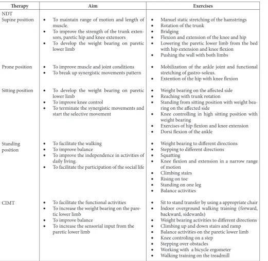
Each program for two groups was executed by a physi-otherapist, who was the first author and had eight years working experience in the neurologic rehabilitation field. mCIMT was conducted for the paretic lower limb in the study group for two weeks, per week five day and each ses-sion 120 minutes. CIMT included three main components: the intensive practice of the functional activities, limited use of the nonparetic lower limb and transferring the ga-ins from the training session to the patient’s real environ-ment with “transfer package” [7]. The intensive practice was applied to the selection of the appropriate functional activities by the “shaping” principles by the feedback, co-aching, modeling, and encouragement of the physiothera-pist. The functional activities described in Table 1. Each of the functional activities was performed intensively by the paretic lower limb, unilaterally, over 20 minutes, repeti-tively. After each 20-minute activity period, a rest period of 5 minutes was given to the patients. The constraint of the nonparetic lower limb was known to be difficult beca-use of the bilateral characteristics of lower limb function. Therefore, the constraint was fulfilled by a combination of 2 different methods; 1) immobilization of the knee of the nonparetic limb with a whole-leg orthosis and 2) use of a shoe insert on the nonparetic lower limb (Figure 1) [15, 36, 37]. This method may be increased the restriction of the nonparetic lower limb. The shoe insert had a 1 cm lift and 5° lateral wedge, and the whole-leg orthosis constrained the knee flexion at the fullest extension position. Training on the use of the orthosis and shoe insert was provided for patients and their relatives. The restriction was used du-ring treatment sessions, including on the treadmill, and for 90% of the participants’ waking hours. When the patient took off the orthosis, the range of motion exercises of the knee was asked to perform to prevent the contracture. The used functional activities during therapy session were set to home as a home program with transfer package, and the participants were strongly encouraged to incorporate the use of their paretic lower limb into their daily activities. The exercises in the training were personalized and adjus-ted during treatment to fulfill the specific needs of each as determined by the degree of impairment and endurance level. The session was immediately interrupted if the blood pressure and heart rate of participants were uncontrolled or the patient felt chest discomfort.
Figure 1: Constraint method of the nonparetic lower limb:
(a) Whole leg orthosis, (b) addition of a shoe insert in the shoe of the nonparetic lower limb. The shoe insert relati-vely changed the weight-bearing symmetry from the non-paretic lower limb to the non-paretic lower limb [15,37].
Statistical Analysis
All statistical analyses were performed with SPSS 20.0 (IBM Corp, Chicago, IL, US). The variables were investi-gated using the Kolmogorov-Smirnov test to determine whether or not they were normally distributed. Descriptive analyses were performed and presented using means and standard deviations for results of the balance, gait parame-ters and symmetry; medians and interquartile range ort he FAC score. The paired t-test was used to compare the chan-ges in the groups within the times. The improvements in the outcome measures were quantified by subtracting the pretreatment scores from the posttreatment scores. Then, Student’s t-test was used to compare the differences in the-se changes between groups. Chi-square analysis was uthe-sed to compare proportions. Values of p<0.05 were deemed statistically significantly different.
Table 1: Details of the exercises in NDT and CIMT.
Therapy Aim Exercises
NDT
Supine position
Prone position
Sitting position
Standing position
CIMT
• To maintain range of motion and length of
muscle.
• To improve the strength of the trunk exten-sors, paretic hip and knee extensors
• To develop the weight bearing on paretic
lower limb
• To improve muscle and joint conditions • To break up synergistic movements pattern
• To develop the weight bearing on paretic
lower limb
• To improve knee control
• To terminate the synergistic movements and start the selective movement
• To facilitate the walking
• To improve balance
• To improve the independence in activities of daily living.
• To facilitate the participation of the social life
• To facilitate the functional activities
• To increase the weight bearing on the
pare-tic lower limb
• To improve balance
• To increase the sensorial input from the paretic lower limb
• Manuel static stretching of the hamstrings • Rotation of the trunk
• Bridging
• Flexion and extension of the knee and hip
• Lowering the paretic lower limb from the bed
with hip extension and knee flexion
• Pushing the wall with both limbs
• Mobilization of the ankle joint and functional
stretching of gastro-soleus.
• Extention of the hip with knee flexion
• Weight bearing on the affected side • Reaching with trunk rotation
• Standing from sitting position with weight bea-ring on the affected side
• Knee controlling in high sitting position with
weight bearing
• Exercises of hip flexion and knee extension
• Dorsi flexion of the ankle
• Weight bearing to different directions
• Stepping to different directions
• Squatting
• Knee flexion and extension in a narrow range
of motion
• Climbing stairs
• Rising on toe
• Standing on one leg • Balance activities
• Sit to stand transfer by using a appropriate chair
• İndoor overground walking training (forward, backward, sidewards)
• Weight bearing activities to different directions
• Climbing up and down stairs and ramp
• Balance activities on the paretic lower limb • Knee controling on a step
• Stepping over obstacles
• Working with a bicycle ergometer
RESULTS
Figure 2 demonstrates the flow of the participants’ enroll-ment throughout the study. A total of 30 participants inc-luded 16 females and 14 males with a mean age of 56.4 ±
13.45 and average time poststroke of 6.7 ± 2.94 months.
Participants were randomly assigned into the study (n = 15) or control group (n = 15). The clinical characteristics of the participants are summarised in Table 2. There were
no significant differences between the groups at baseline, including demographic data and motor recovery of the lower extremity of Brunnstrom stages.
Figure 2: Study design and sample flowchart.
Table 2: Participant Characteristics.
Study group Control group pa pb
No. of subjects, n
Age in yearsc
Poststroke monthsc
BMI in kg/m2c
Gender, n (%) Male Female
Affected side, n (%) Left
Right
Type of stroke, n (%) Ischemia
Hemorrhage Brunnstrom reco-very stage, n (%) III
IV V
15
55.13 ± 14.70 6.80 ± 2.70 26.26 ± 3.49 8 (53.3) 7 (46.7) 10 (66.7) 5 (33.3) 11 (73.3) 4 (26.7) 12 (80) 3 (20) -15
57.67 ± 12.20 6.63 ± 3.18 29.71 ±7.56 6 (40) 9 (60) 10 (66.7) 5 (33.3) 12 (80) 3 (20) 11 (73.3) 4 (26.7) .61 .88 .12 .46 1.0 .66 .66
aStudent’s t test. bChi Square test.
cValues are given as mean ± standart deviation.
Each therapy methods significantly improved the Berg balance score, walking velocity, cadence and ratio of step length and postural symmetry. When the participants were generally at level 2 and three at pretreatment according to FAC, they developed functionally and progressed to level 4
and 5. Although the median score of FAC level remained at 3 in the control group and changed from 3 to 4 in the study group, the both improvement were meaningful (P < 0.001). Table 3 shows the effects of these therapies.
Table 3: Effects of a Modified Constraint Induced Mo-vement Therapy and a Neurodevelopmental Therapy on
Berg Balance Scale, Functional Ambulation Scale, Wal-king Velocity, Cadance, Step Length Ratio and Postural
Symmetry Ratio in Patients With Stroke.
Study group(n=15) p Control group(n=15) p
BBSa FACb Walking velocity (m/s)a Cadence (step/min)a Step length ratioa Postural symmetry ratioa Pretreat-ment 32.67± 9.64 (15-44) 3 (2-4) 0.33 ± 0.13 (0.09-0.51)
64.33 ± 15.14 (36-91)
1.50 ± 0.56 (0.93-2.56)
16.58 ± 5.07 (6.52-27.96)
Posttreat-ment 43.47 ± 7.60 (24-52)
4 (2-5) 0.44 ± 0.18 (0.11-0.69)
76.40 ± 16.83 (40-104)
1.13 ± 0.29 0.72-1.73)
7.15 ± 4.00 (1.36-15.35) <0.001 <0.001 <0.001 <0.001 0.001 <0.001 Pretreat-ment 34.13 ± 9.12 (11-45)
3 (2-4) 0.34 ± 0.13 (0.13-0.56)
64.33 ± 12.48 (36-76)
1.15 ± 0.45 (0.73-2.65)
13.60 ± 7.46 (2.77-33.37) Posttreat-ment 39.80± 8.89 (20-53) 3 (2-5) 0.38 ± 0.15 (0.15-0.71)
69.87 ± 12.62 (40-82)
1.07 ± 0.24 (0.72-1.66)
10.28 ± 6.40 (0.34-26.39) <0.001 <0.001 <0.001 <0.001 0.040 <0.001
Abbreviations: BBS, berg balance scale; FAC, functional ambulation classification.
aValues are given as mean ± standard deviation
(mini-mum- maximum).
bValues are given as median (minimum- maximum).
Bold values: p<0.05.
The comparison of the improvements on the motor fun-ction score among two groups are shown in Table 4. All motor function domains were found significantly greater improvement in the study group when compared with the control group. Motor function significantly improved the following scores: balance (net change of control group vs study group, respectively: 5.7 vs 10.8; P = 0.002), ambulati-on (0.40 vs 0.93; P = 0.005), walking velocity (0.04 vs 0.11; P = 0.010), cadance (5.5 vs 12.1; P < 0.001), step length ratio (-0.1 vs -0.4; P= 0.035) and postural symmetry ratio (3.3 vs 9.4; P < 0.001).
Table 4: Comparison of therapy effects on berg balan-ce scale, functional ambulation scale, walking velocity, cadance, step length ratio and postural symmetry ratio
between groups.a
Changes in motor func-tion Study group (n=15) Control group (n=15) 95% confidence
interval pb
Lower Upper
BBS FAC Walking velo-city (m/s) Cadence (step/min) Step length ratio Postural sym-metry ratio
10.80 ± 4.54 0.93 ± 0.46
0.11 ± 0.08
12.07 ± 5.52
-0.37 ± 0.43
-9.43 ± 3.66
5.67 ± 3.39 0.40 ± 0.51
0.04 ± 0.04
5.53 ± 3.23
-0.08 ± 0.26
-3.32 ± 1.60
2.14 8.13 0.17 0.89
-0.02 0.12
3.15 9.91
-0.55 -0.02
-8.27 -3.95
0.002 0.005 0.010 <0.001 0.035 <0.001
body mass index.
aValues are given as mean ± standard deviation. b Student’s t Test.
Bold values: p<0.05. DISCUSSION
In this study, we investigated the effectiveness of the mCIMT versus NDT on the motor function of the pare-tic lower limb during stroke rehabilitation. These results suggest that mCIMT can be an efficacious rehabilitation method for patients with stroke. mCIMT increased the walking velocity, cadence, score of the BBS, and improved step length symmetry and postural symmetry significantly better when compared with NDT.
For the lower limb, the studies differ regarding the intensi-ve practice period, the presence of the constraint, method of the constraint and duration of the constraint, because of these differences and any consensus on the CIMT protocol of paretic lower limb, the comparison of the studies are dif-ficult [17]. Although Regnaux et al. (2002) and Bonnyaud et al. (2013)have used the same protocol for one session, 20 minutes, they have found different results [38, 39]. Cor-responding with these studies, their constraint method or duration of their treatment can be considered to be ina-dequate. Hase et al. (2011) have used a below-knee prost-hesis as the constraint method and applied CIMT tech-niques during three weeks, a 3-5 session in a day and each session lasted 5 minutes. They have shown that the stance phase of the paretic limb improved, but gait parameters did not change [16]. Wang et al. (2012)studied the comparison between the Bobath therapy and CIMT without the const-raint of the non-paretic limb [40]. As a result of this study, they suggested that the walking velocity and the balance ability increased.
It is known that the loss of loadperceptionin the paretic limb likely impacts the control of walking in stroke survi-vors [41], the present study has been applied two different constraint method together to increase this perception. One of this constraint method have been used by Rodrigu-ez et al. (2002) found that the symmetrical weight bearing increased walking speed and step length [37]. Kallio et al. (2014) applied the other constraint method which is also used in this study., Numata et al., (2008) Marklund and Klassbo,(2006) and according to them the motor functi-on, balance, walking and weight bearing ability improved significantly [17, 42, 36]. Similarly, with these studies, the present study showed also significant improvements in the walking velocity, cadence, balance ability, postural sym-metry, step length symmetry and functional ambulation. It is known that the minimal clinically important difference in the walking velocity is 0.06 m/s [43]. Thus, the impro-vement observed in the study group suggest functional im-provements in the walking velocity. Also, Perry et al. (1995) reported that ambulation ability had been correlated with walking velocity and they categorized the ambulation th-ree subgroups [44]. According to this classification, while the participants in the study groups were more likely to be
household ambulators at pretreatment and progressed to limited community ambulators at the end of the treatment, the participants in the control group remained at the same household ambulators level. Because the walking velocity changed from 0.28 m/s to 0.44 m/s in the study group and from 0.28 m/s to 0.38 m/s in the control group. Similarly, with these results, the change of FAC level suggested the functional improvement and decreased the requirement of the assistance of the another people. As a result of functi-onal improvements in FAC, while the study group prog-ressed from level 3 to level 4, the control group was still at level 3. Moreover, Nakamura et al. (1988)demonstrated that the walking velocity was improved by increasing the cadence and step length [45]. The results of this study have also suggested Nakamura et al. because in the study group have meaningful changes in the walking velocity, cadence and nonparetic step length.
Berg balance scale was sensitive to change and had excel-lent test-retest reliability in stroke population [46]. The mi-nimal clinical detectable change was found 4.66,and 6.9 in the different studies [46, 47]. The results of the present study also suggested that the change of the balance ability in both groups will be reflected the function. Nevertheless, the control group remained in the acceptable balance le-vel. Also, while the BBS score in the study group increased from acceptable to good balance level, Berg et al. (1992) reported that this improvement (from 22 to 43.5) requires the using of walking assistance device in daily life to avoid the falling [48].
Quadri-ceps activity [54]. This mechanism also may lead to relati-vely declining the compensatory strategies in gait (e.g., stiff knee gait). We showed that mCIMT is effective to increa-se the weight bearing on a paretic limb that is one of the major problems to avoid the sufficient gait pattern. We are thinking of the developing of the gait symmetry constitu-tively by limb loading. Therefore, the primary goal of the rehabilitation to develop the gait symmetry should be to increase the weight bearing following a stroke.
The main strengths of this study are the randomized cont-rolled study, and the comprehensive assessment of outco-mes related to motor function (objective measures of gait, balance, symmetry, and ambulation level). Moreover, the present therapy was transferred into the patient’s daily life outside of training session. The transferring the gains from the training session reinforced the motor learning. Last-ly, two different constraint method was used firstly in the current study to increase the weight bearing on an affected lower limb and the representation area of the paretic limb. However, this study also has some limitations. First, despi-te the sample size calculation, the sample size was relatively small. Demonstration of these results is required with a lar-ger sampling. Second, the results can only be generalized to patients who met the inclusion criteria. Third, the effect of the therapy is not clear whether arise from the const-raint method. Therefore, further study would evaluate the efficacy of using different constraint settings in patients with stroke. Fourth, the physiotherapist was not blind to the application of the treatment program, but the assessor physiotherapist was blind to groups. We think that this fa-ctor may strengthen the study. In the present study, two different constraint method is used firstly, and their effect on the brain structure has not been reported. Therefore, further studies are needed to investigate whether this met-hod causes the changes in brain structure.
CONCLUSION
mCIMT for lower limb greater improved the balance, gait performance, ambulation level and symmetry in patients with mild to moderate stroke when compared with NDT. The therapy program should practice for many patients with different stages of stroke (acute, subacute, chronic). Further studies of these effects in a long-term follow-up are needed to determine the effectiveness period.
Acknowledgements
The authors would like to show their gratitude the partici-pating hospitals, all the patients, their families and physio-therapists who attended to this work.
Declaration of Conflicting Interests
The authors have no conflict of interest to declare of this article.
REFERENCES
[1] Ferrarello F, Baccini M, Rinaldi LA, Cavallini MC, Mossello E, Masotti G, et al.Efficacy of physiotherapy interventions late after stroke: a meta-analysis. J Neurol Neurosurg Psychiatry. 2011;82(2):136-43.
[2] de Wit DC, Buurke JH, Nijlant JM, Ijzerman MJ, Hermens HJ. The effect of an ankle-foot orthosis on walking ability in chronic stroke patients: a randomized controlled trial. Clin Rehabil. 2004;18(5):550-7. [3] Esquenazi A, Ofluoglu D, Hirai B, Kim S. The effect of
an ankle-foot orthosis on temporal spatial parameters and asymmetry of gait in hemiparetic patients. Pm r. 2009;1(11):1014-8.
[4] Taub E, Uswatte G, Pidikiti R. Constraint-Induced Movement Therapy: a new family of techniques with broad application to physical rehabilitation--a clinical review. J Rehabil Res Dev. 1999;36(3):237-51.
[5] Woolley SM. Characteristics of gait in hemiplegia. Top Stroke Rehabil. 2001;7(4):1-18.
[6] Mark VW, Taub E. Constraint-induced movement therapy for chronic stroke hemiparesis and other disabilities. Restor Neurol Neurosci. 2004;22(3-5):317-36.
[7] Morris DM, Taub E. Constraint-induced therapy approach to restoring function after neurological injury. Top Stroke Rehabil. 2001;8(3):16-30.
[8] Taub E, Uswatte G, Mark VW, Morris DM. The learned nonuse phenomenon: implications for rehabilitation. Eura Medicophys. 2006;42(3):241-56.
[9] Ro T, Noser E, Boake C, Johnson R, Gaber M, Speroni A, et al.Functional reorganization and recovery after constraint-induced movement therapy in subacute stroke: case reports. Neurocase. 2006;12(1):50-60. [10] Duncan PW. Synthesis of Intervention Trials To
Improve Motor Recovery following Stroke. Top Stroke Rehabil. 1997;3(4):1-20.
[11] Gray CK, Culham E. Sit-to-Stand in People with Stroke: Effect of Lower Limb Constraint-Induced Movement Strategies. Stroke Res Treat. 2014;2014:683681.
[12] Mishra S, Chitra J. Effect of modified constraint induced movement therapy (mCIMT) for lower limb on weight bearing symmetry and balance in stroke patients: a pre-post experimental study. International Journal of Scientific Research. 2014;3(6):485-8. [13] Uswatte G, Taub E, Morris D, Barman J, Crago J.
Contribution of the shaping and restraint components of Constraint-Induced Movement therapy to treatment outcome. NeuroRehabilitation. 2006;21(2):147-56. [14] Vearrier LA, Langan J, Shumway-Cook A, Woollacott
M. An intensive massed practice approach to retraining balance post-stroke. Gait Posture. 2005;22(2):154-63. [15] Aruin AS, Rao N, Sharma A, Chaudhuri G. Compelled
body weight shift approach in rehabilitation of individuals with chronic stroke. Top Stroke Rehabil. 2012;19(6):556-63.
[16] Hase K, Suzuki E, Matsumoto M, Fujiwara T, Liu M. Effects of therapeutic gait training using a prosthesis and a treadmill for ambulatory patients with hemiparesis. Arch Phys Med Rehabil. 2011;92(12):1961-6.
in elderly persons with chronic stroke: single-subject experimental design study. Top Stroke Rehabil. 2014;21(2):111-9.
[18] Hüseyinsinoğlu BE. İnmeli hastalarda üst ekstremite iyileşmesi üzerine kısıtlayıcı zorunlu hareket tedavisi ve Bobath tedavi yaklaşımının etkileri [Dissertation]. Istanbul University; 2010.
[19] Bayona NA, Bitensky J, Foley N, Teasell R. Intrinsic factors influencing post stroke brain reorganization. Top Stroke Rehabil. 2005;12(3):27-36.
[20] Balasubramanian CK, Neptune RR, Kautz SA.
Variability in spatiotemporal step characteristics and its relationship to walking performance post-stroke. Gait Posture. 2009;29(3):408-14.
[21] Collen FM, Wade DT, Bradshaw CM. Mobility after stroke: reliability of measures of impairment and disability. Int Disabil Stud. 1990;12(1):6-9.
[22] Wolf SL, Catlin PA, Gage K, Gurucharri K, Robertson R, Stephen K. Establishing the reliability and validity of measurements of walking time using the Emory Functional Ambulation Profile. Phys Ther. 1999;79(12):1122-33.
[23] Mehrholz J, Wagner K, Rutte K, Meissner D, Pohl M.
Predictive validity and responsiveness of the functional ambulation category in hemiparetic patients after stroke. Arch Phys Med Rehabil. 2007;88(10):1314-9. [24] Pohl M, Mehrholz J, Ritschel C, Ruckriem S.
Speed-dependent treadmill training in ambulatory hemiparetic stroke patients: a randomized controlled trial. Stroke. 2002;33(2):553-8.
[25] Berg K, Wood-Dauphinee S, Williams JI. The Balance Scale: reliability assessment with elderly residents and patients with an acute stroke. Scand J Rehabil Med. 1995;27(1):27-36.
[26] Geiger RA, Allen JB, O’Keefe J, Hicks RR. Balance and mobility following stroke: effects of physical therapy interventions with and without biofeedback/force plate training. Phys Ther. 2001;81(4):995-1005.
[27] Kwakkel G, Kollen B, Twisk J. Impact of time on improvement of outcome after stroke. Stroke. 2006;37(9):2348-53.
[28] Balasubramanian CK, Bowden MG, Neptune
RR, Kautz SA. Relationship between step length asymmetry and walking performance in subjects with chronic hemiparesis. Arch Phys Med Rehabil. 2007;88(1):43-9.
[29] Baker R. Gait analysis methods in rehabilitation. J Neuroengineering Rehabil. 2006;3:4.
[30] Wilkinson MJ, Menz HB, Raspovic A. The
measurement of gait parameters from footprints. The Foot. 1995;5(2):84-90.
[31] Zverev Y, Adeloye A, Chisi J. Quantitative analysis of gait pattern in hemiparetic patients. East Afr Med J. 2002;79(8):420-2.
[32] Wilkinson MJ, Menz HB. Measurement of gait parameters from footprints: a reliability study. The Foot. 1997;7(1):19-23.
[33] Zverev YP. Spatial parameters of walking gait and
footedness. Ann Hum Biol. 2006;33(2):161-76. [34] Wong AM, Lee MY, Kuo JK, Tang FT. The development
and clinical evaluation of a standing biofeedback trainer. J Rehabil Res Dev. 1997;34(3):322-7.
[35] Mansfield A, Danells CJ, Inness E, Mochizuki G, McIlroy WE. Between-limb synchronization for control of standing balance in individuals with stroke. Clin Biomech (Bristol, Avon). 2011;26(3):312-7. [36] Marklund I, Klassbo M. Effects of lower limb intensive
mass practice in poststroke patients: single-subject experimental design with long-term follow-up. Clin Rehabil. 2006;20(7):568-76.
[37] Rodriguez GM, Aruin AS. The effect of shoe wedges and lifts on symmetry of stance and weight bearing in hemiparetic individuals. Arch Phys Med Rehabil. 2002;83(4):478-82.
[38] Bonnyaud C, Pradon D, Zory R, Bussel B, Bensmail D, Vuillerme N, et al.Effects of a gait training session combined with a mass on the non-paretic lower limb on locomotion of hemiparetic patients: a randomized controlled clinical trial. Gait Posture. 2013;37(4):627-30.
[39] Regnaux JP, Pradon D, Roche N, Robertson J, Bussel B, Dobkin B. Effects of loading the unaffected limb for one session of locomotor training on laboratory measures of gait in stroke. Clin Biomech (Bristol, Avon). 2008;23(6):762-8.
[40] Wang W, Wang A, Yu L, Han X, Jiang G, Weng C, et al. Constraint-induced movement therapy promotes brain functional reorganization in stroke patients with hemiplegia. Neural Regen Res. 2012;7(32):2548-53.
[41] Bohannon RW. Evaluation and Treatment of Sensory and Perceptual Impairments Following Stroke. Topics in Geriatric Rehabilitation. 2003;19(2):87-97.
[42] Numata K, Murayama T, Takasugi J, Oga M. Effect of modified constraint-induced movement therapy on lower extremity hemiplegia due to a higher-motor area lesion. Brain Inj. 2008;22(11):898-904.
[43] Perera S, Mody SH, Woodman RC, Studenski SA.
Meaningful change and responsiveness in common physical performance measures in older adults. J Am Geriatr Soc. 2006;54(5):743-9.
[44] Perry J, Garrett M, Gronley JK, Mulroy SJ.
Classification of walking handicap in the stroke population. Stroke. 1995;26(6):982-9.
[45] Nakamura R, Handa T, Watanabe S, Morohashi I. Walking cycle after stroke. Tohoku J Exp Med. 1988;154(3):241-4.
[46] Hiengkaew V, Jitaree K, Chaiyawat P. Minimal detectable changes of the Berg Balance Scale, Fugl-Meyer Assessment Scale, Timed “Up & Go” Test, gait speeds, and 2-minute walk test in individuals with chronic stroke with different degrees of ankle plantarflexor tone. Arch Phys Med Rehabil. 2012;93(7):1201-8.
Citation
ACARÖZ CANDAN, S., & LİVANELİOĞLU, A. (2017). EFFECTS OF MODIFIED CONSTRAINT-INDUCED MOVEMENT THERAPY FOR LOWER LIMB ON MOTOR FUNCTION IN STROKE PATIENTS: A RANDOM-IZED CONTROLLED STUDY. International Journal of Physiotherapy, 4(5), 269-277.
2001;47(1):29-38.
[48] Berg K, Wood-Dauphinee SL, Williams JI, Maki B.
Measuring balance in the elderly: validation of an instrument. Can J Public Health. 1992;83 Suppl 2:S7-11.
[49] Roth EJ, Merbitz C, Mroczek K, Dugan SA, Suh WW.
Hemiplegic gait. Relationships between walking speed and other temporal parameters. Am J Phys Med Rehabil. 1997;76(2):128-33.
[50] Hsu AL, Tang PF, Jan MH. Analysis of impairments influencing gait velocity and asymmetry of hemiplegic patients after mild to moderate stroke. Arch Phys Med Rehabil. 2003;84(8):1185-93.
[51] Chu VW, Hornby TG, Schmit BD. Perception of lower extremity loads in stroke survivors. Clin Neurophysiol. 2015;126(2):372-81.
[52] Patterson KK, Parafianowicz I, Danells CJ, Closson V, Verrier MC, Staines WR, et al.Gait asymmetry in community-ambulating stroke survivors. Arch Phys Med Rehabil. 2008;89(2):304-10.
[53] Dietz V, Duysens J. Significance of load receptor input during locomotion: a review. Gait Posture. 2000;11(2):102-10.