Original Article
The small dorsolateral incision approach for surgical
treatment of sanders type III intra-articular
fractures of the calcaneus
Mu Hu, Xiang-Yang Xu
Foot and Ankle Disease Diagnosis and Treatment Center, Department of Orthopedics, Ruijin Hospital North, School of Medicine, Shanghai Jiaotong University, Shanghai 201801, China
Received October 15, 2015; Accepted March 9, 2016; Epub June 15, 2016; Published June 30, 2016
Abstract: Objective: To investigate the effects of the small dorsolateral incision approach in operative treatment of Sanders type III calcaneal intra-articular fractures. Methods: 90 patients with intra-articular calcaneal fractures
of sanders III type were treated with open reduction and internal fixation through a small dorsolateral incision ap -proach from October 2006 to April 2012. The patients include 69 men and 21 women, aged from 17-61 years with average age 39 years, consist of 49 right calcaneal fractures and 41 left calcaneal fractures. According to Sanders
classification, there were 26 type IIIAB fractures, 36 type IIIAC and 28 type IIIBC fractures, and all articular surfaces
displaced more than 1 mm. The functions of forefoot hindfoot were postoperative evaluated before and after the operation with Ankle and hindfoot score of the American Orthopedic Foot and Ankle Society (AOFAS). Results On average, this group of patients required 4~6 hollow pins (4.0 mm diameter) and 2 full screws (6.5 mm diameter) per patient. 76 patients were followed up for 10 to 56 months (average 20.5 months). All patients showed bony
fusion at 8 to 12 weeks (average 9.5 months), no incision infection, skin necrosis, posterior tibial nerve damage,
vascular damage or other complications was found. At the time of the latest follow-up, Lateral and axial X-rays of the
calcaneus showed good reduction and fixation, and that the length, width, and height of the calcaneus, as well as the Böhler angle and Gissane angle improved after the surgery (P < 0.05). AOFAS ankle/hindfoot scores were 70 to 100 points with the good rate 90.8%, while 45 out of 90 patients were excellent, 24 were good, 5 were acceptable
and 2 were poor. There were no incidences of serious complications like the incision infection, nonunion of bones,
or osteomyelitis. Conclusion The small dorsolateral incision combined with open reduction and internal fixation can
gain good clinical results in treatment of Sanders type III calcaneal intra-articular fractures associated with displace-ment more than 1 mm.
Keywords: Calcaneus fracture, fracture fixation, internal, bone nails
Introduction
The calcaneus which has a complicated ana-tomical structure and biomechanical character-istic, also has a complex and diverse fracture morphology. Calcaneal fractures, 60~75% of which involve the subtalar joint, was a very seri-ous injury with 20~30% disability rate, accounts for 2% of all bone fractures and 60% of all tar -sal bone fractures [1]. While the calcaneal frac-tures mainly occur in the peak of adulthood (20~50 years of age), improper treatment may result in severe functional disabilities, patients’ incapacity to work, and severe social and eco-nomic problems. Currently in China, calcaneal fractures are often treated with open reduction
fractures has been gaining serious clinical attention. The incision runs parallel to the line connecting the base of the fourth metatarsal to the outside tip of the ankle, located just above the tarsal sinus, for which it was also called tar-sal sinus incision. As there are no uniform nam-ing in China yet, this type of incision were uni-fied to small dorsoloateral incisions for simplicity in this article. 90 patients with Sanders type III complex intra-articular frac-tures of the calcaneus from October, 2006 to April, 2012 were treated with internal fixation with hollow tension screws through the small dorsolateral approach, and all achieved satis-factory results, as reported below.
Materials and methods
General Information
90 patients including 69 men and 21 women, aged from 17-61 years with average age 39 years, were selected as subjects of this study. 75 out of 90 patients got fracture because of high falling injury and 15 because of traffic accidents with 49 in right calcaneus and 41 in left calcaneus. All cases were judged Sanders type III including 26 type IIIAB, 36 type IIIA, and 28 type IIIBC. The time interval from injury ranged from 5 to 20 days with average time 7.5 days.
Standard preoperative X-ray images were taken for all patients (lateral ankle and axial calcane-us views). After hospital admission, 3D CT reconstructions were performed. All patients have serious fractures involved posterior artic-ular surface, with displacement more than 1 mm, with or without ankle fractures and inter-nal deltoid ligament injuries. Most patients were unable to walk, while some were able to walk into the examination room by themselves with chief complaint of pain around the ankle joint. Calcaneal fractures can be easily missed diagnosis as lack of careful physical examina-tion. Body examination results: petechiae and tenderness of heel lateral, heel medial and planta, widened heel, diminished arch, severe limited eversion and inversion of the foot, crepi-tus and palpable sense of bone rubbing in the back of the heel.
Preoperative preparation
Detailed medical history especially history of peripheral vascular disease, smoking, diabetes
have been were recorded after admission. Take positive measures to control swelling of soft tis-sue, elevated and immobilized the limb before operation. Perform the surgery after swelling subsides, local skin wrinkles appeared, and the soft tissue condition was stable.
Surgical methods
After general or continuous epidural anesthe-sia, patient was placed in the lateral position with flexion of the hips and knees. Con-ventionally, a proximal thigh tourniquet was used. A straight line incision was made from the lateral malleolus to the base of the fourth metatarsal. Dissect the subcutaneous tissues carefully, identify sural nerve, peroneal tendon and calcaneofibular ligament and retracted them downwards. Expose the extensor digito-rum brevis tendon, separate part of the exten-sor digitorum brevis, which would be retracted to the distal of the incision for exposure of the subtalar joint cavity (If the fracture line extend-ed to the anterior process of the calcaneus, then insertion of the muscle was incised to expose the calcaneocuboid articular surface). Clean up the blood clots and soft tissue, wash the incision, and then the subtalar joint cavity should be clearly visible. Several bone frag-ment of posterior articular surface were dis-placed and collapsed. Pried them loose one by one from the medial to lateral, then checked the remaining calcaneal fractures before reduc-ing the bone fragments. Calcaneocuboid artic-ular surface was usually displaced, but remained intact after calcaneal fractures. Most of them collapsed, but may defected in a few of serious fracture. Even the articular surface was shattered, it remained intact. As soon as the fracture was reduced, defects would occur in the cancellous tissue in the core of the calca-neus. The better the bone was reduced, the bigger the defects would be. Fewer defects indicated that the fracture was not properly reduced. These types of core defects do not usually require grafts, but grafts are accept-able for large defects.
through small dorsoloateral incision with trac-tion from inside to outside, from from front to back. Temporarily fixate the calcaneal thala -mus with Kirschner pins as soon as the reduc-tion was satisfactory, stabilize the posterior articular surface, squeezed the fractured bones of the lateral wall by hand, stretch the posterior tuberocity with Steinmann pins to make sure the calcaneus in the right position from a varus or valgus position, fixate posterolateral and anterointernal bones, anterolateral and antero-internal bones quickly with guiding pins, drilled in the Guiding pins from both sides of the Achiles tendon to the anterior part of the calca-neus and under the surface of the subtalar joint, respectively, along the long axis of the cal-caneus. A C-arm X-ray was used to check the reduction of the calcaneus. The above proce-dure was repeated as needed until satisfactory reduction of the fractured bones was achieved. Hollow tension screws of appropriate length fol-lowed the guide pins, effectively reaming out bigger holes. Be careful not to overpress in case of compression deformation of the width and length of the calcaneus. Another intraop-erative C-arm X-ray ensured that screws haven’t entered the subtalar cavity or broken through the internal wall. The screw along the long axis of the calcaneus should be the full thread screw with 6.5 mm diameter. The screw should reach cortical bone of the anterior part of the calca-neus and the surface of the subtalar joint, too short (reach the porotic bones in middle of cal-caneus) or too long (break through the surface of the subtalar joint and damage the cartilage there) were not suitable. The hollow tension screws with smaller diameter (3.5~4.5 mm) were used to fixate the subtalar articular facet. They arranged in parallel under the subtalar articular surface, strongly fixating the displaced bone fragment of posterior articular surface. The screw should reach solid bone of the sus-tentaculum tali, too long or under the susten-taculum tali might damage the posterior tibial vessels, nerve, and tendon in the tarsal tunnel. After fixation, another set of lateral and axial C-arm X-ray images of the calcaneus was per-formed to check reduction of the fracture and the internal fixation condition. Cleaned all bone fragments out of the articular cavity, washed the incision, place the drainage tube routinely, and then closed the incision layer by layer, and wrapped in a compressive bandage.
Postoperative treatment
Forbid the patients and people around to smoke. Fixed the foot in the functional dorsi-flexion position with a brace for 2~4 weeks. Antibiotics were used routinely within 24 hours. Remove the drainage tube till the volume was less than 10 ml per day. The patients were encouraged to exercise the toe joint and ankle joint on 3th day to reduce stiffness of the sub-talar joint. Every other week after surgery, lat-eral and axial X-rays of the calcaneus were taken. If the X-ray image showed that the frac-ture line in the sustentaculum tali had disap-peared, and there was continuous trabecular bone across the fracture line, then progressive loading exercises might be started. Hot com-press and other physical therapy were used to promote rehabilitation if the patient still had pain or other discomfort.
Efficacy evaluation
Postoperative efficacy were evaluated with Ankle and hindfoot score of the American Orthopedic Foot and Ankle Society (AOFAS). This evaluation score was based on three crite-ria: pain, function, and alignment. A full score was 100 points. Excellent was 90~100 points. Good was 80~89 points. Acceptable was 70~79 points. Poor was less than 70 points.
Statistical analysis
Statistical analyses were performed using the SPSS software (version10.0). Preoperative and postoperative measurement data were com-pared using the paired t test. P values < 0.05 were considered statistically significant. P val-ues < 0.05 were considered strong statistically significant.
Results
which relieved after hot compress treatment and oral administration of NSAIDs. 8 patients presented with moderate stiffness of hindfoot, which improved after physiotherapy. All patients restored to the previous working position and have no special requirements for shoes and ground surface. 45 patients didn’t remove the internal fixation screws without any symptoms. 31 patients who were young removed the inter-nal fixation screws at 10 to 15 month upon their request, and the wounds recovered well. At the time of the latest follow-up, Lateral and axial X-rays of the calcaneus showed good re- duction and fixation, and that the length, width, and height of the calcaneus, as well as the Böhler angle and Gissane angle improved after the surgery (P < 0.05, Table 1). AOFAS ankle/ hindfoot scores were 70 to 100 points with the good rate 90.8%, while 45 out of 90 patients were excellent, 24 were good, 5 were accept-able and 2 were poor. There were no incidences of serious complications like the incision infec-tion, nonunion of bones, or osteomyelitis. Typical cases were showed in Figures 1-3. Discussion
History of calcaneal fracture treatment and advantages/disadvantages of the convention-al surgicconvention-al approach
Calcaneal fractures not involving the posterior facet, or fractures without displacement of the articular surface can be treated conservatively with good prognosis. For intraarticular calca-neal fracture with displacement and some extra- articular fracture, conservative treatment could not restore the calcaneal form, the heel axis, and the smoothness of the articular surface, re- sult in malunion, collapse of the arch, abnormal force line of the hindfoot, ankylosis, collision be- tween the fibula and calcaneus bones, trau -matic arthritis, serious decline of the walking function, and even physical disability. Open
Calcaneal surgery can easily lead to skin com-plications such as marginal necrosis or infec-tion of the incision wound. However, due to its excellent prognosis, open reduction and inter-nal fixation has already become the standard method to treat displaced intra-articular frac-tures of the calcaneus. A great deal of clinical researches [4] showed that the ratio of good/ excellent results of open reduction and internal fixation surgery is over 75%. Based on biome -chanical and clinical researches [5], it is neces-sary to restore the overall appearance, length, width, height, Böhler angle, and Gissane angle parameters of the calcaneus during reduction of calcaneal fractures. It is also necessary to restore the smoothness of the subtalar articu-lar facet, the normal anatomic relationships at the three articular surfaces, and the weight-bearing axis of the hindfoot. The fixation meth -od should be stable and reliable, allowing early physical training. Therefore, as long as there are no systemic or local contraindications, severely displaced extra-articular fractures and stepped intra-articular fractures should all be treated with surgery.
The treatment of displaced intra-articular frac-tures of the calcaneus remains controversial. Currently, the Sanders classification scheme is the most widely used typing method for calca-neal fractures. For Sanders type II and III frac-tures of the calcaneus, open reduction and internal fixation surgery (ORIF) is clearly superi -or to conservative treatment. F-or Sanders IV, though it remains unclear whether ORIF is bet-ter than conservative treatment, the studies have demonstrated that surgery can restore the external shape of the calcaneus, reduce the incidence of most complications, make sec-ondary surgery easier.
[image:4.612.90.374.97.175.2]There are four main surgical approaches to treat calcaneus fractures: the extensile lateral L-shaped incision approach, the small dorsolat-eral incision approach, the internal approach, Table 1. Comparing Preoperative and Postoperative Length, Width,
Height, Bohler Angle and Gissane Angle (x±s, n=76)
Time Length (mm) Width (mm) Height (mm) angle (°)Bohler angle (°)Gissane Preoperative 69.0±3.5 40.2±2.9 47.2±1.9 16.4±5.4 95.1±10.2
Last follow-up 73.3±2.9 33.3±1.7 49.9±2.4 25.3±3.3 122.1±6.1
T value 3.912 6.272 3.523 10.893 17.591
P value < 0.01 0.000 0.000 0.000 0.000 0.000
and the sustentaculum tali approach. Though the internal approach can expose the internal wall of the calcaneus very well, it cannot expose of the posterior articular facet, and may
[image:5.612.88.525.68.650.2]dam-age blood vessels and nerve bundles on the inside of the hindfoot. As the incision of the sustentaculum tali approach is comparatively small [6], and it is only used for simple fractures
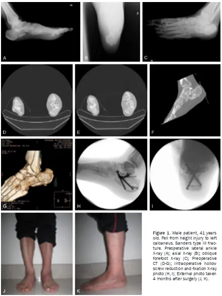
Figure 1. Male patient, 41 years old. Fell from height injury to left calcaneus. Sanders type III frac-ture. Preoperative lateral ankle
X-ray (A); axial X-ray (B); oblique
forefoot X-ray (C); Preoperative
CT (D-G); Intraoperative hollow screw reduction and fixation X-ray
of the sustentaculum tali or as an auxiliary method in the small dorsolateral approach for complex intra-articular fractures associated with sustentaculum tali fracture or fractures of the internal articular facet. Currently, the most widely used approach is the extensile L-shaped incision approach. It allows direct exposure and reconstruction of the entire lateral calcaneal wall, the posterior surface of the subtalar joint, and the calcaneocuboid articulation. This approach is suitable for over 90% of intra-artic -ular calcaneal fracture involving the posterior articular facet [7]. Its advantages were signifi
-cant as well as the disadvantages. Incidence of incision complications of the surgery performed by inexperienced surgeons or at primary hospi-tals was up to 30%. Other disadvantages of the extensile lateral L-shaped approach include the following: ① Exposure of the articular surface is not ideal. ② Soft tissues are retracted for long periods of time. This can lead to ischemia and damage to the sural nerve. ③ There is some unavoidable skin necrosis at the corner during the wound healing period. ④ Extensive devascularization can cause ischemia of the lateral wall of the calcaneus. ⑤ The same risks are present again during extraction of fixation plates. ⑥ Some patients experience severe traumatic arthritis. This incision is not the best choice for subtalar arthrodesis surgery.
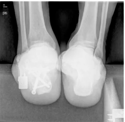
[image:6.612.90.290.70.274.2]The ideal pathway was the incision which makes the displaced articular surface clearly visible for convenient reduction of the fracture, offers sufficient space for fixation, and reduces the damage to soft tissue. Therefore, the sur-geons are developing a preference for the small dorsolateral incision approach. Its advantages are: ① The subtalar joint surface is directly exposed for convenient reduction. ② The use of hollow tension screws makes internal fixa -tion materials simple. ③ There is little damage to soft tissue. There is no risk of skin flap necro -sis because lateral skin flap is not required dur -ing the surgery. ④ It will not irritate the sheath of the peroneal tendon and the cutaneous nerves of the sural nerve. ⑤ It does not affect blood supply to the lateral wall of the calcaneus by avoiding the injury of the lateral calcaneal arteries. ⑥ Postoperative management of the incision wound is simple, and the wound heals quickly. ⑦ A skin flap is not required in the sec -ondary surgery during plate extraction. Compared to the extensile lateral L-shaped inci-sion approach, the small dorsolateral inciinci-sion approach greatly decreases the risks associat-ed with the incision wound, could avoid the occurrence of the skin flap necrosis, metal plate exposure. However, this approach also has obvious drawbacks, including lack of expo-sure of he lateral wall of the calcaneus, and higher requirements on the installation of fixa -tion materials, which require the surgeon to have more clinical experiences and better sur-gical skills. However, these disadvantages can be overcome with proper screening of patients and intensified clinical training for surgeons. Figure 2. Lateral ankle radiograph 30 months after
surgery while patient was standing.
[image:6.612.90.290.323.519.2]Biomechanical characteristics of calcaneal fractures and indications for the small dorso-lateral incision approach
Displaced intra-articular calcaneal fractures result from high-energy injuries, happen fre-quently to patients with high falling injury, dur-ing which the weight concentrates on the ankle and cause the fracture. The position of the foot during the injury, magnitude of the force, and osseous condition are all important factors determining the degree of bone comminution and the location of the fracture lines. Although the exact mechanisms of injury are still in dis-pute, the mechanisms described by Essex-Loprest [8] and Carr [9] et al. are basically the same. Essex-Lopresti believed that the first fracture line begins where the outer edge of the talus impacts the lateral calcaneus, and then extends inward [8].
In a fall, the posterior subtalar joint is immedi-ately forced into eversion, and the sharp outer taloid spur is driven like an axe into the Gissane angle, splitting it and the outer wall of the bone along its grain. The remainder of the force then descends through the anterior subtalar on to the sustentaculum tali, which may be sheared off the inner side of the body together and with the medial one-third or one-half of the posterior subtalar surface. If the force continues, it can cause fracture lines extending all the way to the leading edge of the calcaneus or even to the calcaneocuboid articulation causing anterolat-eral bone fragmentation. If the foot falls hori-zontally, and the force is directed to the hind-foot, the fracture line will extend rearward and upward, extending to the posterior articular sur-face, causing posterior intra-articular bone fragmentation and downward collapse of the foot. Carr [9] et al. produced the experimental calcaneus fractures by axially loading 18 the amputated lower limb specimens, make two constant primary fracture lines with varied loads on the tibia. One type of fracture line split the calcaneus into internal and lateral parts, roughly parallel to the sagittal plane, while the other type extended inward from the vertex of the Gissane angle, splitting the calcaneus into anterior and posterior parts, roughly in the cor-onal plane. The lateral fracture line proceeded downward, pointing to the plantar surface or forward. These two types of fractures jointly form various tongue-shaped fractures or articu-lar compression type fractures, and anterolat-eral, posterolatanterolat-eral, and anterointernal bone
fragments. The other type of bone fracture extended inward from the vertex of the Gissane angle, splitting the calcaneus into anterior and posterior parts, roughly in the coronal plane. The lateral fracture line proceeded downward, pointing to the plantar surface or forward. These two types of fractures jointly form vari-ous tongue-shaped fractures or articular com-pression type fractures, and anterolateral, pos-terolateral, and anterointernal bone frag- ments.
After fully understanding the biomechanical fracture mechanisms described above, we know that calcaneal fracture usually result in anterolateral, anterointernal (sustentaculum tali portion), and posterolateral fragments. On this basis, the subtalar articular surface may be fragmented into lateral, central, and internal parts in the vertical plane, and collapse down-ward. Reviewing the CT scans of the patients in this study wity the standard as mentioned, we can conclude that comminuted calcaneal frac-tures are not contraindications for the use of the small dorsolateral incision approach, which is also suitable as long as the comminuted frac-tures are coincide with the biomechanical mechanisms described above. Specific criteria include the following: anterior calcaneus is comminuted but uncompressed form an anteri-olateral fragment. The internal portion must be relatively intact. It would be better if the part of internal wall attaching to the sustentaculum was intact, because it offers the possibilities for screw fixation. The posterior subtalar articu -lar surface fracture line permits 2~4 screws, but each bone fragment should be relatively large and collapsed wholly, so the fragments can be fixated with screws. The posterior calca -neal tuberosity was complete, only the thala-mus was collapsed and comminuted. The Achilles tendon insertion was intact, and there is no upturn tongue-shaped fragment. There was a bulge on the lateral wall. The bottom of the foot was relatively intact. In summary, as long as the calcaneal fractures are consistent with the charateristics described above, a trained surgeon can treat Sanders type III com-plex intra-articular calcaneal fractures with the small dorsolateral incision approach combined with hollow tension screws.
Surgical skills
thin screws) could be used for fixation. As for collapsed subtalar joint surface, the fracture line in the sagittal plane and the bone frag-ments break into lateral, central, and internal parts and collapse downward. The subtalar joint becomes open as the assistant puts for-ward, backfor-ward, and downward traction on the calcaneal body, and then the primary surgeon can then reduce the posterior subtalar articular surface by lightly poking upward. Special atten-tion should be paid for the reducatten-tion of the anteriointernal bone fragments, which are the only reference for reduction of the lateral frag-ments. After all fractured bone fragments have been reduced, screw in 1~2 hollow tension screws with diameter of 3.5~4.0 mm from out-side to inout-side, parallel to the subtalar articular surface. This will strongly fixate the fragmented bones of the subtalar articular surface.
We used big diameter hollow tension screws to fixate the three main parts of the body of the calcaneus, the anteriolateral, anteriointernal (the sustentaculum portion), and the poteriolat-eral parts. After intraoperative traction assis-tance to lever and reduce the bones; ① The screws (4.5 mm) were screwed from anterior side of the calcanous towards the internal sus-tentaculum tali to fixate the anteriolateral and internal bone fragments. ② The screws (4.5 mm) were screwed posteriolateral side of the calcanous towards the internal sustentaculum tali to fixate the posteriolateral and internal bone fragments. ③ The screws (6.5 mm) were screwed from the Achilles tendon of the poste-rior calcaneal tubercle back to front along the long axis of the calcaneus to fixate the calca -neal body. While maintain full traction lever to restore the height of the calcaneal thalamus, squeeze the bulge of the lateral wall to restore the width, while correct the alignment of the calcaneal tubercle. Through the above process, the calcaneal body anteriolateral, anteriointer-nal, and posteriolateral portions were firmly fixated.
In addition, clean all bone fragments out of the articular cavity and check whether the screws have entered the subtalar articular cavity and broken through the internal wall before closing the incision.
In summary, Though the skin of the hindfoot is tough, the soft tissues are very fragile because of poor blood supply. Extensile lateral L-shaped incision method requires operating in an
isch-emic region, with large incision wound. Intraoperative stretching, clamping, folding of the skin flap may result in skin flay necrosis, infection, and even plate exposure. Small dor-solateral incision combined with hollow tension screw internal fixation surgery is a good way to avoid these disadvantages. Through training and a good understanding of the indications for this type of surgery, it is definitely possible to achieve the same clinical effect as with exten-sile L-shaped incisions using steel pins for inter-nal fixation of the calcaneus. Even for rather complex Sanders III type calcaneal fractures, it not only can achieve excellent results, but also completely avoid the occurrence of postopera-tive complications from a weakened lateral skin flap. Although high loss rate in this group and short follow-up time, the follow-up results have showed that small dorsolateral incision com-bined with hollow tension screw internal fixa -tion treatment for Sanders III type calcaneal fractures can achieve satisfactory results for both patients and doctors.
Disclosure of conflict of interest
None.
Address correspondence to: Xiang-Yang Xu, Foot and Ankle Disease Diagnosis and Treatment Center, Department of Orthopedics, Ruijin Hospital North, School of Medicine, Shanghai Jiaotong University,
Shanghai 201801, China. Tel: +86-18121263171; Fax: +86-18121263171; E-mail: xu664531@hot-mail.com
References
[1] Sanders R. Displaced intraarticular fracture of
the calcaneus. J Bone Joint Surg Am 2000; 82:
225-250.
[2] Zwipp H, Rammelt S and Barthel S. Calcaneal fractures: open reduction and internal fixation
(ORIF). Injury 2004; 35: 46-54.
[3] Buckley R, Tough S, McCormack R, Pate G, Leighton R, Petrie D and Galpin R. Operative
compared with nonoperative treatment of dis-placed intra-articular calcaneal fractures: a prospective, randomized, controlled
multi-center trial. J Bone Joint Surg Am 2002; 84:
1733-1744.
[4] Rammelt S and Zwipp H. Calcaneus fractures: facts, controversies and recent developments. Injury 2004; 35: 443-461.
[5] Benirschke SK and Kramer PA. Wound healing
complications in closed and open calcaneal
[6] Della Rocca GJ, Nork SE, Barei DP, Taitsman LA and Benirschke SK. Fractures of the susten -taculum tali injury characteristics and surgical technique for reduction. Foot Ankle Int 2009; 30: 1037-1041.
[7] Benirschke SK and Sangeorzan BJ. Extensive
intraarticular fractures of the foot. Surgical management of calcaneal fractures. Clin
Or-thop Relat Res 1993; 292: 128-134.
[8] Essex-Lopresti P. The mechanism, reduction, technique, and results in fractures of the os
calcis. British J Surg 1952; 39: 395-419.
[9] Carr JB, Hamilton LL and Bear LS. Experimen -tal intra articular calcaneal fractures: