Original Article
Occlusion of lateral aneurysm in common carotid artery
of rabbits with flow-diverting stent
Daohua He1, Ming Yang2, Mingjun Cai2, Peng Liu2, Li Pan2, Bo Diao2, Lei Feng2, Guang Feng2, Lianting Ma2 1Department of Neurosurgery, Panyu Central Hospital, Guangzhou 511400, China; 2Department of Neurosurgery,
Wuhan General Hospital of Guangzhou Military Command, Wuhan 430070, China
Received November 27, 2015; Accepted February 15, 2016; Epub June 15, 2016; Published June 30, 2016
Abstract: Objective: To assess the feasibility of flow-diverting stent in the treatment of lateral aneurysm models in common carotid artery of rabbit. Methods: Ten rabbits were included in our study. Six of nine successful experi-mental aneurysms were treated with flow-diverting stents, the other three aneurysms were untreated as controls. Angiography was performed immediately and 28 days after the procedure. Aneurysm and stented arteries were removed with animal alive for histopathological examination. Results: Before stent placement angiography of the common artery showed round saccular side-wall aneurysms and complex pattern of flow. Immediately after stent placement the aneurysms were still visible and the stented common carotid arteries remained patent, but the time for the contrast agent in the aneurysm was longer, the complex pattern of flow disappeared. 28 days after stent placement the aneurysms were no longer visible and the stented common carotid arteries remained widely patent. All controlled aneurysms and common carotid arteries have been patent and unchanged for 28 days. Histological analysis indicated that all treated aneurismal pouches were almost filled with thrombus as well as fibrotic reactive scar tissue. Stent wires were found to be located within the vessel wall and encased by an extension of the tunica intima. The endothelium was almost mature at 28 days, and various degrees of degenerate cells were seen under the transmission electron microscopy. Conclusions: The occlusion of lateral aneurysm in common carotid artery of rabbit with flow-diverting stent is safe and effective.
Keywords: Rabbit, common carotid artery, lateral aneurysm, flow-diverting stent
Introduction
Intracranial aneurysm is the main cause of and accounting for 85% of spontaneous subarach-noid hemorrhage. It has a high mortality and morbidity, so it is important in issues of Neurosurgery. The rapid development of mate-rials for neurological intervention provides a new choice for the treatment of intracranial aneurysms.
Endovascular treatment of intracranial aneu-rysm is minimally invasive and show quick recovery and good efficacy, providing the pos-sibility of timely treatment for patients who can’t receive surgery or have difficulty in sur-gery. Currently, the most widely used treatment is Guglielmi detachable coils (GDC) emboliza-tion, which provides a timely and effective treatment for patients with certain intracranial
aneurysms. But it has been observed in clinical applications that GDC technology has also dis-advantages, such as the risk of intraoperative rupture in operation, aneurysm recurrence caused by blood flow compressing coils, diffi-culty in dealing with small, wide-neck dissecting and other special types of aneurysms. Recently, an increasing number of studies focus on the blood flow-diverting stent without assisted coils in the treatment of in intracranial aneurysms [1-4]. It blocks blood flow into the tumor cavity, resulting in complete thrombosis in the tumor cavity.
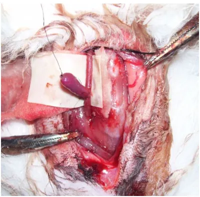
sia. After skin preparation of the surgical region, rabbits were fixed on the operating table. After routine disinfection and draping, a verti-cal incision about 6 cm in length was made horizontally along the middle of the neck below the thyroid cartilage for the blunt dissection of the subcutaneous tissue. One side of the exter-nal jugular vein was dissociated for its distal and proximal ligation, venous sac about 2 cm in length was clipped, then, heparinized saline (12500 U heparin was added into 500 ml nor-mal saline) was collected with an injector to wash it to be clean. One dissociated end was trimmed and the other end was ligated to a dead-end. Then, it was placed into heparinized saline for preparation and blunt dissection was conducted on sternoclavicular papillary mus-cle, sternohyoid muscle and other anterior cer-vical muscle groups, trachea was exposed to find out paratracheal carotid sheath, one side of the common carotid artery was dissociated, after, two atraumatic vascular clamps were used to clamp the proximal and distal ends for occlusion, the vascular adventitia was stripped, in its sidewall, a spindle-shaped incision slight-er longslight-er than the diametslight-er of the extslight-ernal jugu-lar vein was made and manicured, the lumen was washed with heparinized saline. Under the surgical microscope, end-to-side anastomosis was conducted on dissociated end of venous sac and common carotid artery with 9-0 micro-surgical suture using the lock-edge continuous eversion suture, after, hemostatic clamp was loosen to completely fill the venous sac, and the presence of blood leaking was carefully checked around the tumor neck, more dense suture would be conducted if necessary (Figure
2). Muscle and skin were sutured layer by layer. After surgery, a single subcutaneous injection was conducted for heparinization (1 mg/kg) after surgery.
in hemodynamics, blood flow velocity, the pres-sure and stress within the aneurysm, achieved the occlusion of the aneurysm cavity, and gained good results, which are reported below.
Materials and methods
Animals
Ordinary male rabbits were purchased from Wuhan Wanqian Jiahe Laboratory Animal Breeding Co., Ltd. [License No: SCXK (Hubei) 2007-0006]. The body weight was 2.5-3.5 kg. They were all fed according to strict experimen-tal standards.
Production of local mesh flow-diverting stent
We designed and produced a new-type local mesh flow-diverting stent in cooperation with Hunan Aipu Te Medical Devices Co., Ltd. The stent is made of nickel-titanium alloy. On its
structure configuration, it is a mesh tube woven densely in the middle and sparsely on both sides (Figure 1).
Establishment of animal models of side wall aneu-rysms
Fasting was conducted 12 h before surgery. 3% pentobar-bital (1 ml/kg) was injected by ear vein for general
[image:2.612.92.376.72.186.2]anesthe-Figure 1. Local mesh flow-diverting stent.
[image:2.612.89.289.222.419.2]After surgery, 800,000 units of potassium peni-cillin were injected intramuscularly for 3 d; sub-cage feeding was conducted on animals, and their indications of life and wound bleeding sit-uation were closely observed after regaining consciousness.
Arterial angiography 14 d after establishment of models (OEC96000) showed nine aneu-rysms and parent arteries were patent, one aneurysm was occluded, but the parent artery was unobstructed. It was manifested by the gathering of contrast agent within the aneu-rysm, vortex and smooth filling saccular app- earance.
Placement of local-mesh flow-diverting stent
14 d after the establishment of the model, flow-diverting stents were placed in 6 aneurysms which have been confirmed unobstructed by arteriography, and the other 3 aneurysms were untreated as controls. The specific operations are as follows:
During 3 d before stent placement, aspirin (3 mg/d) and Ticlid (6 mg/d) were administered orally. Fasting was conducted 12 h before sur-gery. ② After general anesthesia and the suc-cess of puncturing one side of the femoral artery using the Seldinger technique, 4F cathe-ter sheath was placed in animals, and common carotid artery catheterization was conducted
for superselective angiography. The angle of X-ray tube was adjusted to clearly show the aneurysm location. ③ Through 4F angiographic catheter, appropriate models of new local mesh flow-diverting stents were selected for outside wetness, then, they were sent into animals by micro-catheter. Under the guidance of videoflu-oroscopy and trace graph, the middle compact segment of the stent and the aneurysm entrance were exactly aligned. The stent was released, completely covering the aneurysm neck. Then, the stent release system was with-drawn. Carotid angiography was conducted through 4F angiographic catheter in the original working angle. Angiographic catheter was with-drawn after angiography and stent placement, and catheter sheath was removed immediately, the femoral artery was pressed to stop bleed-ing, and puncture point was compressed and bandaged. Penicillin sodium (800000 U) was injected intramuscularly daily for 3 d after sur-gery; Aspirin (3 mg/d) and Ticlid (6 mg/d) were administered orally after surgery until they were sacrificed.
Imaging reexamination immediately and 28 d after stent placement: angiography reexamina-tion was conducted on the common carotid artery immediately and 28 d after stent place-ment to understand the situation on the parent artery patency and aneurysm occlusion.
Histopathological examinations
28 d after stent placement, after the CCA angi-ography of the stent placement side, the aneu-rysm and the parent artery were anatomized for light microscopy and electron microscopy examinations.
Results
All animals were survived and healthy after sur-gery, with no apparent neurological complica-tions. Ten rabbits were used to establish 10 cervical saccular aneurysm models. 14 d after surgery, angiography confirmed 9 models showed unobstructed aneurysms and parent arteries. The success rate of model establish-ment was 90%.
Before stent placement, the imaging of aneu-rysm cavity was clear, the residence time of contrast agent was very short in the aneurysm cavity, flow speed was rapid, vortex could been
[image:3.612.90.290.69.277.2]seen in chaotic state, parent artery was unob-structed (Figure 3); After stent placement, angi-ography conducted immediately showed aneu-rysm cavity could develop images, the time of aneurysm filling was prolonged, residence time of the contrast agent in the cavity was pro-longed, flow rate slowed down, no vortex was observed, parent arteries remained patent
without stenosis, stent position was accurate (Figure 4); 28 d after stent placement, the aneurysm cavity was occluded, stent showed no displacement, and parent arteries remained patent without stenosis (Figure 5). In control group, the aneurysm cavities of 3 cases devel-oped clear images 28 d after surgery, the resi-dence time of contrast agent was very short in the aneurysm cavity, flow speed was rapid, vor-tex could been seen in chaotic state, parent artery was unobstructed.
Light microscopy showed six aneurysms pre-sented complete embolism 28 d after stent placement, showing thrombosis (Figure 6), stent was embedded in the vessel wall, and parent arteries showed intimal thickness; Electron microscopy showed the endothelial-ization of the stent surface was basically com-pleted and endothelial cells arranged loosely. In control group, no thrombosis appeared in aneurysmal cavity, and parent arteries were unobstructed.
Discussion
In 1954, German and Black9 [5] proposed the idea of establishing aneurysm model. Rabbits are used as subjects in this study because: firstly, rabbit carotid artery diameter is very sim-ilar with human proximal middle cerebral artery, very suitable for the research on embolism [6, 7]. Secondly, it is a non-primate which process-es of thrombosis and fibrinolysis are most simi-lar to those of human [8, 9]. Thirdly, the cost is low and the operation is simple.
Endovascular treatments of intracranial aneu-rysms mainly include parent artery occlusion, tumor cavity balloon embolization, coil emboli-zation, liquid embolic agents and vascular tis-sue engineering. GDC embolization technology is the most widely used treatment currently, which provides a timely and effective treatment for some of patients with intracranial aneu-rysms. But it has been observed in clinical applications that GDC technology has also dis-advantages as follows: firstly, there is risk of intraoperative rupture as the treatment behav-ior occurred in the aneurysm cavity; secondly, the low filling rate of the tumor cavity may cause aneurysm recurrence; thirdly, it has difficulty in dealing with small, wide-neck dissecting and other special types of aneurysms; Fourthly, the gravity pressure due to the filling of the huge
[image:4.612.89.290.68.277.2]Figure 4. After stent placement, angiography con-ducted immediately showed the time of aneurysm filling was prolonged, residence time of the con-trast agent in the cavity was prolonged and flow rate slowed down.
[image:4.612.89.290.354.556.2]aneurysm cavity could easily lead to parent artery occlusion, especially for anterior com-municating artery aneurysms and middle cere-bral artery aneurysms; fifthly, in aortic aneu-rysms and giant aneuaneu-rysms, the degree and time of thrombosis are varying; sixthly, in the combination of micro-coil and ordinary stents, the micro-catheter has large difficulty in pass-ing the stent mesh, and the coil may be tangled with stent when removing micro-catheter, thus causing stent migration or coil invading into the parent artery; seventhly, some scholars believe that, the incidence of thrombosis is relatively high in the GDC treatment of intracranial aneu-rysms. This may be related to the electrolytic detachment mechanism of GDC in which embo-lism occurs when current generates bubbles; eighthly, expensive GDC also limits its applica-tion in the domestic market. Therefore, to explore a more ideal treatment material has been a focus of clinical attention.
Theoretically, covered stent is the most reason-able approach for the treatment of aneurysms. But there are more branches of intracranial vessels, covered stent can’t protect branch vessel, and covered stents developed currently have hard texture and poor flexibility so that they are difficult to be bent. Therefore, its appli-cation has been limited because there are cer-tain difficulties to send it to the tortuous intra-cranial arteries. Another concern after covered stent placement is the increased number of foreign body in contact with the artery wall,
which will cause endometrial hyperplasia- in- ducted stenosis due to more pronounced inflammatory response.
In the mid-1990s, some scholars [10, 11] implanted a simple mesh stent in the tumor neck of canine carotid artery side wall aneu-rysm model. In follow-up visits, we found stable thrombosis within the aneurysm which was gradually replaced by fiber scar tissue, and finally, the tumor neck was covered by new endangium, and aneurysms were completely isolated from circulation, being thoroughly cured. In clinical practice, it has also been observed that some aneurysms are gradually occluded and cured after single stent implanta-tion without further filling in coils [12]. Some clinicians also have used overlapping release of several stents to treat intracranial aneurysms, gaining a few good follow-up results. However, thrombus is difficult to form in tumors because the large mesh of the existing simple mesh stent. It is particularly not suitable for ruptured and bleeding aneurysms.
[image:5.612.90.289.69.270.2]The metal coverage of intracranial aneurysm stent is 6.5%-9%, while that of the local mesh flow-diverting stent is 30% [13, 14]. Meanwhile, using closed-loop design, the single mesh size of local mesh flow-diverting stent is quite small. Theoretically, flow-diverting stent should have better clinical outcomes. It can restore aneu-rysm parent artery to normal, avoid the “point effect” of coil, has no “stickiness” and “toxicity” of liquid embolic materials at risk [15, 16], and reduce the risk of covered stent blocking vascu-lar branches. In the coil treatment of wide-neck aneurysms and giant aneurysms, it needs to release assisted stent for aneurysms first. Then, different sizes of coils are released into the aneurysm cavity. One aneurysm often fills more than 10 coils. And the operation is cum-bersome, equipments are very expensive. Comparatively, local mesh flow-diverting stent need no coils to complete aneurysm treatment, simplifying the operation. So people put for-ward ideas that local mesh flow-diverting stent is used in treatment of intracranial aneurysms, they believe that after placement into the par-ent artery and covering aneurysm neck, stpar-ent can change hemodynamics within aneurysms, including flow rate, vortex intensity and aneu-rysm wall shear stress, to stabilize hemody-namics within the aneurysm, leading to the
thrombus formation and organization within the aneurysm, and the stent surface will be covered by a thin layer of neointima, vessels of tumor section are completely rebuilt [17, 18]. Using this technology, metal stent is sent to the tumor entrance of the parent artery by propel-ler intravascularly, tumor entrance is closed to block blood into the tumor cavity, and then the aneurysm is excluded, changing hemodynam-ics of tumor cavity. It is followed by blood stasis, promoting thrombosis, fibrous scar tissue forms after organization, thus embolizing the tumor cavity.
In this study, we used local mesh flow-diverting stent in the treatment of rabbit side wall sac-cular aneurysm. The middle dense braid covers aneurysm neck. It has been observed experi-mentally: Before stent placement, the aneu-rysm cavity develops clear images, the reten-tion time of contrast agent is very short in the aneurysm cavity, blood flow entered the aneu-rysm at the proximal neck through parent artery and flow out from the proximal end, vortex forms at end-systole; Immediately after the stent placement, vortex which dominates previ-ously disappears immediately, although the aneurysm cavity can develop images, the time of aneurysm filling is prolonged, residence time of the contrast agent in the cavity is prolonged; 28 d after stent placement, the aneurysm cav-ity is occluded, and parent arteries remain pat-ent without stenosis. While in control group, the aneurysm cavities of 3 cases develop clear images 28 d after surgery, and parent artery is unobstructed. Light microscopy shows six aneurysms present complete embolism 28 d after stent placement, showing thrombosis, stent is embedded in the vessel wall, and par-ent arteries show intimal thickness; Electron microscopy shows the endothelialization of the stent surface is basically completed and endo-thelial cells arranged loosely. This fully proves that after the placement of local mesh flow-diverting stent, the middle dense braid covers aneurysm neck, and the stent surface present endothelialization, blocking blood flow to enter the tumor cavity mostly, reducing the blood mass and momentum transfer between aneu-rysm and parent artery, changing their hemody-namics and blood flow velocity, pressure and stress within aneurysms, reconstructing the parent artery blood flow, and gradually promot-ing thrombosis in aneurysm cavity to reach the satisfactory results aneurysm occlusion and
parent artery patency. This operation is rela-tively simple. It leads to less intraoperative and postoperative complications. The large mesh of the sparse part on both sides of the stent may maintain unobstructed vessel branches in the treatment of aneurysms, providing a new way of thinking for the treatment of intracranial aneurysms. But presently, stent imaging has not yet been clear after the release of the stent we developed. The flexibility of stent materials and mobility of delivery device needs yet to be further tested for the application to human tor-tuous blood vessels. The safety of stent releas-ing in branch vessels needs to be assessed. The issue that the stent causes thrombosis and stenosis needs to be further assessed. More importantly, more prolonged pre-clinical obser-vation and studies need to be conducted fur-ther on tissue and blood compatibility.
Disclosure of conflict of interest None.
Address correspondence to: Daohua He, Depart- ment of Neurosurgery, Panyu Central Hospital, Guangzhou 511400, China. Tel: 13926117165; E-mail: whzyyhdh@126.com
References
[1] Kallmes DF, Ding YH, Dai D, Kadirvel R, Lewis DA and Cloft HJ. A new endoluminal, flow-dis-rupting device for treatment of saccular aneu-rysms. Stroke 2007; 38: 2346-52.
[2] Kallmes DF, Ding YH, Dai D, Kadirvel R, Lewis DA and Cloft HJ. A second-generation, endolu-minal, flow-disrupting device for treatment of saccular aneurysms. AJNR 2009; 30: 1153-8. [3] Appelboom G, Kadri K and Hassan F. Infectious
aneurysm of the cavernous carotid artery in a child treated with a new-generation of flow-di-verting stent graft: case report. Neurosurgery 2010; 66: E623-4.
[4] Lubicz B, Collignon L, Raphaeli G, Pruvo JP, Bruneau M, De Witte O and Leclerc X. Flow-diverter stent for the endovascular treatment of intracranial aneurysms: a prospective study in 29 patients with 34 aneurysms. Stroke 2010; 41: 2247-53.
[5] German WJ and Black PW. Experimental pri-duction of carotid aneurysms. J N Eng Med 1954; 250: 104-6.
[6] Meng H, Natarajan SK, Gao L, Ionita C, Kolega J, Siddiqui AH and Mocco J. Aneurysmal chang-es at the basilar terminus in the rabbit elas-tase aneurysm model. AJNR 2010; 31: E35-6. [7] Ding Y, Dai D, Kadirvel R, Lewis DA and Kallmes
an-eurysms in rabbits. AJNR Am J Neuroradiol 2010; 31: 1236-9.
[8] Dai D, Ding YH, Kadirvel R, Lewis DA and Kallmes DF. Experience with microaneurysm formation at the basilar terminus in the rabbit elastase aneurysm model. AJNR Am J Neu- roradiol 2010; 31: 300-3.
[9] Chang DW, Kim BK, Shin JH, Yoon YM, Oh SH, Yoon YS, Hong SH, Lee KC, Lee YW, Seo KM, Kweon OK, Yoon JH, Shin NS, Lee KH, Suh JG and Seong JK. Assessment of experimental saccular aneurysm using selective angiogra-phy in common carotid artery of rabbits. Anat Cell Biol 2010; 43: 118-24.
[10] Geremia G, Haklin M and Brennecke L. Embolization of experimentally created aneu-rysms with intravascular stent devices. AJNR Am J Neuroradiol 1994; 15: 1223-1231. [11] Wakhloo AK, Schellhammer F, de Vries J,
Haberstroh J and Schumacher M. Self-ex- panding and balloon-expandable stents in the treatment of carotid aneurysms: An experi-mental study in a canine model. AJNR Am J Neuroradiol 1994; 15: 493-502.
[12] Mase M, Banno T, Yamada K and Katano H. Endovaseular stent placement for multiple an-eurysms of the extracranial internal carotid artery: Technical case report. Neurosurgery 1995; 37: 832-5.
[13] Kallmes DF, Ding YH, Dai D, Kadirvel R, Lewis DA and Cloft HJ. A new endoluminal, flow-dis-rupting device for treatment of saccular aneu-rysms. Stroke 2007; 38: 2346-52.
[14] FD and Kelly ME. Endovascular treatment of cerebral Aneurysms. Endovascular Today 2008; 53-65.
[15] Rasskazoff S, Silvaggio J, Brouwer PA, Kauf- mann A, Nistor A and Iancu D. Endovascular treatment of a ruptured blood blister-like aneu-rysm with a flow-diverting stent. Interv Neuroradiol 2010; 16: 255-8.
[16] van-Rooij WJ and Sluzewski M. Perforator in-farction after placement of a pipeline flow-di-verting stent for an unruptured A1 aneurysm. AJNR Am J Neuroradiol 2010; 31: E43-4. [17] Turowski B and Macht S. Early fatal
hemor-rhage after endovascular cerebral aneurysm treatment with a flow diverter (SILK-Stent): do we need to rethink our concepts? Neuro- radiology 2011; 53: 37-41.