Original Article
Effect of ginkgo biloba extract on splenic immune
function of chronic ulcerative colitis in mice
Hongwei Zhao1, Hua Han2, Hongtao Hou1, Jianquan Chen3, Xiaomeng Lang4, Yuehong Yue5, Haixiang Gao6, Jinsheng Kang7, Yeshan Zhu8, Ziqian Dun9, Qilu Hu10
Departments of 1Gastroenterology, 2Gynaecology, 5Neurology, 6Respiratory Medicine, 7Neurosurgery, 10
Oncol-ogy, Hebei General Hospital, Hebei Province, China; 3Department of Traditional Chinese Medicine, Yutian County
Hospital of Tangshan City, Tangshan, China; 4Hebei Provincial Hospital of Traditional Chinese Medicine, Hebei
Province, China; 8Department of Spleen and Stomach, Tangshan City Hospital of Traditional Chinese Medicine,
Tangshan, China; 9Department of General Medicine, The Fifth Hospital of Shijiazhuang City, Hebei Province, China Received December 4, 2016; Accepted December 30, 2016; Epub May 15, 2017; Published May 30, 2017
Abstract: Objective: Ulcerative colitis (UC) is a chronic nonspecific inflammatory bowel disease. Ginkgo biloba ex-tract, EGB761, has therapeutic action on colitis in animals. In this study, we discussed effect of EGB761 on splenic immune function of chronic UC in mice. Methods: Dextran sulfate sodium-induced chronic UC in mice was used to test the effect of EGB761 on regulation of immunologic splenic function. Disease activity index (DAI), body weight (BW) changes, colon length, and pathological changes in colon were monitored. Changes in spleen shape, length, weight and pathological condition were observed. Single karyocyte from spleen was separated and enumerated then tested for positive rate using flow cytometer. Real-time quantitative PCR and immunohistochemistry were ap-plied to test mRNA and protein expression of inflammatory factor IL-7, IL-6, TNF-α, and IFN-γ. Results: EGB761 could improve the general condition of mice with chronic UC; increase their body weight, colon length and colon weight; and slow down the pathology changes in chronic colitis. The amount of karyocytes and positive rate of CD4, CD8, and CD45R on karyocytes were reduced after applying EGB761. Real-time quantitative PCR further demonstrated that EGB761 could decrease the mRNA and protein expression of inflammatory factor IL-7, IL-6, TNF-α, and IFN-γ. Conclusion: EGB761 can ease inflammatory reaction on mice with chronic UC through depressing the activity of splenic immune cells and through down regulating the expression of splenic inflammatory factors.
Keywords:Ginkgo biloba extract, ulcerative colitis, spleen, inflammatory factor, dextran sulfate sodium
Introduction
Ulcerative colitis (UC) is a chronic nonspecific inflammatory bowel disease (IBD) with increas -ing morbidity in recent years [1-3]. Studies of UC have been focused on its mechanism. The onset of IBD is considered to be the result of interaction among genetic effects, environment factors, infection and self-immunity. The disor -der of immunoregulation is the major cause of IBD. As the largest peripheral immune organ, spleen is an important locality for immunore-sponses. There are various of immunocytes in spleen, including T lymphocytes, B lymphocy-tes, dendritic cells, macrophages, natural killer cells, and monocytes, that involve in variety of immunoresponses [4]. Spleen is the locality for T lymphocytes residing and responding
mediators, such as IL-1, IL-6, TNF-α, et al, thus causing inflammation [7]. The function of spleen is closely related to UC. Both medica- tion and surgery can improve the function of spleen in UC [8]. But this improvement is still depended on extent of UC.
Ginkgo biloba extract, EGB761, is extracted and purified from Ginkgo biloba leaves, con-taining 24% Ginko flavone glycosides and 6% terpene lactones. EGB761 has various biolo- gical and pharmacological functions, such as cleaning free radicals, anti-inflammation, regu -lating immune system, and so on [9]. Study showed that EGB761 can regulate the deve- lopment of immune organs, cellular immunity and humoral immunity. EGB761 can effectively activate monocyte-macrophage to start non-specific immune response and promote anti -gen passed to T lymphocytes and B lympho -cytes [10]. T lympho-cytes play an important role in cellular immunity. EGB761 can increase T lymphocytes counts and its concentration in blood serum and strengthen T lymphocytes functions. The ratio of mature T cells (CD3+) and helper T cells (CD4+) can also be regulated by EGB761 [10]. Cytokines (CK) are proteins or peptides secreted by immune cells that pass information between cells, and regulate immu- ne responses and effector function [11]. CK is one of important factors to assess one’s im- muno-competence. EGB761 can also improve one’s immunocompetence by regulating the level of CK (for example, interleukins, tumor
Materials and methods
Ethics statement
The mice and the protocol involved in the study had been approved by Institutional Animal Care and Use Committee (IACUC) of Hebei General Hospital. Approval ID: I07-038-3. All the mice were housed under standard condi-tions per protocols of IACUC and Hebei me- dical university vivarium in a barrier facility (GB14925-2001).
Animals and reagents
Male C57BL/6 mice (weight: 18-22 g; 7-12 weeks) were purchased from Vital River La- boratory Animal Technology Co. Ltd.. Dextran sulfate sodium (DSS) was bought from Sigma. Ginkgo biloba extract standard, Tebonin, was acquired from Dr. Willmar Schwabe Gmblt & Co. KG. CD3, CD4, CD8, CD45R and anti-mou- se CD45R antibodies were purchased from Beckmen Coulter. Anti-mouse CD3e antibody, anti-mouse TNF-α antibody, anti-rabbit IL-17 antibody, anti-mouse IFN-γ antibody, anti-goat IL-6 antibody, and anti-goat GAPDH polyclonal antibody were bought from Santa Cruz Bio- technology, inc..
Modeling method
[image:2.612.91.521.84.165.2]Three model groups were included in this study: control group; chronic UC model group (Model group), and EGB761 intervention group
Table 1. DAI score criteria
Score Weight loss (%) Stool consistency Stool occult blood
0 <1 Low amount, hard, dry, non-sticky, non-dispersive Colorless
1 1--5 Low amount, hard, wet, sticky Spotted color
2 6--10 High amount, soft, very sticky Continuous blue
3 11--15 High amount, soft, dispersive Dark stool, blue occult blood
4 >15 Loose stool Bloody stool, dark blue
*DAI = (score of Weight Loss + score of Stool Consistency + Stool Occult Blood)/3.
Table 2. Macroscopical colon scoring criteria
Macroscopical appearance Score
Normal colon appearance 0
Colonic wall mildly thicken, no congestion 1
Colonic wall moderate thicken, congestion 2
Colonic wall obviously thicken and harden, congestion 3 Colonic wall obviously thicken and harden, congestion, adhesion 4
[image:2.612.90.367.212.294.2](EGB761 group). Each group randomly got 10 male C57BL/6 mice and was observed for 28 days. Control group was fed with deioni- zed water all the time. For Model group and EGB761 group, 2% DSS (10 g DSS dissolved in 500 mL DI water) was fed to replace DI water
at Day 1 to Day 5, Day 8 to Day 12, Day 15 to Day 19, and Day 22 to Day 26. From Day 14, EGB761 group was intervened with EGB761 gavage at dosage of 100 mg/kg, one time per day, which lasted for 14 days. As control, model group and control group were intervened with phosphate buffer solution (PBS) gavage at the same volume. On Day 29, mice were sacrifi-ced by dislocate cervical vertebrae for tests. Disease activity index (DAI) measurement
Mental state, weight, activity, hair glossiness, appetite, and stool consistency of mice were monitored each day. According to DAI scoring criteria (Table 1), scores were recorded based on the weight, stool consistency and occult blood of mice [12].
Colon length and weight change measurement
Colon was dissected and its length and weight were measured and recorded. Scores were given based on the criteria given in Table 2. Hematoxylin-eosin staining (H-E staining)
Paraffin-embedded colon tissue was sectioned to 5 μm-thick slices. Paraffin was washed off using dimethylbenzene. Gradient ethanol was used to dehydrate tissue. Rinse dehydrated tis-sue with DI water for 1 min. Stain the tistis-sue with hematoxylin for 7 min then rinse it with water for 2 min, with 1% HCl-ethanol for 2 s, with water for another 2 min, with 1% ammonia for 30 s, with water for 4 min. Stain the tissue with 1% Eosin in ethanol for 30 s. Dehydrate the tissue with gradient ethanol, make the tis-sue transparent using dimethylbenzene. Seal the tissue with neutral balsam and observe it under microscope. Histopathological chang-es of colon tissue were scored based on H-E staining observation under microscope [13] (Table 3).
Spleen length and weight measurement
[image:3.612.90.306.96.469.2]Anesthetize mouse by intraperitoneal injecting 10% chloral hydrate. Collect peripheral blood at 1500 rpm for 15 min and collect serum. Test calcium concentration in blood serum. Open ab- domen at center and harvest spleen. Observe the macroscopical appearance of spleen and measure its length and weight. Calculate spl- een index (SI) using weight of spleen ×100/
Table 3. Histopathological changes scoring crite-ria
Histological appearance Score
Inflammation (I)
Non 0
Mild 1
Moderate 2
Severe 3
Depth (E)
Non 0
Mucosa 1
Mucosa and submucosa 2
Through mucosa 3
Hyperplasia (R)
Complete 0
Almost complete 1
With crypt 2
Incomplete epidermis 3
No tissue repair 4
Crypt damage (C)
Non 0
1/3 base crypt damaged 1
2/3 base crypt damaged 2
Only complete surface epithelium 3
Crypt and epithelium completely damaged 4 Scope of lesions (P, %) S
1--25 1
26--50 2
50--75 3
75--100 4
*Histological score = I+E+R+C+P.
Table 4. Immunohistochemistry analysis scoring criteria
A-score for percentage
of positive cells B-score for color intensity of positive cells
0 Negative Negative
1 1%--25% positive Slightly positive
2 26%--50% positive Positive
3 51%--75% positive Strongly positive 4 76%-100% positive
[image:3.612.91.306.525.616.2]weight of mouse [12]. H-E stain spleen tissue and observe the slide under microscope. Splenic monocytes separation and enumera-tion
Anesthetize mouse by intraperitoneal injection of 10% chloral hydrate and fix it on plate, expos -ing abdominal area for surgery. Sterilize surgi-cal area with 70% ethanol then cut skin from bottom transversely. Blunt dissect peritoneum and open abdominal cavity along linea alba transversely. Cut ligaments around spleen and take out spleen, put it on ice. Place spleen in a petri dish and immerse it in 10 mL cold com-plete RPMI medium (1 bag of RPMI medium powder dissolved in 1 L sterile water, adding 2.0 g sodium bicarbonate, glutamine, sodium pyruvate, HEPES, 100 U/mL penicillin, and 100 U/mL streptomycin), then crush it with two glass slides rinsed ahead with the same medi-um. Filter tissue with 70 μm mesh and trans-fer filtrate into a 50 mL centrifuge tube. Repeat filtration several times until the 50 mL centri -fuge tube is full. Centri-fuge cell suspension 3000 rpm for 10 min at 20°C. Discard super- natant and add 2 mL red blood cell lysis buf- fer to sediment. Stand for 2 min then add 10 mL complete RPMI medium. Centrifuge above mentioned cell suspension 3000 rpm for 10 min and discard supernatant. Resuspend se- diment with complete RPMI medium until the cell concentration is 106 cells/mL.
CD8, 2.5 μL for each. The third tube of cell suspension was labelled with FITC-anti-CD45R and IgG-Cy5-anti-CD3, 2.5 μL for each. The fourth tube of cell suspension was labelled as control without any incubation with fluores -cence-conjugated antibodies. Each tube was incubated for 30 min in dark and rinse with saline once. Resuspend cells with 100 μL saline for flow cytometry analysis.
Immunohistochemistry (ICH) analysis
Spleen tissue was embedded in paraffin and sectioned into slices. Slices were dehydrated, dewaxed, followed by repairing antigens, then sealed. Primary antibodies, IL-7, IL-6, TNF-α, and IFN-γ were incubated at 37°C for 1.5 h, respectively, then rinsed with PBS five times. Secondary antibodies were incubated at 37°C for 30 min then rinsed with PBS for three times. The expression of IL-7, IL-6, TNF-α, and IFN-γ were observed under microscope. The percent -age of positive staining area was analyzed us- ing multifunctional true color cell image analy-sis system. IHC score was obtained according to following criteria (Table 4).
Real time quantitative PCR (Real-time Q-PCR) analysis of mRNA expression of IL-7, IL-6,
TNF-α, and IFN-γ
[image:4.612.91.390.86.241.2]All the procedures were conducted aseptically at low temperature. Grind spleen tissue in a mortar with liquid nitrogen and collect powder
Table 5. Genes for PCR and primers sequences
Detected
gene PCR primer sequence DNA fragmentPropagated
TNF-α Sense: 5’ GGT TCT GTC CCT TTC ACT CAC T 3’ 169 bp Antisense: 5’ GAG AAG AGG CTG AGA CAT AGG C 3’
IFN-γ Sense: 5’ ATG AAC GCT ACA CACC TGC ATC TT 3’ 139 bp Antisense: 5’ TTT CTT CCA CAT CTA TGC CAC TT 3’
IL-6 Sense: 5’ CAT GTT CTC TGG GAA ATC GTG 3’ 141 bp
Antisense: 5’ TCC AGT TTG GTA GCA TCC ATC 3’
IL-17 Sense: 5’ TAT CCC TCT GTG ATC TGG GAA G 3’ 161 bp Antisense: 5’ ATC TTC TCG ACC CTG AAA GTG A 3’
GAPDH Sense: 5’-GAGACCTTCAACACCCCAGC-3’ 263 bp
Antisense: 5’-ATGTCACGCACGATTTCCC-3’
The results of Real-time Q-PCR were concluded from threshold comparison. The amplification of target gene = 2-ΔΔC(t), C(t) is the cycle number of thermal cycler to reach fluorescence threshold. The amplification of target gene compared to control group was calculated as: ΔΔC(t) = ΔC(t)experiment - ΔC(t)control, Where ΔC(t)experiment = C(t)target gene - C(t)GAPDH, and ΔC(t)control = C(t)target gene - C(t)GAPDH.
Flow cytometry analysis of splenic immunocytes posi-tive rate
PE-anti-mouse-into a 1.5 ml centrifuge tube. Add 1 mL of Trizol isolation reagent (Roche, USA), vortex rapidly then place it on ice. Add 0.2 mL of chloroform and homogenized mix for 15 s. Stand on ice for 5 min then centrifuge the tube at 1200 g for 15 min. Transfer supernatant to another 1.5 mL centrifuge tube and add 0.5 mL of pre-cooled isopropanol, then store the tube at -20°C for 1 h. Centrifuge the tube at 1200 g for 15 min and discard supernatant. Wash RNA sediment immediately with 75% ethanol and centrifuge it at 7500 g for 5 min. Discard supernatant and air dry RNA sediment for 15-20 min. Dissolve dried RNA in 20 μL diethypyrocarbonate (DEPC) processed MiliQ water and take 1 μL of RNA solution for integrity, purity, and concentration test. Keep the rest at -70°C. PCR primers of IL-7, IL-6, TNF-α, and IFN-γ were designed using Primer 5 (Table 5) (ABI, USA) and synthesized by Sango Biotech.
Statistical method
Quantitative data was expressed as “Average ± Standard deviation” (_x ± s) and analyzed by SPSS 13.0 software. Comparison between mul- ti groups was achieved using one-way ANOVA and inter-group comparison was analyzed us- ing SNK, P<0.05 was considered to be statis- tically significant.
Results
Effects of EGB761 on chronic UC mice
General condition: After fed with 2% DSS for 1 day, mice in model group all gained weight and increased water and food intake. After 3 days of feeding with 2% DSS, mice in both model group and EGB761 group began to lose weight and water and food intake decreased, along with loosened and increased stool. Between Day 13 to Day 15, mice in both model group and EGB761 group showed significant weight loss and lack of spirit. Their hair was messy and stool was loose. With time come by, these symptoms become more obvious. For EGB761 group, intervention with EGB761 gavage began on Day 14. Weight loss, loose stool and low spirit last at the beginning of intervention. But on Day 17, mice in EGB761 group had stable weights and better spirit. From Day 18, mice in EGB761 gained weights, although not much. On Day 29, the weight of mice in EGB761 re- turned to original status and mental state was good (Figure 1A).
DAI score
DAI score of control group was 0.00 ± 0.00. Compared to control group, DAI score (1.31 ± 0.65, P<0.01) of Model group increased after feeding them with 2% DSS on Day 5. On Day 13, DAI score of model group reached peak value (4.00 ± 0.43, P<0.01). On Day 16, DAI score of EGB761 group was 3.30 ± 0.65. From Day 19, DAI score of EGB761 group (3.00 ± 0.85, P<0.01) began to be lower than it of model group. On Day 29, DAI score of EGB761 decreased to 0.30 ± 0.46, P<0.01, which was a little higher than it of control group but much lower than it of model group (Figure 1B). Thus it can be concluded that intake of EGB761 can lower DAI score.
Colon length and weight
Compared to control group, which had an average colon length of 5.90 cm ± 0.27 cm (P<0.01), average colon length of model group was shorter, 4.65 cm ± 0.63 cm (P<0.01). Com- pared to model group, average colon length of EGB761 group was lower, 5.56 cm ± 0.42 cm (P<0.01).
Compared to control group, which had an aver-age colon weight of 0.73 g ± 0.12 g (P<0.01), average colon weight of model group was heavier, 0.92 g ± 0.15 g (P<0.01). Compared to model group, average colon weight of EGB761 was lighter, 0.81 g ± 0.10 g (P<0.01) (Figure 1C-E).
Pathological changes of colon
For control group, after H-E staining, complete intestinal mucosa epithelium was observed. Single epithelial and enteraden in lamina intes-tine arranged orderly. For model group, intesti -nal mucosa was impaired and enteraden was damaged or disappeared. Amount of goblet cells was low. Lymphocytic infiltration to mu-cosa, submumu-cosa, even muscular layer was ob- served. After treatment with EGB761, patho-logical changes were alleviated compared to model group (Figure 1F).
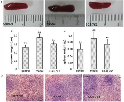
Effect of EGB761 on mice colon general condi-tion
model group swelled up with dark color. Spl- eens in EGB761 group had smaller size com-pared to model group (Figure 2A).
Average length of spleens in control group was 1.217 cm ± 0.12 cm, (P<0.01). Average length of spleens in model group was obviously larger (1.767 cm ± 0.17 cm, P<0.01). After EGB761 intervention, average length of spleen shortens to 1.417 cm ± 0.12 cm, (P<0.01), compared to model group (Figure 2B).
Average weight of spleens in control group was 0.059 g ± 0.017 g, (P<0.01). Average weight of
[image:6.612.91.527.74.451.2]had orderly structure. Red pulps had malacic hemorrhage. Congestion and lymphocytes infil -tration were less severe compared to model group (Figure 2D).
Effect of EGB761 on immune cells of UC mice
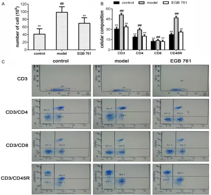
The concentration of monocytes in control group was 56×106/mL ± 15.2×10⁶/mL. Model group had increased monocytes concentra- tion as 108×106/mL ± 20.3×10⁶/mL. After EG- B761 intervention, monocytes concentration in EGB761 group was lower (76.8×10⁶/mL ± 18.6×10⁶/mL) compared to model group (Fig-
ure 3A).
In control group, the positive rate of CD3, CD4, CD8 and CD45R in splenic monocytes was
30.50% ± 0.90%, 18.22% ± 0.70%, 14.00% ± 0.73%, and 20.02% ± 0.81%, respectively. After intervention with EGB761, the positive rate of CD3, CD4, CD8 and CD45R was 50.01% ± 0.67%, 29.00% ± 0.68%, 15.82% ± 0.59%, and 48.40% ± 0.67%, respectively. All of them were lower than them in model group (Figure 3B,
3C).
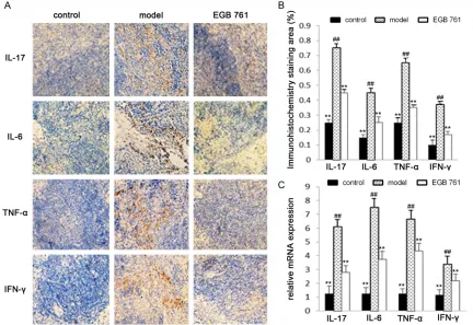
Effect of EGB761 on splenic cytokines of UC mice
[image:7.612.90.524.71.428.2]tokines. The expressions of IL-17, IL-6, TNF-α, and IFN-γ were not much in control group but increased in model group at level of 0.75 ± 0.02 vs 0.25 ± 0.02, 0.45 ± 0.03 vs 0.15 ± 0.02, 0.65 ± 0.03 vs 0.25 ± 0.03, and 0.37 ± 0.02 vs 0.10 ± 0.03, (P<0.001), respectively. The expression of IL-7, IL-6, TNF-α, and IFN-γ in EGB761 group were 0.45 ± 0.02; 0.25 ± 0.04, 0.35 ± 0.02, and 0.17 ± 0.02, (P<0.001), re- spectively, which were lower than them in mo- del group (Figure 4B).
mRNA of above mentioned 4 types of cytokin- es were expressed more in Model group than them in control group, at a level of 6.11 ± 0.50 vs 1.25 ± 0.56 for IL-17 mRNA, 7.50 ± 0.65 vs
1.25 ± 0.44 for IL-6 mRNA, 6.65 ± 0.65 vs 1.25 ± 0.34 for TNF-α mRNA, and 3.37 ± 0.60 vs 1.15 ± 0.40 for IFN-γ mRNA (P<0.001). The expression of IL-17 mRNA, IL-6 mRNA, TNF-α mRNA, and IFN-γ mRNA were lower in EGB761 group compared to model group, at a level of 2.78 ± 0.49, 3.75 ± 0.56, 4.35 ± 0.55, and 2.17 ± 0.50, respectively (Figure 4C).
Effect of EGB761 on colon macroscopic ap-pearance
[image:8.612.92.526.74.476.2]colonic wall and shorten colon length. Macro- scopic score for model group was 2.17 ± 0.98. In EGB761 group, colon mucosa was more
[image:9.612.92.524.77.374.2]smooth, less congestion, without edema and less shorten length compared to model group, with a score of 1.33 ± 0.52 (Figure 5A). Figure 4. The expressions of IL-17, IL-6, TNF-α and IFN-γ examined by immunohistochemistry staining in the spleen (×400). A: Immunohistochemistry staining of IL-17 and IL-6, TNF-α, IFN-γ in spleen specimen. B: Immunohistochem-istry score of IL-17 and IL-6, TNF-α, IFN-γ in the spleen. IL-17 and IL-6, TNF-α, IFN-γ expression increased significantly in the model group than that in the control group, and decreased in the EGB761 group than the model group. C: The relative expressions of IL-17, IL-6, TNF-α, IFN-γ mRNA in the spleen. The relative expressions of IL-17 and IL-6, TNF-α, IFN-γ mRNA were significantly increased in the model group compared with that in the Control group, and decreased in the EGB761 group. **P<0.05 vs model group, ##P<0.01 vs control group.
[image:9.612.93.524.478.631.2]Compared to control group, the histological score of Model group increased from 5.00 ± 0.00 to 8.33 ± 0.89. With EGB761 treatment, histological changes were alleviated and num-ber of inflammatory cells decreased. The histo -logical score of EGB761 group (6.89 ± 1.01) was lower than it of model group (Figure 5B).
Discussion
Ulcerative colitis (UC) is a chronic non-speci- fic inflammation with an unclear mechanism. Study showed that UC may be caused by inter-actions among susceptibility genes, environ-mental factors, infections, and disorder of im- mune system [14]. In other words, environmen-tal factors act on people with susceptibility gens; then intestinal immune and non-immune system begin to fight with the help from intesti -nal flora, which results in hyper-functio-nal and non-controllable immunological inflammatory reactions [8]. In recent years, the occurrence of UC in China is increasing. Thus there is a need to study the mechanism of UC and final suitable medication to control UC development.
There is a network of inter-collaboration and inter-constrains among immune organs, cells and molecules. Once the balance broken, the immune response will be abnormal, which in- cludes unbalanced ratio among T lymphocy-tes subgroup, disordered expression of cyto-kines, suppressed macrophage functions, da- maged microstructure or immune organs, etc [15]. Especially, the uncontrollable release of cytokines can result in local even general le- sion. Spleen is the largest immune organ and lymphatic organ in human body, with vast num-ber of lymphocytes, macrophages, and various immunocytes and cytokines. It takes 25% of total lymphatic tissues [16]. The amount and function of T lymphocytes in spleen is positive related to immune responses. Spleen also is the location where T lymphocytes and B lym -phocytes reside, proliferate and accept immu-no-stimulation from antigens. Spleen consists of white pulp, red pulp and marginal zone. Marginal zone is the first place to contact anti -gens and respond to immune-stimulation. Red pulp consists of cord and sinus. It is the major location of phagocytosis. White pulp consists of periarterial lymphatic sheath and corpus-cles. It is the major location of specific immune responses [17]. Spleen is directly involved in cellular immunity and regulates distribution of
T lymphocyte subgroups in peripheral blood. T lymphocytes can be classified into two sub -groups based on its surface antigen expres-sion, CD4+ subgroup and CD8+ subgroup. The unbalanced ration between these two groups can lead to immunologic inadequacy and im- munological diseases [18].
is the fourth generation preparation with ac- tive ingredients of ginkgolides and flavonoids. It can promote blood circulation and stimula- te immune cells. Studies showed that EGB761 strengthen immunocompetence by regulating the ratio between CD4+ and CD8+ T lympho -cytes thus promote recovery of damaged immu-nofunctions [23]. EGB761 cleared oxygen free reagents and reduced expression of TNF-α, and IFN-γ to alleviate colon inflammation of experi -mental mice [24]. Pretreatment with EGB761 can enforce proliferation activity of lymphocy- tes and improve immunosuppression in acute ischemic stroke. The degree of cerebral in-fraction can be lowered by fine controlling the ratio of CD4+ and CD8+ T lymphocytes in cer -tain range. EGB761 down-regulates express- ion of IL-6 and TNF-α in a normal range, which is good for human body to maintain anti-infec-tion ability in immunologic homeostasis. There-fore, on one hand EGB761 can treat and sup-press excessive inflammation; on the other hand, EGB761 can maintain and promote im- munological defense. EGB761 can also incre- ase the weight of thymus and spleen, and im- prove the thymus- and spleen- index of immu-nological suppressed mice, thus to protect im- mune organs. In summary, EGB761 can improve nonspecific immunity, specific immunity (includ -ing cellular immunity and humoral immunity), and phagocytic index of immunological sup-pressed mice, indicating its improvement in phagocytosis of monocytes. EGB761 can also regulate the development of immune organs and differentiation of monocytes and natural killer cells.
In this study, 2% DSS was used to stimulate immune system response in mice to induce UC. Results showed that after 14 days of in- take 2% DSS, experimental mice showed a series of inflammatory symptoms, such as low spirit, decreased water and food intake, luster-less and caducous hair, loose and bloody stool, etc. Meanwhile, the weight of mice decreased to the lowest value of 80% original weight. Further DAI evaluation showed that model group has apparently higher score than con- trol group, indicating successful model build-ing. Different degrees of diarrhea, bloody stool, weight loss and shortened colon appeared on model group mice. Histopathological chang- es included shortened and twisted intestinal glands and submucosa. With experiment time
compared to Control group. After intervention with EGB761, all the indexes mentioned above decreased significantly, which indicated that EGB761 could regulate cellular and humoral immunity. This might be one mechanism for spleen participating in UC development. Recent study showed that Th17 type cellular immunity was a major cause of UC [19]. The function of IL-17 is regulated by IL-6, IL-1β, TNF-α and chemokine. Th17 can stimulate epi -theliums, endothelium, monocytes and fibro -cytes to function in immunoregulation. IL-17 has powerful ability to recruit and activate neu-trophils, induced activate T cells, and stimulate secretion of various proinflammatory factors by fibrocytes, macrophages and epitheliums. The expression of IL-17 mRNA, IL-6 mRNA, TNF-α mRNA, and IFN-γ mRNA were higher in model group than it in control group, which indicated changes in Th17 subgroup.
Conclusions
In conclusion, our study demonstrate that EGB761 regulates immune responses by re- ducing amount of immunocytes, restraining ac- tivity of immunocytes and suppressing secre-tion of cytokines, and further alleviate mucosa changes caused by inflammatory factors.
Acknowledgements
The study was supported by Scientific research project of Hebei Provincial Administration of traditional Chinese Medicine (2016180). Disclosure of conflict of interest
None.
Address correspondence to: Hongwei Zhao, Depart- ment of Gastroenterology, Hebei General Hospital, No. 348 Heping West Road, Shijiazhuang 050051, Hebei Province, China. Tel: +8613303039680; Fax: +8631185989696; E-mail: [email protected]
References
[1] Marin AC, Gisbert JP and Chaparro M. Immu- nogenicity and mechanisms impairing the re-sponse to vaccines in inflammatory bowel dis-ease. World J Gastroenterol 2015; 21: 11273-11281.
[2] Takedatsu H, Mitsuyama K and Torimura T. Nanomedicine and drug delivery strate- gies for treatment of inflammatory bowel
dis-ease. World J Gastroenterol 2015; 21: 11343-11352.
[3] Lord JD. Promises and paradoxes of regulatory T cells in inflammatory bowel disease. World J Gastroenterol 2015; 21: 11236-11245. [4] Lee S, Khang D and Kim SH. High dispersity of
carbon nanotubes diminishes immunotoxicity in spleen. Int J Nanomedicine 2015; 10: 2697-2710.
[5] Lim MA, Lee J, Park JS, Jhun JY, Moon YM, Cho ML and Kim HY. Increased Th17 differentiation in aged mice is significantly associated with high IL-1beta level and low IL-2 expression. Exp Gerontol 2014; 49: 55-62.
[6] deLeeuw RJ, Kost SE, Kakal JA and Nelson BH. The prognostic value of FoxP3+ tumor-infiltrat-ing lymphocytes in cancer: a critical review of the literature. Clin Cancer Res 2012; 18: 3022-3029.
[7] Nam YS, Kim N, Im KI, Lim JY, Lee ES and Cho SG. Negative impact of bone-marrow-derived mesenchymal stem cells on dextran sulfate sodium-induced colitis. World J Gastroenterol 2015; 21: 2030-2039.
[8] Gong Y, Liu L, He X, Zhao H, Yang J, Li L, Lu A, Lin Y and Jiang M. The th17/treg immune balance in ulcerative colitis patients with two different Chinese syndromes: dampness-heat in large intestine and spleen and kidney yang deficiency syndrome. Evid Based Complement Alternat Med 2015; 2015: 264317.
[9] Serrano-Garcia N, Pedraza-Chaverri J, Mares-Samano JJ, Orozco-Ibarra M, Cruz-Salgado A, Jimenez-Anguiano A, Sotelo J and Trejo-Solis C. Antiapoptotic effects of EGB761. Evid Ba- sed Complement Alternat Med 2013; 2013: 495703.
[10] Takuma K, Mizoguchi H, Funatsu Y, Kitahara Y, Ibi D, Kamei H, Matsuda T, Koike K, Inoue M, Nagai T and Yamada K. Placental extract im-proves hippocampal neuronal loss and fear memory impairment resulting from chronic re-straint stress in ovariectomized mice. J Phar- macol Sci 2012; 120: 89-97.
[11] Sochocka M, Zaczynska E, Tabol A, Czarny A, Leszek J and Sobczynski M. The influence of donepezil and EGB761 on the innate im- munity of human leukocytes: effect on the NF-kappaB system. Int Immunopharmacol 2010; 10: 1505-1513.
[12] Turcotte K, Gauthier S, Mitsos LM, Shustik C, Copeland NG, Jenkins NA, Fournet JC, Jolicoeur P and Gros P. Genetic control of myeloprolifera-tion in BXH-2 mice. Blood 2004; 103: 2343-2350.
[14] Hakansson A, Tormo-Badia N, Baridi A, Xu J, Molin G, Hagslatt ML, Karlsson C, Jeppsson B, Cilio CM and Ahrne S. Immunological altera-tion and changes of gut microbiota after dex-tran sulfate sodium (DSS) administration in mice. Clin Exp Med 2015; 15: 107-120. [15] Ma W, Gilligan BM, Yuan J and Li T. Current
sta-tus and perspectives in translational biomark-er research for PD-1/PD-L1 immune check-point blockade therapy. J Hematol Oncol 2016; 9: 47.
[16] Suarez-Vilela D, Izquierdo FM, Mendez JR, Escobar J, Urdiales G and Junco P. Diffuse lymphangiomatous hyperplasia of the spleen with hyaline bodies. A pseudotumoral prolifera-tion arising from the lymphatic vessels of the periarteriolar lymphatic sheath. Virchows Arch 2011; 458: 505-509.
[17] Teixeira LG, Leonel AJ, Aguilar EC, Batista NV, Alves AC, Coimbra CC, Ferreira AV, de Faria AM, Cara DC and Alvarez Leite JI. The combination of high-fat diet-induced obesity and chronic ul-cerative colitis reciprocally exacerbates adi-pose tissue and colon inflammation. Lipids Health Dis 2011; 10: 204.
[18] Churlaud G, Pitoiset F, Jebbawi F, Lorenzon R, Bellier B, Rosenzwajg M and Klatzmann D. Human and Mouse CD8(+)CD25(+)FOXP3(+) Regulatory T Cells at Steady State and during Interleukin-2 Therapy. Front Immunol 2015; 6: 171.
[19] Ueno A, Ghosh A, Hung D, Li J and Jijon H. Th17 plasticity and its changes associated with inflammatory bowel disease. World J Gas- troenterol 2015; 21: 12283-12295.
[20] Huo M, Han H, Sun Z, Lu Z, Yao X, Wang S and Wang J. Role of IL-17 pathways in immune priv-ilege: A RNA deep sequencing analysis of the mice testis exposure to fluoride. Sci Rep 2016; 6: 32173.
[21] Nagashima H, Okuyama Y, Hayashi T, Ishii N and So T. TNFR-associated factors 2 and 5 dif-ferentially regulate the instructive IL-6 receptor signaling required for Th17 development. J Immunol 2016; 196: 4082-4089.
[22] Park J, Goergen CJ, HogenEsch H and Kim CH. Chronically elevated levels of short-chain fatty acids induce T cell-mediated ureteritis and hydronephrosis. J Immunol 2016; 196: 2388-2400.
[23] Kotakadi VS, Jin Y, Hofseth AB, Ying L, Cui X, Volate S, Chumanevich A, Wood PA, Price RL, McNeal A, Singh UP, Singh NP, Nagarkatti M, Nagarkatti PS, Matesic LE, Auclair K, Wargovich MJ and Hofseth LJ. Ginkgo biloba extract EGB761 has anti-inflammatory pro- perties and ameliorates colitis in mice by driv-ing effector T cell apoptosis. Carcinogenesis 2008; 29: 1799-1806.
[24] Mustafa A, Medany A, Hagar HH and El-Medany G. Ginkgo biloba attenuates mucosal damage in a rat model of ulcerative colitis. Pharmacol Res 2006; 53: 324-330.