Original Article
Effect of hyperbaric oxygen on bone mesenchymal
stem cells transplant in spinal cord injury rats
Yanbing Sheng1, Yumiao Zhao1, Rongmei Xu2, Yandong Yuan2, Long Zhang1, Aiguo Xu1
1Department of Respiration and Intensive, The First Affiliated Hospital of Zhengzhou University, Zhengzhou
450052, Henan Province, China; 2The Center of Physical Health, Henan Polytechnic University, Jiaozuo 454000,
Henan Province, China
Received September 22, 2015; Accepted January 30, 2016; Epub February 15, 2016; Published February 29, 2016
Abstract: Purpose: To observe the effect of bone mesenchymal stem cells (BMSCs) transplant on spinal cord injury (SCI) rats with the intervention of hyperbaric oxygen (HBO). Method: 60 SCI rats are divided into an Experimental Group, a BMSCs Group and a Control Group, with each group consisting of 20 rats. After the model is set up, the Experimental Group are given HBO treatment for 2 times a day; BMSCs are transplanted into the BMSCs Group and the Experiment Group on the 7th days and the 28th day after the model, while the same amount of normal saline is
injected into the Control Group on the same dates. After the model is set up, the motor functions of the rear limbs of all the rats will be assessed applying BBB motor function evaluation. On the 57th day after the model is built, all the
rats are killed to compare the nerve growth factor (NGF) in the injured spinal cord of the rats and the expression of brain-derived neurotrophic factor (BDNF). Result: At different points after the model is built up, the motor function of the Experimental Group is better than the other two groups (P<0.05); on the 57th day, the expression of NGF and
BDNF protein of the Experimental Group is obviously higher than that of the BMSCs Group and the Control Group (P<0.05). Conclusion: Combination of HBO with BMSCs can evidently promote the recovery of the motor functions of SCI rats probably because HBO can promote the survival of BMSCs and enhance the expression of NGF and BDNF in the injured spinal cord.
Keywords: HBO, BMSCs transplant, spinal nerves, repair
Introduction
The relevant fundamental researches have
confirmed that, after the mammals suffer spi -nal cord injury, the new nerve cells created by endogenous repair are very few and unable to start the regeneration of functional axons,
therefore, their recovery is difficult [1, 2].
Me-senchymal stem cells (MSC) which exist in many organs like spinal cord and can differenti-ate cells with multiple mesoderm sources in an agreeable situation; in recent years, BMSCs transplant has provided a new way for SCI treat-ment, however, the SCI repair is not satisfacto-ry only by means of BMDCs transplant, so, other treatment methods shall be combined Materials and methods
Experiment materials
70 Wistar healthy rats (male, clean, 8-10 weeks
(Japanese Sanyo); Dulbecco’s Modified Eagle
medium (DMEM) (made by American Gibco
Company); pipettes; fluorescence microscope;
Fetal Bovine Serum (FBS) (bought from Hang- zhou Sijiqing company); SP kit (bought from Beijing ZSGB-BIO company); BDNF and NGF antibody (bought from American Santa Cruz company).
Separation, culture and mark of MSCs
adherent cells. Afterwards, change the liquid once for every 3~5 days, and when 80% of the cells are fused, use 2.5 g/L trypsin for absorp-tion and passage. Repeat the said process until the 7th generation of the cells, then put in 10 mg/L 5-Bromodeoxyuridine and mark it. Building and grouping of SCI models
Take the rest 69 Wistar rats (male and clean) and record their age (in terms of weeks) and weight. Use special laminectomy rongeur to bite T8 and T9 spinal process and vertebral plate to reveal the dura mater. Use a Microscopic tweezer to directly clip the T9 spinal cord (about 0.5 s). The spastic wag of the tails and paralysis of the rear limbs of the rats is deemed as the successful building of the models. Then, ch- oose 60 rats for which the models are success-fully build and randomly divide them into Con- trol Group (n=20), BMSCs Group (n=20) and Experimental Group (n=20). There are no obvi-ous differences among the basic indicators of the three groups of rats like their weight, age and the extent of paralysis of their rear limbs (P>0.05), therefore, they are comparable. Therapeutic intervention
On the 7th and the 28th day after the models of BMSCs rats are built, conduct operation on them to expose their spinal cord and slowly inject 5 μL BMSCs (about 5×10⁴ pieces/μL) DMEM into the center of the injured spinal cord within 3 minutes, retain the needle for 5 minutes, then use the medical biogum to seal the outflow of DMEM and suture the wound layer by layer. The same operation is done on the Control Group except that what is injected into the rats is the same quantity of normal saline. While for the Experimental Group, the rats are placed into the hyperbar-ic oxygen chamber one hour after their mod-els are built. The chamber is washed with pure oxygen for 10 minutes, then increase its pressure to 0.2 MPa at the constant rate of 0.01 MPa/min, keep the pressure stable for 30 minutes, during which time, put in pure oxy-gen at intervals to keep the volume fraction of oxygen over 70%, then reduce the pressure of the chamber at a constant rate to a normal pressure for 10 minutes and take out the rats. Treat the same rats 2 times a day in the hyper-baric oxygen chamber for a consecutive peri-od of 28 days; during this periperi-od, transplant
BMSCs into such rats in the same method and for the same duration as those of BMSCs Group.
Evaluation of motor functions of rear limbs Use the Basso-Beatlie-Bresnahan (BBB) Sys- tem for Motor Functions Evaluation to evaluate the motor functions of the rear limbs of the two groups of rats respectively on the 7th, 14th, 28th, 42nd and 56th day after the models are built, with zero mark representing total paralysis and 21 marks representing total recovery of the motor functions of the rear limbs. Each rat is evaluated for 3 times and the average of the 3 results of evaluation is adopted for analysis. Inspection of NGF and BDNF protein
On the 57th day after the models after built, use cervical dislocation method (press the head of the rats on one hand and the tail with the other hand to cause disconnection of the spinal cord at the cervical vertebra) to kill the rats of all the groups and inspect the expression of NGF and BDNF protein. In the western blot aseptic oper-ation, take 15 mg spinal cord with T9 as the core from the rats of the groups and preserve it in liquid nitrogen. When inspection is conduct-ed, freeze the specimen and extract its protein for measurement of its concentration, then cryopreserve the same under -70°C for future use. Then make a casting of sodium dodecyl sulfate (SDS)-polyacrylamide separation gel and stacking gel, and add Tris-Glycine electro-phoresis buffer to it, then pull off the comb to
add 20 μ the specimen to each hole. Then add
pre-dyed Molecular low weight markers
pro-teins to the first electrophoresis. The initial volt -age which is 8 V/cm is increased to 15 V/cm after the specimen enters the separation gel. After the electrophoresis, transfer the protein to nitrocellulose membranes and seal the membranes with skim milk powder before TBS with NGF and BDNF primary antibodies (1:500) are added. Keep the membrane in a surround-ing at the temperature of 4°C, then wash it before adding secondary antibodies for reac-tion for 1 hour. After the membrane is washed, add DAB for color development. In the inspec-tion of protein, PBS is used for negative control,
while β-actin is used for positive control. The
gel imaging and analysis system is adopted to compare the gray value of the electrophoretic
Statistical analysis
Conduct statistical treatment with SPssl3.0 software and the measurement data shall be expressed with (_x±s). Variance analysis is used to make inter-group comparison of the relevant data, if the difference showed by such compari-son has statistical difference, then make pair-wise comparison (also called multiple compari-son) of the average data of the samples. This article employs the Dunnett-t test for pairwise comparison between a number of control group and one experimental group and P<0.05
dem-onstrates the statistical significance of the
di-fference. Result
Comparison of BBB ratings of motor functions of the three groups of rats
Before the models are built, all the rats score 20 in BBB ratings of their motor functions, and after the models are built, they are paralyzed and score zero in BBB rating; on the 7th days after the models are built, the rear legs of all the rats show retraction reaction to acupunc-ture, but the inter-group difference has no
sta-tistical significance (P>0.05); 14 days after the
models are built, the rats in all the three groups begin slight movement which becomes obvious 21 days after the models are built; 42 days after the models are built, the motions of their rear legs appear harmonious. The comparison
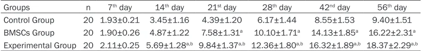
of their scores in BBB rating at different times shows that, from the 28th day of the model, the score of BBB rating of the BMSCs Group is evi-dently higher than that of the Control Group (P<0.05); while, from the 28th day of the model, the score of BBB rating of the Experimental Group is evidently higher than those of both the Control Group and the BMSCs Group (P<0.05), implying that the motor function of the Experimental Group is better than that of the Control Group and the BMSCs Group See Table 1.
Comparison of NGF and BDNF protein expres-sions of the three groups on the 57th day
See Table 2 for NGF and BDNF protein expres-sions of part of the injured spinal cord of the three groups of rats on the 57th day, which shows that, the NGF and BDNF protein expres-sions of the Experimental Group and the BMSCs Group is obviously higher than that of the Control Group (P<0.05); and the increase of the Experimental Group is most obvious, providing
a statistical significance to the difference
be-tween the Experimental Group and the BMSCs Group and the Control Group (P<0.05).
Discussion
According to the traditional view, after the nerve system is injured, new nerve cells are hard to generate to build new synaptic connection, thus making it hard for the injured nerves to recover their function, so, how to prompt the recovery of the central nervous system disease
has always been a globally knotty problem [6, 7]. With the improvement of medical technolo -gy, some academicians found in recent years that, stem cells have the great potential for treatment of the central nervous system. For
example, Brazelton [8] has proved that, MSC of
the mouse can be differentiated in to nerve cells in its brain. Some academicians, by study-ing the cerebral ischemic animal model, found that, the transplanted MSC can survive in the Table 1. Comparison of BBB ratings of motor functions of the three groups of rats (_x±s)
Groups n 7th day 14th day 21st day 28th day 42nd day 56th day
Control Group 20 1.93±0.21 3.45±1.16 4.39±1.20 6.17±1.44 8.55±1.53 9.40±1.51 BMSCs Group 20 1.90±0.26 4.87±1.22 7.58±1.31a 10.10±1.71a 14.13±1.85a 16.22±2.31a
Experimental Group 20 2.11±0.25 5.69±1.28a,b 9.84±1.37a,b 12.36±1.80a,b 16.32±1.89a,b 18.37±2.29a,b
[image:3.612.89.524.88.141.2]Note: compared with the Control Group, aP<0.05; compared with BMSCs Group, bP<0.05.
Table 2. Ratio of positive cells of NGF and BDNF protein expression of the injured spinal cord of the three groups of rats on the 57th day (%, _x ±s)
Group n NGF BDNF
Control Group 20 0.24±0.04 0.35±0.06 BMSCs Group 20 0.51±0.05a 0.57±0.07a
Experimental Group 20 0.72±0.07a,b 0.80±0.09a,b
Note: compared with the Control Group, aP<0.05; compared
brain and move to the ischemic part of the brain, and that the recovery of the nervous sys-tem of that group is obviously better than that
of the control group [9]. Another example is Park [10] who injected MSC into the healthy
female rat before building it into a Parkinson disease (PD) model to observe the protective function of MSC for nigral cells. The result was that the number of nerve cells with tyrosine hydroxylase positive immune activity is obvi-ously larger than that of the control group. As for the work mechanism of MSC transplant, most researchers believe that, the stem cells can build new synaptic connection by connect-ing the injured part of the spinal cord while gen-erating GDNF at the injured part to improve the micro-condition of the injured spinal cord so that the generation of myelin sheath is acceler-ated to prompt the recovery of the nerve
con-duction [11-13]. At present, transplant of stem
cells has become the focus of the international medical circle and brings hope to cure the dis-ease of nervous system of human beings. A lot of fundamental and clinical studies show
that HBO has the confirmed treatment effect
on SCI and has become one of the most com-mon therapies in nerve rehabilitation. HBO can speed up the recovery of the functions of the injured spinal nerves by increasing the content of oxygen and the distance of oxygen diffusion and improving the aerobic metabolism and
microcirculation [14, 15]. Other researches
show that, HBO can promote the survival of endogenous and exogenous BMSCs, increase the expression of various GDNF and re-genera-tion-related genes in the spinal cord, speed up the differentiation of endogenous and exoge-nous stem cells into nerve cells and induce them to move the injured part of the spinal cord
[16]. The experiment conducted by Dayan shows that [17], after SCI occurs to the rat, the
reaction of the endogenous stem cells prolifer-ates which is further promoted and induced by HBO treatment to the injured part of the spinal cord, thereby, accelerating the recovery of the injured spinal cord.
The present study shows that, NGF is a special protein that can promote and maintain the growth, survival, differentiation and function execution of the nerves and protect the injured
nerve cells [18]; the relevant study on animals
proves that, NGF induce the esthesioneure and the noradrenergic neuron of SCI rats to extend
to prompt the sprouting of the fibers of the
nerve cells of the focus; the injection of NGF into the focus of cross-sectional SCI rat
increas-es the concentration of Spinal cord axon [19, 20]. NDGF is an important member of GDNF
family and has 50% of homology with NGF. It not only plays an important role in maintaining the normal physiological functions of the nerve cells, but induces the oriented growth of ner-vous process and adjusts the growth directi-
on of sensory and sympathetic nerve fibers.
Meanwhile, BDNF can provide nutrition to the
nerve fibers and protect the injured nerve cells. As the study of Jakeman et al. [21] pointed out,
BDNF can promote the development, differen-tiation and re-generation of various nerve cells; prevent the death of motor neuron in case of trans-section of sciatic nerves, and save the red nucleus in case of Brown-Sequard. The results of the said studies and researches show that, the increase of NGF and BDNF protein expression at the focus is essential to the recovery of functions of the injured nerves in case of SCI.
This study, after treatment of SCI rats by
com-bining BMSCs cells transplant with HBO, finds
that, after treatment for 56 days, the motor functions of the rear legs of the BMSCs Group and the Experimental Group of rats are obvi-ously better than that of the Control Group, and the NGF and BDNF expression of the motor functions and injured spinal cord of the Ex- perimental Group is evidently better than that of the BMSCs Group. This study implies that the combination of BMSCs transplant with HBO has a synergistic treatment effect which can further increase the motor functions of the rear limbs of the rats. This may be linked to the fact that HBO can promote the survival of exoge-nous BMSC and increase the NGF and BDNF
protein expression. However, the definitive
tre-atment mechanism still calls for further ex- ploration.
Acknowledgements
Colleges and universities in henan province key
scientific research project, No. 16B310008.
Science and Technology Department of Henan Province, No. 201503061.
Disclosure of conflict of interest
Address correspondence to: Dr. Aiguo Xu, De- partment of Respiration and Intensive, The First
Affiliated Hospital of Zhengzhou University, 2001
Jianshe Road, Zhengzhou 450052, Henan Province, China. Tel: +86-13523510886; E-mail: aiguoxu@ zzu.edu.cn
References
[1] Ramer LM, Ramer MS, Bradbury EJ. Restoring function after spinal cord injury: towards clini-cal translation of experimental strategies. Lan-cet Neurol 2014; 13: 1241-56.
[2] Song Q, Xu R, Zhang Q, Ma M, Zhao X. Thera-peutic effect of transplanting bone mesenchy-mal stem cells on the hind limbs’ motor func-tion of rats with acute spinal cord injury. Int J Clin Exp Med 2014; 7: 262-7.
[3] Dietz V, Fouad K. Restoration of sensorimotor functions after spinal cord injury. Brain 2014; 137: 654-67.
[4] Xia LP, Fan F, Tang AL, Ye WQ. Effects of elec-troacupuncture combined with bladder train-ing on the bladder function of patients with neurogenic bladder after spinal cord injury. Int J Clin Exp Med 2014; 7: 1344-8.
[5] Liu X, Zhou Y, Wang Z, Yang J, Gao C, Su Q. Ef-fect of VEGF and CX43 on the promotion of neurological recovery by hyperbaric oxygen treatment in spinal cord-injured rats. Spine J 2014; 14: 119-27.
[6] Mackinnon SE, Yee A, Ray WZ. Nerve transfers for the restoration of hand function after spi-nal cord injury. J Neurosurg 2012; 117: 176-85.
[7] Kamradt T, Rasch C, Schuld C, Böttinger M, Mürle B, Hensel C, Fürstenberg CH, Weidner N, Rupp R, Hug A. Spinal cord injury: associa-tion with axonal peripheral neuropathy in se-verely paralysed limbs. Eur J Neurol 2013; 20: 843-8.
[8] Brazelton TR, Rossi FM, Keshet GI, Blau HM. From marrow to brain: Expression of neuronal pheno types in adult mice. Science 2000; 290: 1775-1779.
[9] Kozić D, Njagulj V, Gaćeša JP, Semnic R,
Prvulović N. Perineural tumor spread-Intercon
-nection between spinal and cranial nerves. J Neurol Sci 2012; 323: 254-6.
[10] Park K, Eglitis MA, Mouradian MM. Protection
of nigral neurons by GDNF-engineered marrow cells transplantation. Neurosci Res 2001; 40: 315-323.
[11] Neuhuber B, Timothy Himes B, Shumsky JS,
Gallo G, Fischer I. Axon growth and recovery of function supported by human bone marrow stromal cells in the injured spinal cord exhibit donor variations. Brain Res 2005; 1035: 73-85.
[12] Martinez AM, Goulart CO, Ramalho Bdos S,
Oliveira JT, Almeida FM. Neurotrauma and mesenchymal stem cells treatment: From ex-perimental studies to clinical trials. World J Stem Cells 2014; 6: 179-94.
[13] Cui B, Li E, Yang B, Wang B. Human umbilical
cord blood-derived mesenchymal stem cell transplantation for the treatment of spinal cord injury. Exp Ther Med 2014; 7: 1233-1236.
[14] Lu PG, Hu SL, Hu R, Wu N, Chen Z, Meng H, Lin
JK, Feng H. Functional recovery in rat spinal cord injury induced by hyperbaric oxygen pre-conditioning. Neurol Res 2012; 34: 944-51.
[15] Huang H, Xue L, Zhang X, Weng Q, Chen H, Gu
J, Ye S, Chen X, Zhang W, Liao H. Hyperbaric oxygen therapy provides neuroprotection fol-lowing spinal cord injury in a rat model. Int J Clin Exp Pathol 2013; 6: 1337-42.
[16] Lin SS, Ueng SW, Niu CC, Yuan LJ, Yang CY,
Chen WJ, Lee MS, Chen JK. Hyperbaric oxygen promotes osteogenic differentiation of bone
marrow stromal cells by regulating
Wnt3a/β-catenin signaling--an in vitro and in vivo study. Stem Cell Res 2014; 12: 260-74.
[17] Dayan K, Keser A, Konyalioglu S, Erturk M,
Aydin F, Sengul G, Dagci T. The effect of hyper-baric oxygen on neuroregeneration following acute thoracic spinal cord injury. Life Sci 2012; 90: 360-4.
[18] Park KW, Lin CY, Lee YS. Expression of
sup-pressor of cytokine signaling-3 (SOCS3) and its role in neuronal death after complete spinal cord injury. Exp Neurol 2014; 261: 65-75.
[19] Luo Y, Zou Y, Yang L, Liu J, Liu S, Liu J, Zhou X, Zhang W, Wang T. Transplantation of NSCs with OECs alleviates neuropathic pain associ-ated with NGF downregulation in rats following spinal cord injury. Neurosci Lett 2013; 549: 103-8.
[20] Zhang H, Wu F, Kong X, Yang J, Chen H, Deng L,
Cheng Y, Ye L, Zhu S, Zhang X, Wang Z, Shi H, Fu X, Li X, Xu H, Lin L, Xiao J. Nerve growth fac-tor improves functional recovery by inhibiting endoplasmic reticulum stress-induced neuro-nal apoptosis in rats with spineuro-nal cord injury. J Transl Med 2014; 12: 130.
[21] Jakeman LB, Wei P, Guan Z. Brain-derived
neu-rotrophic factor stimulates hindlimb stepping
and sprouting of cholinergic fibers after spinal