Original Article
Prognostic significance of lymph node metastasis in
triple negative ductal carcinoma of the breast: a
retrospective cohort study
Hongliang Chen, Ang Ding, Maoli Wang, Chuyang Yin, Zheng Zhang
Department of Breast Surgery, Obstetrics and Gynecology Hospital of Fudan University, Shanghai, China Received February 3, 2016; Accepted August 3, 2016; Epub February 15, 2017; Published February 28, 2017
Abstract: Objective: The purpose of this study was to identify the prognostic significance of lymph node (LN) me -tastasis in triple negative breast cancer (TNBC). Methods: A retrospective analysis of 206 TNBC was conducted. Clinicopathological characteristics between different LN statuses were compared by Chi-square test. Survival differ-ences among pT stages, pN stages, lymphovascular invasion (LVI) etc. were compared by Log-rank test. All variables with statistical significance in the univariate analysis were investigated in multivariate analysis by COX regression. Results: The mean age of 206 TNBC patients was 49.80 ± 10.06 years old. The mean size of tumor was 2.96 ± 2.01 cm. There were altogether 83 cases (40.3%) with positive LNs, among which 50 cases in pN1 stage, 24 cases in pN2 stage and 9 cases in pN3 stage. Tumor size and LVI were correlated with LN metastasis. Tumor size (P = 0.023 for DFS, 0.012 for OS), LVI (P = 0.001 for DFS, 0.003 for OS) and LN status (negative or positive) (P = 0.017 for DFS, 0.008 for OS) rather than pN stage (P = 0.090 for DFS, 0.061 for OS) were independent prognostic factors of TNBC. There were no statistically significant differences in the prognosis among pN1-pN3 regardless of tumor size or LVI status. Conclusions: For TNBC, LN status was an independent prognostic factor. But for cases with LN metastasis, the prognosis was not greatly affected by the number of positive LNs. Perhaps apart from lymphatic metastasis, hematogenous metastasis played an important role in TNBC progression, even at an early stage.
Keywords: Triple negative breast cancer, lymph node metastasis, prognosis
Introduction
Currently, breast cancer is the most common cancer among women worldwide. Axillary lymph
node (LN) metastasis is one of the most impor -tant prognostic factors for the survival of breast cancer [1, 2]. The 5-year disease-free survival
(DFS) for breast cancer patients with LN metas -tasis is 40%, lower than that of patients without
LN metastasis [3].
With the advances in molecular biology and microarray analysis, breast cancer can be seg-regated into different “intrinsic subtypes”, which enhanced our understanding of breast cancer heterogeneity. Different molecular sub-types have distinct biological behavior, sug-gesting that molecular subtype has important value in comprehensive evaluation of prognosis [4].
Triple negative breast cancer (TNBC) is defined by the lack of expression of the estrogen recep
-epidermal growth factor receptor 2 (HER2), which accounts for 15-20% of breast cancer patients. It is the most aggressive subtype of invasive ductal carcinoma of the breast (IDC), characterized by occurrence at young age, early
relapse especially visceral metastasis, and lack
of targeted therapy [5, 6]. Most studies
consid-er the TNBC subtype as an independent markconsid-er
of a poor prognosis [7-9].
In spite of poor prognosis, TNBC is not
associ-ated with increased likelihood of LN metasta -ses. Gangi A et al. pointed out that patients with
TNBC are not more likely to have involved nodes
than those with non-TNBC [10]. And early dis-tant metastasis can be observed at relatively high frequency in TNBC patients with negative
LN or few positive LNs.
Although TNBC subtype and LN metastasis
have both been independently demonstrated as prognostic factors, there is little data
-LN metastasis prognostic significance in TNBC
tasis on TNBC patients. Therefore, we conduct-ed an extensive retrospective study to provide a more complete overview of the subject. Patients and methods
A retrospective study was conducted, in which 206 breast cancer patients in TNBC-IDC sub-type treated in Obstetrics and Gynecology Hospital of Fudan University between June 2007 and June 2014 were enrolled. Patients were excluded for the following reasons: male gender, in situ lesion, curative resection was not conducted or distant metastasis was
con-firmed before surgery.
The baseline data included demographic char-acteristics (e.g., age, menopause), and tumor
characteristics (e.g., tumor size stage, LN stage, lymphovascular invasion (LVI), histologic grade). Tumor size and LN staging in this study was
based on the AJCC Staging 7th edition [11]. All
patients received mastectomy or
breast-con-serving surgery (BCS) plus axillary LN dissec
-tion/sentinel LN biopsy, adjuvant/neoadjuvant
chemotherapy composed anthracycline and/or taxane followed by radiotherapy (if required). The status of the ER, PR, and HER2 were deter-mined by immunohistochemical (IHC) staining.
death as a result of any cause. Those without any evidence of relapse or lost to follow-up we- re censored at the date of their last follow-up. Statistical analyses
Data were expressed as mean ± standard devi-ation (SD) for continuous variables. Categorical variables were normally tested with Chi-square test (Pearson statistic) when appropriate. Sur- vival curves were constructed using the Kaplan-Meier method, and univariate survival
differ-ence was determined with the log-rank test. All variables with statistical significance in the uni -variate analysis were investigated by multivari-ate analysis. Adjusted hazard ratios (HRs) with
95% confidence intervals (95% CI) were calcu -lated using Cox proportional hazards model. All the statistical analysis was performed using
SPSS 19.0 software package (SPSS, Chicago, IL, USA). Two-sided P < 0.05 was considered statistically significant.
Results
Clinicopathologic characteristics according to lymph node status
[image:2.612.91.357.95.359.2]The mean age of 206 TNBC patients was 49.80 ± 10.06 years old (range 22-70). The mean size of tumor was 2.96 ± 2.01 cm (range 1.0-15.0). Table 1. Clinicopathological characteristics according to lymph
node status
Lymph node
negative Cases (%) positive Cases (%)Lymph node P value
Menopause status 0.133
Pre-menopausal 61 (49.6%) 50 (60.2%) Post-menopausal 62 (50.4%) 33 (39.8%)
Histological grade 0.503
Median 41 (33.3%) 24 (28.9%)
low 82 (66.7%) 59 (71.1%)
Tumor size 0.002
≤ 2 cm 56 (45.5%) 20 (24.1%)
> 2 cm 67 (54.5%) 63 (75.9%)
LVI < 0.001
Negative 91 (74.0%) 41 (49.4%) Positive 32 (26.0%) 42 (50.6%)
Surgery 0.752
Mastectomy 87 (70.7%) 57 (68.7%)
BCS 36 (29.3%) 26 (31.3%)
Chemotherapy 0.617
Adjuvant 97 (78.9%) 63 (75.9%) neoadjuvant 26 (21.1%) 20 (24.1%)
The cut-off value for ER positivity and PR positivity was 1% of tumor cells with positive nuclear stain-ing. Tumors with an IHC score of 3+ based on circumferential mem-brane-bound staining or with
amplification confirmed by flores -cent in situ hybridization (FISH)
were defined as HER2-positive.
Tumors with an IHC score of 2+ were recommended FISH test. Follow-up information regarding tu- mor relapse and survival status was available through outpatient departmental records and person-al contact with the patients via mail and telephone calls until June 2015. The follow-up duration was calculated from the date of diag-nosis until the date of death or la- st contact. Disease-free survival (DFS) was the time between
diag-nosis and confirmation of disease
There were 76 cases in T1 stage, 112 cases in T2 stage, and 18 cases in T3 stage. There were
altogether 83 cases (40.3%) with positive LNs,
among which 50 cases in pN1 stage, 24 cases in pN2 stage and 9 cases in pN3 stage. The
median number of positive LNs was 2 (range 1-24). The mean number of LNs removed was
15.96 ± 5.04 (range 8-35). Median follow-up duration was 58 months (range 8-96). 5-year DFS rate was 75.6% and 5-year OS rate was 85.1%.
The clinicopathologic characteristics according
to LN status were summarized in Table 1. It was
shown that the tumor size and LVI were corre
-lated with LN status. Tumor size > 2 cm (P = 0.002) and positive LVI (P < 0.001) were more likely to be observed in cases with positive LNs.
Univariate analysis of possible factors corre-lated with prognosis
Univariate analysis by Log-rank test revealed that larger tumor size (P < 0.001 for DFS and OS), higher pN stage (P < 0.001 for DFS and
OS) (Figure 1A, 1C), and positive LVI (P < 0.001
for DFS and OS) were correlated with decreased
[image:3.612.92.520.73.536.2]LN metastasis prognostic significance in TNBC
for DFS, 0.494 for OS), histologic grade (P =
0.909 for DFS, 0.609 for OS), surgery mode (P
= 0.589 for DFS, 0.331 for OS) and chemother
-apy mode (P = 0.354 for DFS, 0.539 for OS)
were not correlated with prognosis.
Further analysis showed that there was a
sta-tistically significant separation between the Kaplan-Meier survival curves of LN statuses (P < 0.001 for DFS and OS) (Figure 1B, 1D) rather
than of pN1-pN3 (P = 0.443 for DFS, 0.091 for
OS).
There was a trend of DFS and OS benefit in
patients receiving neoadjuvant chemotherapy compared with those receiving adjuvant che-motherapy, although it did not reach statistical
significance. (P = 0.354 for DFS, 0.539 for OS)
(Figure 2).
As tumor size was correlated with LN status
(Table 1), survival analysis was conducted
according to LN stage in tumor size ≤ 2 cm and
tumor size > 2 cm respectively (Figure 3).
Among cases in tumor size ≤ 2 cm, none was in pN3 stage. For cases with tumor size ≤ 2 cm, pN stage was not correlated with DFS (P = 0.125) or OS (P = 0.145) (Figure 3A, 3C). Further analysis showed that there was a
sta-tistically significant difference between the Kaplan-Meier survival curves of LN status (P =
0.045 for DFS, 0.042 for OS) (Figure 3B, 3D)
rather than of pN1-pN2 (P = 0.917 for DFS,
0.788 for OS). For cases with tumor size > 2 cm,
pN stage was correlated with DFS (P = 0.006) and OS (P = 0.002) (Figure 3E, 3G). Further analysis showed that there was a statistically
significant difference between the Kaplan-Meier survival curves of LN status (P = 0.001
for DFS, 0.002 for OS) (Figure 3F, 3H) rather
than of pN1-pN3 (P = 0.641 for DFS, 0.187 for
OS).
As LVI status was also correlated with LN status
(Table 1), survival analysis was conducted
according to LN stage in positive or negative LVI
respectively (Figure 4). Among cases with
nega-tive LVI, none was in pN3 stage. For cases with negative LVI, pN stage was correlated with DFS (P < 0.001) and OS (P = 0.003) (Figure 4A, 4C). Further analysis showed that there was a
sta-tistically significant difference between the Kaplan-Meier survival curves of LN status (P <
0.001 for DFS, 0.005 for OS) (Figure 4B, 4D)
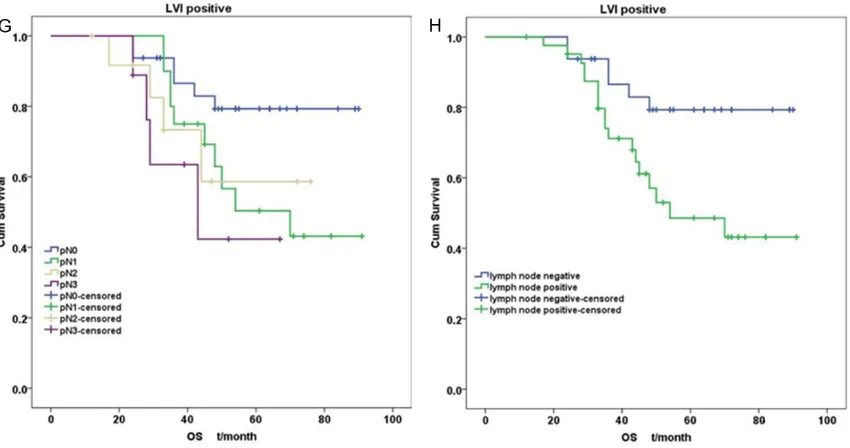
rather than of pN1-pN2 (P =0.216 for DFS, 0.211 for OS). For cases with positive LVI, pN stage was not correlated with DFS (P = 0.288) or OS (P = 0.083) (Figure 4E, 4G). Further anal-ysis showed that there was a statistically
sig-nificant difference between the Kaplan-Meier survival curves of LN status (P = 0.046 for DFS,
0.016 for OS) (Figure 4F, 4H) rather than of
LN metastasis prognostic significance in TNBC
From above, it could be concluded that for
TNBC the prognosis was affected greatly by LN status. But for cases with positive LNs, the
prognosis was not greatly affected by the
num-ber of positive LNs.
Multivariate analysis of prognostic factors for TNBC
When pT, pN and LVI were entered into the Cox
proportional hazards model as covariates, pT
and LVI were both independent prognostic fac
-tors of DFS (pT: P = 0.017, LVI: P = 0.001) and OS (pT: P = 0.018, LVI: P = 0.002), but pN was
not an independent prognostic factor for DFS
or OS (P = 0.090 for DFS, 0.061 for OS). When pT, LN status and LVI were entered into
the Cox proportional hazards model as covari-ates, they were all independent prognostic fac-tors of DFS and OS (Table 2).
So for TNBC, LN status rather than pN stage
was the prognostic factor. For cases with
posi-tive LNs, the survival rate did not decrease lin
-early with the increase of positive LNs.
Discussion
The poor prognosis of TNBC may be due to a higher propensity for distant (rather than regi-
onal) spread [12, 13]. Research into the prog-nostic factors of TNBC may help to instruct the clinical practice and pave the way for mecha-nism study.
The 7th edition of staging in breast cancer has
included tumor size and LN status as the most
important prognostic factors [11]. The size of the primary tumor and the number of positive lymph nodes has an inverse linear relationship with prognosis and survival [14]. But for TNBC, it is not always the same case.
Some recent publications have suggested the
role of molecular subtypes in the risk of LN
involvement [15-17]. Although its poor survival compared with other subtypes, TNBC did not
demonstrate the highest extent of LN involve
-ment [15, 16]. Greater probability of LN involve -ment was in the luminal B and luminal HER2
subtypes [2, 18]. It was reported that the LN
metastasis rate in TNBC was 25-54% [2, 18-20]. In our cohort, the rate was 40.3%, and 60% were in pN1 stage, which was in accord with the literature [9].
Our study revealed that the prognosis was
poorer in cases with positive LN than in nega
[image:6.612.94.522.75.299.2]LN metastasis prognostic significance in TNBC
But in cases with positive LN, the survival rate
did not decrease linearly with the upgrading of
pN stage, regardless of tumor size or LVI status,
either. As the prognosis was not greatly
affect-ed by the number of additional positive LNs,
TNBC had a distinct biologic behavior that dif-fered from other subtypes in which the number
of positive LNs correlated with prognosis [3, 21]. So Park et al. concluded that in TNBC, the AJCC TNM staging system was not
discrimina-tive to reflect the survival rate as the other sub -types [22].
Our study showed that LVI was an independent
prognostic factor, although it correlated with
positive LN status, suggesting that apart from
lymphatic metastasis, hematogenous
metasta-ult that the prognosis was not affected by the
increase of positive LNs.
It was reported that TNBC was associated with a larger tumor size [20]. In our study, 63.1% cases had a tumor size over 2 cm. Our study also demonstrated that larger tumor size occurred more frequently in cases with positive
LNs, and it was in accord with Hernandez-Aya’s research which confirmed the relationship
through a review of 1711 TNBC patients [19]. Multiple studies indicated that tumor size was
important for predicting LN metastasis [24].
In our study, tumor size was an independent
prognostic factor, which agreed with Anders’s
[image:8.612.95.520.73.297.2]report [25]. Although larger tumor correlated
Figure 4. Survival curves according to pN stage and LN status in different tumor LVI status layer. A. DFS according to pN stage in negative LVI; B. DFS according to LN status in negative LVI; C. OS according to pN stage in negative LVI; D. OS according to LN status in negative LVI; E. DFS according to pN stage in positive LVI; F. DFS according to LN status in positive LVI; G. OS according to pN stage in positive LVI; H. OS according to LN status in positive LVI.
Table 2. Multivariate analysis of prognostic factors of TNBC
DFS OS
HR valueP 95% CI HR valueP 95% CI
pT 0.023 0.012
T2:T1 2.604 0.013 1.228-5.522 2.325 0.057 0.974-5.551 T3:T1 3.351 0.013 1.292-8.693 0.003 1.684-12.932 Lymph node status
Positive:negative 2.148 0.017 1.146-4.025 2.741 0.008 1.301-5.774 LVI
Positive:negative 2.708 0.001 1.486-4.936 2.939 0.003 1.453-5.945
sis played an impor-tant role in TNBC pro-gression, even at an ea-
rly stage. Liu et al. de- monstrated higher lev-els of intratumoral and peritumoral lymphangi- ogenesis in early stag-es of TNBC. Patients
with early LN involve-ment may already ha- ve distant metastasis [23]. This may partly
[image:8.612.91.403.377.504.2]res-with positive LN, it was tumor size rather than
positive pN stage that was the independent prognostic factor. Combined with the impor-tance of hematogenous metastasis in TNBC, we hypothesized that tumor cells of TNBC may
be more likely to detach from larger mass to
migrate to distant site through circulation to form a new metastasis.
The result of our study has important clinical
significance. More attention should be paid to
the systemic treatment of early micrometasta-sis through circulation rather than extensive local treatment. Neoadjuvant chemotherapy provides the earliest chance to treat micromet-astatic disease, saving time that could poten-tially be lost to local treatment. And neoadju-vant chemotherapy may enhance the local effect through the intact neovasculature asso-ciated with cancer which could have been altered by surgical excision [26]. Our study showed a tendency of improved survival favor-ing neoadjuvant chemotherapy although
with-out statistical significance, possibly due to
small sample size of cases receiving neoadju-vant chemotherapy. Meanwhile BCS had no
adverse influence on survival, which was in
accord with the literature [27]. Therefore neo-adjuvant chemotherapy and BCS should be rec-ommended for suitable TNBC patients.
Some limitations of this study should be
acknowledged. First, selection bias was not
completely avoided because this was a retro-spective cohort study. Furthermore, Ki-67 path-ological data were not routinely obtained from patients, but it may have an effect on survival. In conclusion, our study indicated that in TNBC,
tumor size and LVI correlated with LN status. Tumor size, LVI, and LN status were all indepen -dent prognostic factors. But once there was
any evidence of LN involvement, the survival
rate did not decrease linearly with the
upgrad-ing of pN stage, regardless of tumor size or LVI
status. Further researches are warranted for the mechanisms of this aggressive behavior. Disclosure of conflict of interest
None.
Address correspondence to: Hongliang Chen, Depa- rtment of Breast Surgery, Obstetrics and Gynecology
Hospital of Fudan University, 419 Fangxie Road, Huangpu District, Shanghai 200011, China. E-mail: hongliangchen1982@163.com
References
[1] Cody HS 3rd and Houssami N. Axillary man-agement in breast cancer: what’s new for 2012? Breast 2012; 21: 411-415.
[2] Howland NK, Driver TD, Sedrak MP, Wen X, Dong W, Hatch S, Eltorky MA and Chao C. Lymph node involvement in immunohisto -chemistry-based molecular classifications of breast cancer. J Surg Res 2013; 185: 697-703.
[3] Dings PJ, Elferink MA, Strobbe LJ and de Wilt JH. The prognostic value of lymph node ratio in node-positive breast cancer: a Dutch nation-wide population-based study. Ann Surg Oncol 2013; 20: 2607-2614.
[4] Perou CM, Sorlie T, Eisen MB, van de Rijn M, Jeffrey SS, Rees CA, Pollack JR, Ross DT, Johnsen H, Akslen LA, Fluge O, Pergamen-schikov A, Williams C, Zhu SX, Lonning PE, Borresen-Dale AL, Brown PO and Botstein D. Molecular portraits of human breast tumours. Nature 2000; 406: 747-752.
[5] Rakha EA, Reis-Filho JS and Ellis IO. Basal-like breast cancer: a critical review. J Clin Oncol 2008; 26: 2568-2581.
[6] Voduc KD, Cheang MC, Tyldesley S, Gelmon K, Nielsen TO and Kennecke H. Breast cancer subtypes and the risk of local and regional re -lapse. J Clin Oncol 2010; 28: 1684-1691. [7] Liedtke C, Mazouni C, Hess KR, Andre F, Tordai
A, Mejia JA, Symmans WF, Gonzalez-Angulo AM, Hennessy B, Green M, Cristofanilli M, Hor- tobagyi GN and Pusztai L. Response to neoad -juvant therapy and long-term survival in pa-tients with triple-negative breast cancer. J Clin Oncol 2008; 26: 1275-1281.
[8] Chen HL, Ding A and Wang FW. Prognostic ef -fect analysis of molecular subtype on young breast cancer patients. Chin J Cancer Res 2015; 27: 428-436.
[9] Braunstein LZ, Niemierko A, Shenouda MN, Truong L, Sadek BT, Abi Raad R, Wong JS, Punglia RS, Taghian AG and Bellon JR. Outcome following local-regional recurrence in women with early-stage breast cancer: impact of bio-logic subtype. Breast J 2015; 21: 161-167. [10] Gangi A, Mirocha J, Leong T and Giuliano AE.
Triple-negative breast cancer is not associated with increased likelihood of nodal metastases. Ann Surg Oncol 2014; 21: 4098-4103. [11] Edge SB and Compton CC. The American Joint
LN metastasis prognostic significance in TNBC
[12] Metzger-Filho O, Tutt A, de Azambuja E, Saini KS, Viale G, Loi S, Bradbury I, Bliss JM, Azim HA Jr, Ellis P, Di Leo A, Baselga J, Sotiriou C and Piccart-Gebhart M. Dissecting the heterogene-ity of triple-negative breast cancer. J Clin Oncol 2012; 30: 1879-1887.
[13] Chen HL and Ding A. Comparison of invasive micropapillary and triple negative invasive duc-tal carcinoma of the breast. Breast 2015; 24: 723-731.
[14] Carter CL, Allen C and Henson DE. Relation of tumor size, lymph node status, and survival in 24,740 breast cancer cases. Cancer 1989; 63: 181-187.
[15] Crabb SJ, Cheang MC, Leung S, Immonen T, Nielsen TO, Huntsman DD, Bajdik CD and Chia SK. Basal breast cancer molecular subtype predicts for lower incidence of axillary lymph node metastases in primary breast cancer. Clin Breast Cancer 2008; 8: 249-256.
[16] Van Calster B, Vanden Bempt I, Drijkoningen M, Pochet N, Cheng J, Van Huffel S, Hendrickx W, Decock J, Huang HJ, Leunen K, Amant F, Berteloot P, Paridaens R, Wildiers H, Van Limbergen E, Weltens C, Timmerman D, Van Gorp T, Smeets A, Van den Bogaert W, Vergote I, Christiaens MR and Neven P. Axillary lymph node status of operable breast cancers by combined steroid receptor and HER-2 status: triple positive tumours are more likely lymph node positive. Breast Cancer Res Treat 2009; 113: 181-187.
[17] Marrazzo A, Boscaino G, Marrazzo E, Taormina P and Toesca A. Breast cancer subtypes can be determinant in the decision making pro -cess to avoid surgical axillary staging: A retro-spective cohort study. Int J Surg 2015; 21: 156-161.
[18] Mazouni C, Rimareix F, Mathieu MC, Uzan C, Bourgier C, Andre F, Delaloge S and Garbay JR. Outcome in breast molecular subtypes accord-ing to nodal status and surgical procedures. Am J Surg 2013; 205: 662-667.
[19] Hernandez-Aya LF, Chavez-Macgregor M, Lei X, Meric-Bernstam F, Buchholz TA, Hsu L, Sahin AA, Do KA, Valero V, Hortobagyi GN and Gonzalez-Angulo AM. Nodal status and clinical outcomes in a large cohort of patients with tri-ple-negative breast cancer. J Clin Oncol 2011; 29: 2628-2634.
[20] Liao GS, Chou YC, Hsu HM, Dai MS and Yu JC. The prognostic value of lymph node status among breast cancer subtypes. Am J Surg 2015; 209: 717-724.
[21] Shen ZZ. [Relation of tumor size, lymph node status and prognosis in breast cancer]. Zhonghua Wai Ke Za Zhi 1991; 29: 554-557, 589.
[22] Park YH, Lee SJ, Cho EY, Choi YL, Lee JE, Nam SJ, Yang JH, Shin JH, Ko EY, Han BK, Ahn JS and Im YH. Clinical relevance of TNM staging system according to breast cancer subtypes. Ann Oncol 2011; 22: 1554-1560.
[23] Liu HT, Ma R, Yang QF, Du G and Zhang CJ. Lymphangiogenic characteristics of triple neg -ativity in node-negative breast cancer. Int J Surg Pathol 2009; 17: 426-431.
[24] Silverstein MJ, Gierson ED, Waisman JR, Colburn WJ and Gamagami P. Predicting axil-lary node positivity in patients with invasive carcinoma of the breast by using a combina-tion of T category and palpability. J Am Coll Surg 1995; 180: 700-704.
[25] Anders CK, Deal AM, Miller CR, Khorram C, Meng H, Burrows E, Livasy C, Fritchie K, Ewend MG, Perou CM and Carey LA. The prognostic contribution of clinical breast cancer subtype, age, and race among patients with breast can-cer brain metastases. Cancan-cer 2011; 117: 1602-1611.
[26] Yeh E, Slanetz P, Kopans DB, Rafferty E, Georgian-Smith D, Moy L, Halpern E, Moore R, Kuter I and Taghian A. Prospective comparison of mammography, sonography, and MRI in pa-tients undergoing neoadjuvant chemotherapy for palpable breast cancer. AJR Am J Roentgenol 2005; 184: 868-877.