Original Article
The diagnostic value of neutrophil-to-lymphocyte ratio
and platelet-to-lymphocyte ratio in tuberculous
spondylitis
Qitian He, Wenjun Tang, Yan Deng, Yu He, Li Xie, Xue Qin, Shan Li
Department of Clinical Laboratory, First Affiliated Hospital of Guangxi Medical University, Nanning, Guangxi, China Received April 22, 2015; Accepted June 12, 2016; Epub August 15, 2016; Published August 30, 2016
Abstract: The neutrophil-to-lymphocyte ratio (NLR) and platelet-to-lymphocyte ratio (PLR) are useful markers in vari-ous inflammatory diseases, but their diagnostic capability intuberculvari-ous (TB) spondylitishas not been evaluated. Our aim was to investigate the diagnostic capability of NLR and PLR on the diagnosis of TB spondylitis. Patients surgically treated for TB spondylitisin the First Affiliated Hospital of Guangxi Medical University from 2012 to 2015 were retrospectively investigated in this study. Correlations between NLR, PLR and laboratory indices in TB spondy -litis patients were evaluated by the Spearman’s correlation analysis. The sensitivity and specificity of NLR and PLR were assessed by the receiver operating characteristic (ROC) curves. Seventy-four TB spondylitis patients and 54 healthy individuals were analyzed retrospectively. The NLR and PLR between TB spondylitis patients and healthy controls were significant different. The relationship between NLR, C-reaction protein (CRP), and erythrocyte sedi -mentation rate (ESR) in TB spondylitis patients were significant correlation, similarly, the correlation between PLR and CRP and ESR in TB spondylitis patients were significantly correlated. The ROC curves analysis of NLR and PLR in TB spondylitis patients showed statistically significance. In conclusion, NLR and PLR might be valuable markers in diagnosing TB spondylitis.
Keywords: Neutrophil-to-lymphocyte ratio, platelet-to-lymphocyte ratio, diagnosis, tuberculosis spondylitis
Introduction
Inhalation of Mycobacterium tuberculosisbacilli can lead to the occurrence of pulmonary
tuber-culosis (PTB) infection. In terms of the annual death toll, tuberculosis (TB) may no longer be
the most deadly human diseases, but it remains a serious disease threat to human health around the world, causing 1.3 million deaths globally in 2012 alone [1]. China has a high rate
of TB infection [2], and thus the diagnosis and treatment of TB represents a serious health
concern in this region. Of all the
extra-pulmo-nary TB patients, skeletal involvement occurs in approximately 10% of cases [3]. TB spondyli -tis is a chronic progressive development dis-ease and is rarely associated with symptoms like cough, dyspnea, or fever [4]. There are no clear guidelines for diagnosis and treatment of
TB spondylitis patients [5]. In order to prevent -complications such as permanent neurological
disability and the occurrence of spinal
deformi-ty in patients with TB spondylitis,early identifi -cation and timely treatment is necessary [6, 7]. It is well known that elevated neutrophils and
lymphocytes are closely related to inflammato -ry state. Platelets have also been shown to be
associated with inflammation [8, 9]. Recent
studies have indicated that neutrophil to lym-phocyte ratio (NLR) and/or platelets to lympho-cyte ratio (PLR) could be used as biomarkersin many diseases [10-13]. Indicators such as CRP, ESR and/or platelet count have been used to
assess the status of PTB and TB spondylitis
inpatients [6, 14-16]. In some recent studies, NLR and PLR have been evaluated in diseases
such as PTB and ankylosing spondylitis (AS)
[13, 17]. However, hematological
characteris-tics including PLR and NLR in TB spondylitis
patients remains unknown. The aim of this study was thus to assess the variety of NLR and
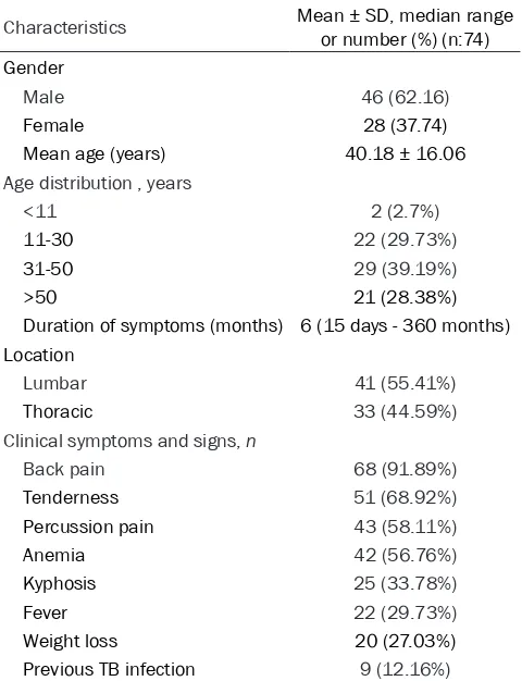
Table 1. Characteristics of patients withTB spondylitis
Characteristics Mean ± SD, median range or number (%) (n:74)
Gender
Male 46 (62.16)
Female 28 (37.74)
Mean age (years) 40.18 ± 16.06 Age distribution , years
<11 2 (2.7%)
11-30 22 (29.73%)
31-50 29 (39.19%)
>50 21 (28.38%)
Duration of symptoms (months) 6 (15 days - 360 months) Location
Lumbar 41 (55.41%)
Thoracic 33 (44.59%)
Clinical symptoms and signs, n
Back pain 68 (91.89%)
Tenderness 51 (68.92%)
Percussion pain 43 (58.11%)
Anemia 42 (56.76%)
Kyphosis 25 (33.78%)
Fever 22 (29.73%)
Weight loss 20 (27.03%)
Previous TB infection 9 (12.16%)
Materials and methods
All patient information was extracted from the
medical records of the First Affiliated Hospital of Guangxi Medical University from 2012 to
2015. During this period, 74 patients were
sur-gically treated for TB spondylitis. The diagnosis of TB spondylitis is achieved through a compre -hensive evaluation of the patients' clinical fea-tures and radiological, intraoperative, and
his-tological examinations. Fifty-four normal con -trols from Health Examination Centers were also included. Patients and healthy controls who met any of the following criteria were excluded: (1) current use of antibiotics or other
drugs that affect white blood cell (WBC) or
platelet count; (2) autoimmune disorders or other infectious diseases; (3) serious liver or renal disease; (4) hematologic diseases; (5) neoplasms; and/or (6) thyroid disease. Patients with active pulmonary tuberculosis in addition
to TB spondylitis, as well as patients with
incomplete data and laboratory test results in their medical records, were also excluded.
statistical software was used to analyze the data. P<0.05 was considered significant.
This study was approved by the local ethics committee, and informed consent was obtained from all participants.
Results
Characteristics of TB spondylitis patients
The characteristics of TB spondylitis patients
are presented in Table 1. Seventy-four Patients
(mean age: 40.18 ± 16.06 years) with TB spon -dylitis were included in the study. The percent-age of males and females were 62.2% and 37.8%, respectively. The 31-50 age group (39.2%) accounted for the largest proportion of
all TB spondylitis patients. The mean duration
of symptoms was 6 month (range, 15 days - 360 months). The proportion of lumbar and thoracic tuberculosis was 55.4% and 44.6%, respectively. Sixty-eight (91.9%) patients had back pain, 51 (68.9%) had tenderness, 43 (58.1%) had percussion pain, 42 (56.8%) had
Clinical and laboratory characteristics were extracted before treatment. De-
mographic characteristics of TB spondyli -tis patients such as age and gender, were
also gathered. Blood samples were col
-lected from all TB spondylitis patients
and healthy controls before treatment.
The results of total WBC count, neutro -phils, lymphocytes, platelets, CRP, and ESR were extracted from participants’ medical records.
Statistical analysis
The Kolmogorov-Smirnov normality test was performed for continuous variables. The normal distribution variables were expressed as mean ± standard devia-tion. Two independent sample t-test X2
test were utilized to compare data
between TB spondylitis patients and con -trols. The spearman correlation tests were done between two continuous vari-ables. The performance of NLR and PLR was detected by ROC curves. MedCalc statistic software (version 11.3.8.0,
Mariakerke, Belgium) was used to com
anemia, 25 (33.8%) had kyphosis, 22 (29.7%) had fever, and 20 (27.0%) had weight loss.
Laboratory results of TB spondylitis patients and healthy individuals
As shown in Table 2, the age and gender of TB
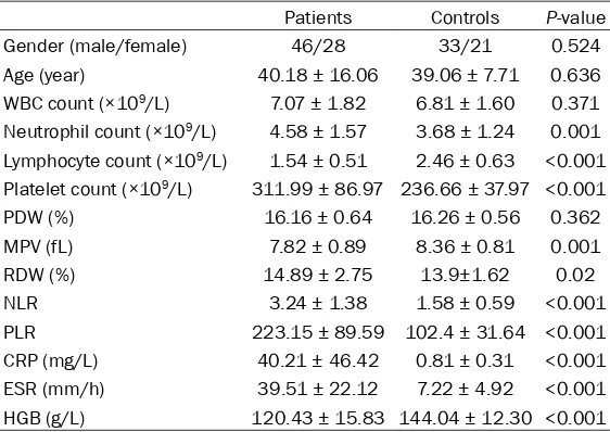
spondylitis patients were matched to the age and gender of healthy controls. Neutrophil and
platelet levels in patients with TB spondylitis were increased significantly, yet lymphocyte levels were decreased significantly. The NLR, PLR, CRP and ESR in patients with TB spondyli
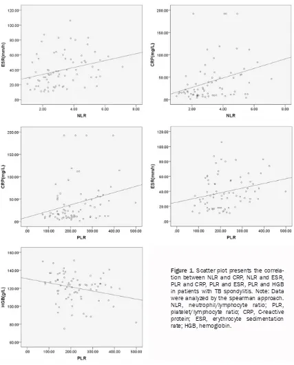
-tis were all significantly higher than in the con -trols. As shown in Figure 1, the results of Spearman’s analysis showed the correlation
coefficients between NLR and CRP and ESR
were0.412 (P<0.001) and 0.362 (P = 0.002)
respectively; the correlation coefficients
be-tween PLR and CRP and ESR were 0.331 (P = 0.004) and 0.301 (P = 0.007), respectively.
NLR and PLR were significantly correlated (r = 0.623, P<0.001). The correlation coefficient
between ESR and platelets was 0.267 (p = 0.022). There is a negative correlation between
PLR and HGB (r = -0.268, P = 0.021). However,
no statistically significant relationship was found between NLR and HGB (data not shown).
As shown in Table 3, no statistically difference in neutrophils, lymphocytes, platelets, NLR and PLR was found between anemia (haemoglobin level: female<120; male<130) and non-anemia
TB spondylitis patients. No statistically dif
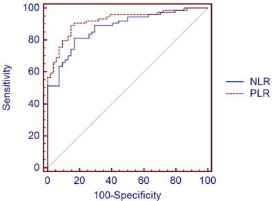
fer-identified, and an 89.2% sensitivity and 85.2% specificity for PLR were observed. However, there were no statistically significant differenc
-es between the AUCs of NLR and PLR (P>0.05). Discussion
Tuberculous spondylitis is characterized by chronic progressive development. When
com-pared to PTB, the diagnosis of TB spondylitis is
more challenging because of its delayed pre-sentation [16]. Early diagnosis and treatment can control the development of the disease, avoid the occurrence of deformity and
paraly-sis, and reduce financial burden.
Inflammation is involved in the pathogenesis of tuberculosis. Abakay et al. considered inflam -matory reaction to be the main
pathophysiolo-gy of PTB. In addition, they found that NLR and PLR might be new markers of inflammation in active PTB [17]. Muzaffar et al. described the hematological changes in TB spondylitis
pati-ents. They found thatanaemia and thrombocy-tosis accounted for the majority of cases [18].
Danielet al. demonstrated that patients with TB
spondylitis had elevated ESR and thrombocyto-sis when compared to other spinal pathologies [16]. In our study, increased NLR, PLR, CRP and
ESR were statistically significant in TB spondyli -tis patients.
[image:3.612.91.372.84.283.2]ESR and CRP are makers commonly used in clinical diagnosis of many diseases. It is known Table 2. Laboratory results of TB spondylitis patients and controls
Patients Controls P-value
Gender (male/female) 46/28 33/21 0.524
Age (year) 40.18 ± 16.06 39.06 ± 7.71 0.636
WBC count (×109/L) 7.07 ± 1.82 6.81 ± 1.60 0.371
Neutrophil count (×109/L) 4.58 ± 1.57 3.68 ± 1.24 0.001
Lymphocyte count (×109/L) 1.54 ± 0.51 2.46 ± 0.63 <0.001
Platelet count (×109/L) 311.99 ± 86.97 236.66 ± 37.97 <0.001
PDW (%) 16.16 ± 0.64 16.26 ± 0.56 0.362
MPV (fL) 7.82 ± 0.89 8.36 ± 0.81 0.001
RDW (%) 14.89 ± 2.75 13.9±1.62 0.02
NLR 3.24 ± 1.38 1.58 ± 0.59 <0.001
PLR 223.15 ± 89.59 102.4 ± 31.64 <0.001
CRP (mg/L) 40.21 ± 46.42 0.81 ± 0.31 <0.001 ESR (mm/h) 39.51 ± 22.12 7.22 ± 4.92 <0.001
HGB (g/L) 120.43 ± 15.83 144.04 ± 12.30 <0.001
Note: Continuous variables are expressed by the mean ± one standard deviation;
NLR, neutrophil/lymphocyte ratio; PLR, platelet/lymphocyte ratio; CRP, C-reactive
protein; ESR, erythrocyte sedimentationrate; HGB, haemoglobin.
ence in neutrophils, lympho-cytes, platelets, NLR and PLR was found between male and female patients (data not
shown). No statistically signifi -cant relationship was discov-ered between NLR, PLR, CRP, and ESR in healthy controls (data not shown).
Characteristics of ROC curves of NLR and PLR in TB spon-dylitis
As shown in Figure 2, the
cut-off values of NLR andPLR in TB
spondylitis patients were 2.1 and 131, respectively. The
area under the curve (AUC) val -ues of NLR and PLR were 0.878 and 0.922, respectively. An 81.1% sensitivity and
that ESR and CRP are elevated in pulmonary tuberculosis [19]. Recently, Daniel K et al. dem-onstrated elevated average ESR in patients
with TBspondylitis [16]. Guo et al. found that changes of ESR and CRP had statistically
sig-nificance for the determination of optimal oper
-ation times, as well as for the prognosis of TB
spondylitis patients [20]. In our study, average ESR and CRP values were increased beyond
the normal range. Once again, these results showed that ESR and CRP might be useful to
the auxiliary diagnosis of TB spondylitis.
Systemic inflammatory response induced by infection and inflammation may result in throm
-bocytosis. Some inflammatory mediators stim -ulate the proliferation of megakaryocytics and
[image:4.612.93.520.69.598.2]acute inflammatory cascade caused by the
release of interleukin-6 (IL-6) is associated with megakaryocytopoiesis, which might result in reactive thrombocythemia [9]. Daniel et al. ob-
serveda significantly increasednumber ofplate
-lets, finding that the correlation coefficient was
0.31 (P<0.01) between platelets and ESR in TB
spondylitis patients. In addition, they found that platelets might be a diagnostic marker had
a index of suspicion for the pathology of TB
spondylitis [16]. Our study found that the
plate-let levels in TB spondylitis patients were higher
than in the controls (P<0.001), the correlation
coefficient between ESR and platelets was
0.267 (P = 0.022) in TB spondylitis patients.
Absoluteneutrophil, and lymphocyte, and plate-let counts are parameters of compplate-lete blood
count (CBC). Neutrophils are associated with the destruction of tissue in the inflammatory
sta-te. Jala et al. showed that a large number of
NLR in autoimmune diseases. They found NLR was higher in rheumatoid arthritis (RA) patients than in controls, and NLR in patients with RA was positively correlated with ESR and CRP. In addition, they considered that NLR might be used as amaker in the evaluation of RA disease activity [28]. Iliaz et al. found that NLR in
tuber-culosis patients was significantly higher than in
sarcoidosis patients. They considered that NLR could be useful in the differential diagnosis of sarcoidosis and tuberculosis [29]. Abakay et al.
described the significantly increased NLR and PLR in patients with PTB; NLR in PTB patients showed a significant correlation with CRP and
ESR. Moreover, by comparing the laboratory
results of PTB patients from different periods,
they determined that NLR may potentially be
useful as a marker of inflammation and disease severity in PTB [17]. As far as this study’s authors are aware, this is the first paper describing NLR and PLR in patients with TB
spondylitis. These hematologic features dem-onstrate that NLR and PLR may be predictors of
TB spondylitis.
Our study had some limitations. This is a retro-spective study and the sample size is relatively small. Nevertheless, the clinical observation shows that the NLR and PLR may be predictors
of TB spondylitis.
In conclusion, NLR and PLR were significantly increased in patients with TB spondylitis. NLR
and PLR were positively correlated with tradi-tional indices such as CRP and ESR, and the
high sensitivity and specificity of NLR and PLR
suggests that these values may be useful
[image:5.612.89.371.103.192.2]mak-ers in the diagnosis of TB spondylitis.
Table 3. The features of parameters between anaemia and no-anaemia patients
Anaemic
pa-tients (n = 42) patients (n = 32)Non-anaemic valueP -Neutrophil count (×109/L) 4.56 ± 1.69 4.59 ± 1.43 0.923
Lymphocyte count (×109/L) 1.52 ± 0.58 1.57 ± 0.46 0.678
Platelet count (×109/L) 325.24 ± 87.30 294.60 ± 84.74 0.134
NLR 3.28 ± 1.35 3.18 ± 1.44 0.765
PLR 237.90 ± 95.06 203.80 ± 79.16 0.105
Note: Continuous variables are expressed by the mean ± one standard deviation;
NLR, neutrophil/lymphocyte ratio; PLR, platelet/lymphocyte ratio; CRP, C-reactive protein; ESR, erythrocyte sedimentationrate.
Figure 2. Comparison of ROC analysis of NLR and PLR in prediction of TB spondylitis. Note: Data were analyzed using the MedCalc statistic software.
inflammatory mediators
releas-ed by neutrophils might con-tribute to tissue injury [22]. Low lymphocyte counts are indica-tive of physiological stress and poor health in general [23]. Recently, the use of NLR and
PLR as parameters of inflam -mation has been evaluated in many diseases [24-26]. Horne et al. showed that NLR may be more effective than leukocyte levels in the prediction of
inflammation [27]. Mercan et
[image:5.612.90.290.249.395.2]Acknowledgements
Thank numerous individuals participated in this study.
Disclosure of conflict of interest None.
Address correspondence to: Shan Li and Xue Qin, Department of Clinical Laboratory, First Affiliated Hospital of Guangxi Medical University, Nanning 530021, Guangxi, China. E-mail: lis8858@126.com (SL); qinxue919@126.com (XQ)
References
[1] Eurosurveillance editorial t. WHO publishes Global tuberculosis report 2013. Euro Surveill 2013; 18.
[2] Wang L, Zhang H, Ruan Y, Chin D P, Xia Y, Cheng S, Chen M, Zhao Y, Jiang S, Du X, He G, Li J, Wang S, Chen W, Xu C, Huang F, Liu X, Wang Y. Tuberculosis prevalence in China, 1990-2010; a longitudinal analysis of national survey data. Lancet 2014; 383: 2057-2064. [3] Weng CY, Chi CY, Shih PJ, Ho CM, Lin PC, Chou
CH, Wang JH, Ho MW. Spinal tuberculosis in non-HIV-infected patients: 10 year experience of a medical center in central Taiwan. J Microbiol Immunol Infect 2010; 43: 464-469. [4] Wang H, Li C, Wang J, Zhang Z, Zhou Y.
Characteristics of patients with spinal tubercu-losis: seven-year experience of a teaching hos-pital in Southwest China. Int Orthop 2012; 36: 1429-1434.
[5] Garg RK, Somvanshi DS. Spinal tuberculosis: a review. J Spinal Cord Med 2011; 34: 440-454. [6] Jain AK. Tuberculosis of the spine: a fresh look
at an old disease. J Bone Joint Surg Br 2010; 92: 905-913.
[7] Jain AK, Dhammi IK. Tuberculosis of the spine: a review. Clin Orthop Relat Res 2007; 460: 39-49.
[8] Mirsaeidi M, Peyrani P, Aliberti S, Filardo G, Bordon J, Blasi F, Ramirez JA. Thrombocytopenia and thrombocytosis at time of hospitalization predict mortality in patients with community-acquired pneumonia. Chest 2010; 137: 416-420.
[9] Unsal E, Aksaray S, Koksal D, Sipit T. Potential role of interleukin 6 in reactive thrombocytosis and acute phase response in pulmonary tuber-culosis. Postgrad Med J 2005; 81: 604-607. [10] Ahsen A, Ulu M S, Yuksel S, Demir K, Uysal M,
Erdogan M, Acarturk G. As a new inflammato-ry marker for familial Mediterranean fever: neutrophil-to-lymphocyte ratio. Inflammation 2013; 36: 1357-1362.
[11] Li L, Xia Y, Chen C, Cheng P, Peng C. Neutrophil-lymphocyte ratio in systemic lupus erythema-tosus disease: a retrospective study. Int J Clin Exp Med 2015; 8: 11026-11031.
[12] Koseoglu S, Ozcan K M, Ikinciogullari A, Cetin M A, Yildirim E, Dere H. Relationship Between Neutrophil to Lymphocyte Ratio, Platelet to Lymphocyte Ratio and Obstructive Sleep Apnea Syndrome. Adv Clin Exp Med 2015; 24: 623-627.
[13] Boyraz I, Koc B, Boyaci A, Tutoglu A, Sarman H, Ozkan H. Ratio of neutrophil/lymphocyte and platelet/lymphocyte in patient with ankylosing spondylitis that are treating with anti-TNF. Int J Clin Exp Med 2014; 7: 2912-2915.
[14] Tozkoparan E, Deniz O, Ucar E, Bilgic H, Ekiz K. Changes in platelet count and indices in pul-monary tuberculosis. Clin Chem Lab Med 2007; 45: 1009-1013.
[15] Sahin F, Yazar E, Yildiz P. Prominent features of platelet count, plateletcrit, mean platelet vol-ume and platelet distribution width in pulmo-nary tuberculosis. Multidiscip Respir Med 2012; 7: 38.
[16] Daniel K, Dunn R. Comparison of platelet count in tuberculosis spine to other spine pa-thology. Eur Spine J 2013; 22: 2810-2814. [17] Abakay O, Abakay A, Sen HS, Tanrikulu AC. The
relationship between inflammatory marker lev -els and pulmonary tuberculosis severity. In- flammation 2015; 38: 691-696.
[18] Muzaffar TM, Shaifuzain AR, Imran Y, Haslina MN. Hematological changes in tuberculous spondylitis patients at the Hospital Universiti Sains Malaysia. Southeast Asian J Trop Med Public Health 2008; 39: 686-689.
[19] Yu CC, Liu YC, Chu CM, Chuang DY, Wu WC, Wu HP. Factors associated with in vitro interferon-gamma production in tuberculosis. J Formos Med Assoc 2011; 110: 239-246.
[20] Guo LX, Ma YZ, Li HW, Xue HB, Peng W, Luo XB. [Variety of ESR and C-reactive protein levels during perioperative period in spinal tuberculo-sis]. Zhongguo Gu Shang 2010; 23: 200-202. [21] Toptas M, Akkoc I, Savas Y, Uzman S, Toptas Y,
Can MM. Novel hematologic inflammatory pa -rameters to predict acute mesenteric isch-emia. Blood Coagul Fibrinolysis 2016; 27: 127-130.
[22] Jala VR, Haribabu B. Leukotrienes and athero -sclerosis: new roles for old mediators. Trends Immunol 2004; 25: 315-322.
[24] Aktar F, Tekin R, Bektas MS, Gunes A, Kosker M, Ertugrul S, Yilmaz K, Karaman K, Balik H, Yolbas I. Diagnostic role of inflammatory mark -ers in pediatric Brucella arthritis. Ital J Pediatr 2016; 42: 3.
[25] Alan S, Tuna S, Turkoglu EB. The relation of neutrophil-to-lymphocyte ratio, platelet-to-lym-phocyte ratio, and mean platelet volume with the presence and severity of Behcet's syn -drome. Kaohsiung J Med Sci 2015; 31: 626-631.
[26] Akbas EM, Demirtas L, Ozcicek A, Timuroglu A, Bakirci EM, Hamur H, Ozcicek F, Turkmen K. Association of epicardial adipose tissue, neu-trophil-to-lymphocyte ratio and platelet-to-lym-phocyte ratio with diabetic nephropathy. Int J Clin Exp Med 2014; 7: 1794-1801.
[27] Horne BD, Anderson JL, John JM, Weaver A, Bair TL, Jensen KR, Renlund DG, Muhlestein JB, Intermountain Heart Collaborative Study G. Which white blood cell subtypes predict in-creased cardiovascular risk? J Am Coll Cardiol 2005; 45: 1638-1643.
[28] Mercan R, Bitik B, Tufan A, Bozbulut U B, Atas N, Ozturk M A, Haznedaroglu S, Goker B. The Association Between Neutrophil/Lymphocyte Ratio and Disease Activity in Rheumatoid Arthritis and Ankylosing Spondylitis. J Clin Lab Anal 2015.