Original Article
Role of miRNA-185 in
cerebral ischemia-reperfusion injury
Yuanchuan Wang, Xiaohong Yin, Shun Li, Long Zhao, Jie Duan, Renzhao Kuang, Junwei Duan
Department of Neurosurgery, Affiliated Hospital of North Sichuan Medical College, Nanchong 637000, P. R. China
Received November 26, 2015; Accepted February 13, 2016; Epub July 15, 2016; Published July 30, 2016
Abstract: Aims: This study is to investigate the role of miRNA-185 in cerebral ischemia-reperfusion injury. Methods: The cerebral ischemia-reperfusion rat model was established by clipping common carotid artery. HE staining was applied to observe the morphological changes of cerebral cortex cells in rats. qRT-PCR was applied to detect the changes of NOS2 mRNA and miRNA-185 expressions in rat cerebral cortex tissue specimens and blood samples. Immunohistochemical staining, Western blot and ELISA were also applied to detect the changes of NOS protein in these two specimens. Results: Compared with those in sham operation group, obvious pathological changes in cerebral cortex cells were shown by HE staining. The expression of NOS2 mRNA and protein in cerebral cortex and
blood were significantly up-regulated. The expression of miRNA-185 was down-regulated in model group. The dif
-ference was statistically significant (P < 0.05). Conclusions: The expression levels of NOS mRNA and protein were
significantly regulated in cerebral cortex tissues and blood in cerebral ischemia-reperfusion rats, and these up-regulations may be related to the down-regulation of miRNA-185 expression. Our findings suggest that miRNA-185
may be involved in the pathogenesis of cerebral ischemia reperfusion injury through the regulation of NOS2.
Keywords: miRNA-185, Cerebral ischemia-reperfusion, NOS2
Introduction
Ischemic cerebrovascular disease, refers to the brain blood supply disorder due to cerebral vas-cular wall lesions or hemodynamic impairment, which further results in a corresponding cere-bral tissue necrosis or softening due to isch-emia and hypoxia [1, 2]. The current treatment method is to restore brain tissue blood supply [3]. However, there are many metabolic chang-es and injurichang-es after ischemia when blood oxy-gen supply is restored, which is called reperfu-sion injury [4]. Many studies [5, 6] have shown that this is due to the generation of oxygen free radicals during reperfusion [7]. Once damage
occurs, it is difficult to reverse [8-10]. Therefore,
it is essential to clarify the pathogenesis of
cerebral ischemia-reperfusion, to find new ther -apeutic gene targets, to improve the early diag-nosis rate, and to implement individualized treatment, which is the key to improve the cura-tive effect and prognosis.
Long term studies have found that a wide vari-ety of mRNA and miRNA are involved in the pathogenesis of cerebral ischemia-reperfusion
injury [11, 12], and many cytokines, such as
TNF-α and NF-κB, are also involved in this pro -cess. Recent researches have focused on the roles [13] and signal transduction pathways [14] of these factors. However, the regulatory mechanism of cerebral ischemia-reperfusion is still not fully understood.
Nitric oxide synthase 2 (NOS2) is a key enzyme in the synthesis of endogenous NO in human body, which plays a vital role in the synthesis of NO in vivo. Studies [15, 16] have found that appropriate amount of NO can relax diastolic cerebral blood vessels in the early cerebral ischemia, thus playing a protective role for cere-bral tissues in maintaining local cerecere-bral blood
flow [17]. However, with the extension of isch -emic time, NOS2 will induce a large number of NO. NO acts as a chemically active oxygen free radical at this time, and will produce more toxic superoxide radicals to damage brain tissues [18].
bioinfor-detect the expressions of NOS2 mRNA and protein in cerebral tissues and blood of cere-bral ischemia-reperfusion rat models. The relationship between NOS2 expression and miRNA-185 expression was also predicted and
verified, in order to explore the mechanism
of miRNA-185 in cerebral ischemia-reperfusion injury.
Materials and methods
Experimental animal
Totally 60 male Sprague Dawley (SD) rats, weighing between 150-200 g, were purchased
from Chongqing Tengxin Bill Experimental
Animal sales Co. Ltd (Chongqing, China). All rats were adaptive fed for a week, with free access to food and drinking water. The 3R animal wel-fare principle was followed in this experiment. All animal experiments were conducted accord-ing to the ethical guidelines of Animal Protection
Committee, Affiliated Hospital of North Sichuan
Medical College.
Reagents and instruments
Chloral hydrate was purchased from Qingdao Yulong seaweed Co. Ltd (Qingdao, China). miR-cute miRNA Isolation Kit, miRmiR-cute miRNA First-strand cDNA Synthesis Kit, miRcute miRNA
qPCR Detection Kit, SuperReal PreMix (SYBR
Green) and TIANScript II First-strandcDNA Synthesis Kit were all purchased from TIANGEN
Biotech (Beijing) Co, Ltd (Beijing, China). Rabbit
anti-rat NOS2 primary antibody (ab15323),
rabbit anti-rat β-actin primary antibody
(ab-129348) and goat anti-rabbit secondary antibody were all purchased from Abcam Inc, (MA, USA). Trizol was purchased from Yeasen
Co, Ltd (Shanghai, China). BCA Protein Assay Kit was purchased from Real-Times (Beijing) Biotechnology Co. Ltd (Beijing, China). miR -Neasy Serum/Plasma Kit was purchased from
Guangzhou Jianlun Biological Technology Co.
Ltd (Guangzhou, China). NOS2 ELISA kit was
purchased from Shanghai Xin Yu Biotechnology
Co., Ltd (Shanghai, China), and HE staining kit
was purchased from Beyotime Biotechnology
Co., Ltd (Shanghai, China). iQ5 real-time PCR
detection systems were purchased from
Bio-Rad Corporation (Hercules, CA, USA). Nano- Drop ND-1000 UV-Vis Spectrophotometer
pur-chased from Thermo Fisher Scientific Inc.
(Wilmington, USA). Automatic tissue
embed-ding machine Leica EG1150 was purchased
from Leica Biosystems (Wetzlar, Germany).
Image lab3.0 software was purchased from
Bio-Rad Corporation (Hercules, CA, USA).
Construction of cerebral ischemia-reperfusion rat model
Rats were fasted 24 hours before surgery with free drinking water. 30 rats were anesthetized by intraperitoneal injection with 10 percent of chloral hydrate at the dose of (weight * 3 + 0.1) ml. Then, the common carotid arteries on both sides were exposed and separated by a glass probe. The common carotid arteries on both sides were clamped to induce cerebral isch-emia, thus establishing the cerebral ischemia rat model. After 2 hours, the clamp was released to make total bilateral carotid artery recanalization, and the common carotid arter-ies were reperfused for 24 hours to form the cerebral ischemia-reperfusion rat model. Rats in the sham operation group received the same procedure without clamping the common carot-id artery. Rectal temperature of rats was main-tained at 37.0 ± 0.5°C in the process of opera-tion, and 4 rats with coma, epilepsy, seizures and other complications were abandoned. Sample collection
Blood was collected from abdominal aorta with
puncture. And, serum was separated and stored at -80°C. The rats were decapitated after anesthesia and the cerebral cortex tis-sues were collected. After washing with pre-cold 0.9% saline, one part of the cerebral cor-tex tissue was stored at -80°C, and the other
part of the cerebral cortex tissue was fixed with 10% formalin and paraffin-embedded
sectioned for HE and immunohistochemical staining.
HE staining
HE staining was performed with HE staining kit.
Briefly, tissues were fixed, embedded in paraf
water. After dehydration and differentiation in alcohol, sections were mounted and observed under ECLIPSE TS100-F microscopy (Nikon Instruments (Shanghai) Inc, Shanghai, China). qRT-PCR
Total RNA was extracted by Trizol according to the manufacturer’s protocol. Then RNA was reverse transcribed into cDNA. The cDNA was used as a template for PCR to detect the expression of NOS2 and miRNA-185. The
expression of β-Actin and U6 were set as inter -nal controls to NOS2 and miRNA-185, respec-tively. The primer sequences were as follows: NOS2: Sense: 5’-ATCCCGAAACGCTACACTT-3’; Anti-sense: 5’-GGTCT GGCGAAGAACAATC-3’.
β-actin: Sense: 5’-CGTGCG TGACATTAAAGAG-3’; Anti-sense: 5’-CTGGAAGGTGGACAGTGAG-3’. U6: Sense: 5’-CTCGCTTCGGCAGCACA-3’; Anti-sense: 5’-AACGCTTCACGAATTTGCGT-3’.
miR-185: Sense: 5’-ACACTCCAGCTGGG TGGA- GAGAAAGGCAGT-3’; Anti-sense: 5’-TGGTGTCG- TGGAGTCG-3’.
Quantitative PCR for NOS2 and β-actin were
performed with the following procedure: 95°C for 10 min, followed by 35 cycles of 95°C for 45 s, 52°C for 45 s and 72°C for 45 s. And quanti-tative PCR for miR-185 and U6 were performed with the following procedure: 95°C for 3 min, followed by 40 cycles of 95°C for 12 s, 62°C for 40 s and 72°C for 20 s. The relative expression was calculated by 2-ΔΔCT method.
Immunohistochemical staining
Immunohistochemical staining was performed
as follows. Paraffin embedded tissues were
de-waxed and rehydrated in graded alcohols. Antigen retrieval was achieved by incubating with Citric acid sodium antigen retrieval
solu-tion P0081 (Beyotime Biotechnology Co., Ltd,
Shanghai, China). And heating at 97°C for 12 min. After washing, sections were incubated with 0.3% hydrogen peroxide at room tempera-ture for 10 min to inactivate endogenous per-oxidase. After blocking, anti-NOS2 antibody (1:50) was added and incubated overnight at 4°C. After washing, the secondary antibody was added and incubated at room temperature
for 1 h. Then sections were developed with DAB
chromogenic reagent. Finally, sections were counterstained with haematoxylin. Sections were mounted with neutral gum. The NOS2 expression positive cerebral cells were observed under ECLIPSE TS100-F microscopy (Nikon Instruments (Shanghai) Inc, Shanghai, China).
The staining positive cells were brown or tan, and NOS2 positive expression was mainly local-ized in the cell membrane, or cytoplasm. Five
fields under high optical magnification (10×40)
were randomly selected in each section. Positive cells were counted, and the mean number of positive cells was calculated.
Western blot
The total protein was extracted and the protein
concentration was determined by BCA protein
assay kit. After boiling in loading buffer for 5 min, 20 µg samples were subjected to 10%
SDS-PAGE for Western Blot analysis. The pri -mary antibodies, including anti-NOS2 antibody
(1:800) and anti-β-actin antibody (1:5000)
were added and incubated overnight at 4°C. Then, secondary goat anti-rabbit antibodies (1:3000) was added and incubated at room temperature for 1 h. Membrane was placed in ECL solution for color development. And, image was obtained by gel imaging system and ana-lyzed by image lab3.0 software. The relative content of NOS2 protein was calculated as the
ratio of NOS2 gray value to β-actin gray value.
ELISA assay
Serum was separated from blood sample by centrifugation at 3000 rpm for 10 min. Then, 50 µg samples (1:4 dilution) or standard refer-ence solutions were added to the correspond-ing well. HRP-conjugated detection antibody (100 µl) was added to each well, sealed and incubated in constant temperature incubator for 1 h. After washing for 5 times, 50 µl
sub-strate A and B was added to each well. After
incubation at 37°C for 15 min, 50 µl termina-tion solutermina-tions were added, and OD value of each well at 450 nm wavelength were deter-mined within 15 minutes.
Bioinformatics prediction
The miRanda, TargetSean, PieTar, MiRanda,
BibiServ and other target gene prediction
upstream regulatory miRNAs, and the possible regulatory sites were also predicted.
Statistical analysis
All statistical analyses were performed by using the Statistical Package for Social Sciences software (SPSS, Windows version release 18.0; SPSS Inc.; Chicago, IL, USA). Data were pre-sented as mean ± standard deviation. All data were analyzed with normality test. One-way ANOVA was applied for multiple sets of mea-surement data analysis. LSD and SNK method were applied when there was homogeneity of
variance, and Tamhane’s T2 or Dunnett’s T3 method was applied when there was not homo-geneity of variance. A P value < 0.05 was con
-sidered statistically significant, and P < 0.01
difference was considered extremely
statisti-cally significant.
Results
Morphological changes of rat cerebral cortex tissues
[image:4.612.93.521.73.269.2]Changes of rat cerebral cortex cells in cell mor-phology were preliminary detected by HE stain-Figure 1. Morphological changes of cerebral cortex tissues in model group rats (B) and sham group rats (A).
[image:4.612.97.517.319.504.2]ing. As shown in Figure 1, cell size was decreased, cytoplasm had a tendency to con-centrate, and nuclear chromatin condensed in the model group. In the sham group, cell mor-phology was more complete, nucleus was in high visibility, and cytoplasm was in normal color. This result indicates that compared with that in sham group, a series of morphological changes occurred in brain cells in model group, which may be an early sign of apoptosis or necrosis.
Changes of NOS2 mRNA expression in cere-bral cortex tissues and blood samples
The qRT-PCR was applied to detect the expres-sion of NOS2 mRNA in different samples. As shown in Figure 2, compared with that in the sham group, NOS2 mRNA expressions in cerebral cortex tissues and blood samples
were significantly increased, and these differ
-ences were statistically significant (P < 0.05).
This result indicates that NOS2 may play a cer-tain role in the regulation of ischemia and reperfusion.
Expression changes and distribution of NOS2 protein in cerebral cortex tissues
Immunohistochemical staining and Western
Blot was applied to detect the expression and
distribution of NOS2protein in cerebral cortex tissues. The number of NOS2 positive cells in cerebral cortex tissues in model group was
sig-nificantly higher than that in sham group (Figure 3A and 3B), and the difference was statistically
significant (P < 0.05). And compared with that
in sham group, the expression level of NOS2 protein in cerebral cortex tissues in model
group was significantly higher than that in sham
group (Figure 3C and 3D), and the difference
was statistically significant (P < 0.05). This
result indicates that consistent with the trend of mRNA, the expression of NOS2 protein in cerebral cortex tissues was also up-regulated. Expression changes of NOS2 protein in blood samples
[image:5.612.94.523.73.354.2]ELISA was applied to detect the expression of NOS2 protein in blood samples. As shown in Figure 3. Relative NOS2 expression positive cell numbers and corresponding expression levels in rat cerebral cortex
tissues. A. Relative NOS2 expression positive cells in rat cerebral cortex tissues. B. Relative NOS2 expression posi
-tive cell numbers in rat cerebral cortex tissues. Compared with sham group, **Represents P < 0.01. C. Rela-tive
NOS2 expression in rat cerebral cortex tissues. D. Relative NOS2 expression levels in rat cerebral cortex tissues.
Figure 4, compared with that in sham group, the expression level of NOS2 protein in blood
sample in model group was significantly higher
than that in sham group, and the difference
was statistically significant (P < 0.05). This
result indicates that consistent with the trend of mRNA, the expression of NOS2 protein in blood was also up-regulated.
Prediction miR-185 targeted regulation of NOS2
The NOS2 upstream regulatory miRNAs were predicted with target gene prediction software. Results showed there may have targeted regu-latory relationship between miR-185 and NOS2,
and the specific regulatory binding sequences
as shown in Figure 5.
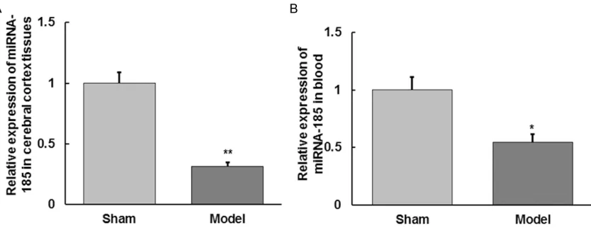
Expression changes of miRNA-185 in cerebral cortex tissues and blood samples
The qRT-PCR was applied to detect the expres-sion of miRNA-185 in cerebral cortex tissues and blood samples. As shown in Figure 6, the expression level of miRNA-185 in cerebral cor-tex tissues and blood samples in model group
were significantly lower than those in sham
group, and these differences were statistically
significant (P < 0.05). This result indicates that
changes in NOS2 mRNA and protein expression
may be caused by the down-regulation of miRNA-185.
Discussion
In this study, we observed the morphological changes of cerebral cells in cerebral ischemia-reperfusion injury rat model, detected the expression levels of NOS2 mRNA and protein and miRNA-185 in cerebral cortex tissues and blood, and preliminary explored the molecular mechanisms of miRNA-185 on cerebral isch-emia-reperfusion injury through regulating NOS2.
Cerebral ischemic stroke accounts for about 75% of all strokes, and the most common cause of cerebrovascular diseases is cerebral atherosclerosis, followed by cerebral arthritis
[19]. Blood supply re-perfusion to cerebral isch -emic areas is the primary treatment to avoid ischemic injury, but reperfusion injury may occur [20]. Experiments show that many patho-logical processes, including oxidative stress, blood-brain barrier damage, excitatory amino
acid toxicity, infiltration of inflammatory cells, medium infiltration and so on, can result in
ischemia-reperfusion injury [21-23]. It has very important theoretical and practical values to investigate the pathogenesis of cerebral isch-emia-reperfusion injury, and to research devel-opment drugs for the protection of ischemic brain damage [24].
As early as 1980, Furchgoll et al found that under the action of acetylcholine, a substance named endothelium-derived relaxing factor (EDRF) was produced in blood vessels, which can cause vascular smooth muscle to relax
[25]. This substance was then confirmed as
NO. NO plays a double role in cerebral isch-emia-reperfusion injury, which can be synthe-sized by the NOS2 catalysis. It is considered that NOS2 is not expressed in physiological state, and is induced expression after sti-
mulation by bacterial lipopolysaccharide, γ-interferon, tumor necrosis factor, interleukin-1β
and some other cytokines [26]. In this study, we have observed that NOS2 was abnormality highly expressed in cerebral cortex tissues and blood in cerebral ischemia-reperfusion injury rat model. And, a certain pathological changes of cell morphology occurred in NOS2 high-expressed cerebral cortex tissues, which suggest that NOS2 plays an important biologi-Figure 4. Relative expression levels of NOS2 protein
in rat blood. Compared with sham group,*Represents
P < 0.05.
Figure 5. The predicted specific regulatory binding
cal role in cerebral ischemia-reperfusion in- jury, and abnormal expression of NOS2 may be a key regulator of cerebral ischemia-reperfu-sion injury. In view of the current study, a con-sensus is that NOS2 has a close relationship with vascular diseases. Some vascular
diseas-es can cause abnormal blood flow and then
leads to a variety of pathological conditions including cerebral ischemia, anoxia, and me- tabolite accumulation [27]. Therefore, it is even more important to study the regulation mechanism of NOS2 in cerebral ischemia-reperfusion injury.
After prediction, we found that NOS2 may be one of the targets of miRNA-185. miRNA-185 genes were differentially expressed in a variety of diseases. For example, Kim et al showed that miRNA-185 played a role in reversal of cardiac hypertrophy through a variety of signaling path-ways [28]. Ma et al showed that miRNA-185 inhibited the proliferation of clear cell, renal cell and carcinoma cell through the targeted gene VEGFA, and also induced apoptosis of these
cells [29]. Bao et al showed that miRNA-185
inhibited the functional disorder of β cells
caused by diabetes through targeted on SOCS3 [30]. Fu et al showed that miRNA-185 inhibited proliferation and metastasis of human breast cancer cells through targeting c-met [31]. Wang et al also found that miRNA-185 had a similar effect in breast cancer, but the target gene was VEGFA [32]. All these results suggest that miRNA-185 is closely related to human diseas-es. In this study, we have observed that
miRNA-185 expression was significantly decreased
in cerebral cortex tissues and blood in cere- bral ischemia-reperfusion injury rat model. Considered that NOS2 was highly expressed in cerebral cortex tissues and blood in cerebral ischemia-reperfusion injury rat model, we speculate that the down-regulation of miRNA-185 is one of the reasons leading to the increase of NOS2. Considered that HE staining
of brain cells in rats, we find that three are
regulatory relationships in miRNA-185-NOS2-morphological changes of rat cerebral cells. Changes of miRNA-185, especially changes in blood, may be used as a strong indicator of cerebral ischemia-reperfusion injury.
In conclusion, one of the mechanisms of cere-bral ischemia-reperfusion injury may be that reduced miRNA-185 expression in cerebral cor-tex tissues and blood resulted in up-regulated expression of NOS2 mRNA and protein, and
finally caused a series of vascular lesions.
Acknowledgements
The authors want to Professor Xiaolan Guo
from Clinical Laboratory, the Affiliated Hospital
of North Sichuan Medical College for her valu-able help.
Address correspondence to: Yuanchuan Wang,
Department of Neurosurgery, Affiliated Hospital of
[image:7.612.99.516.73.234.2]North Sichuan Medical College, No. 63, Wenhua Road, Shunqing District, Nanchong 637000, Sichuan Province, P. R. China. Tel: 86-817-2262420; E-mail: cvr777@163.com
References
[1] von Kummer R, Dzialowski I and Gerber J.
Therapeutic efficacy of brain imaging in acute
ischemic stroke patients. J Neuroradiol 2015; 42: 47-54.
[2] von Kummer R and Gerber J. Treatment of acute ischemic stroke: systemic or local? Ann N Y Acad Sci 2012; 1268: 79-84.
[3] Yin KJ, Hamblin M and Chen YE. Angiogenesis-regulating microRNAs and Ischemic Stroke. Curr Vasc Pharmacol 2015; 13: 352-365. [4] Berger HR, Morken TS, Vettukattil R, Brubakk
AM, Sonnewald U and Wideroe M. No improve-ment of neuronal metabolism in the reperfu-sion phase with melatonin treatment after hy-poxic-ischemic brain injury in the neonatal rat. J Neurochem 2015; [Epub ahead of print]. [5] Wang Y, Yuan L, Liu P and Zhao M. Delayed
hy-peroxic ventilation attenuates oxygen-induced free radical accumulation during early reperfu-sion after global brain ischemia. Neuroreport 2015; 26: 131-138.
[6] Alluri H, Anasooya Shaji C, Davis ML and
Tharakan B. Oxygen-glucose deprivation and
reoxygenation as an in vitro ischemia-reperfu-sion injury model for studying blood-brain bar-rier dysfunction. J Vis Exp 2015; e52699. [7] Schmidley JW. Free radicals in central nervous
system ischemia. Stroke 1990; 21: 1086-1090.
[8] Novgorodov SA and Gudz TI. Ceramide and mitochondria in ischemia/reperfusion. J Car- diovasc Pharmacol 2009; 53: 198-208. [9] Novgorodov SA and Gudz TI. Ceramide and
mi-tochondria in ischemic brain injury. Int J
Biochem Mol Biol 2011; 2: 347-361.
[10] Du J and Zhao H. The cerebral collateral circu-lation and ischemic stroke. Journal of Apoplexy and Nervous Diseases 2012; 29: 760-762. [11] Ni J, Wang X, Chen S, Liu H, Wang Y, Xu X,
Cheng J, Jia J and Zhen X. MicroRNA let-7c-5p protects against cerebral ischemia injury via mechanisms involving the inhibition of
microg-lia activation. Brain Behav Immun 2015; 49:
75-85.
[12] Tao Z, Zhao H, Wang R, Liu P, Yan F, Zhang C, Ji X and Luo Y. Neuroprotective effect of microR-NA-99a against focal cerebral ischemia-reper-fusion injury in mice. J Neurol Sci 2015; 355: 113-119.
[13] Yamaguchi M, Okamoto K, Kusano T, Matsuda Y, Suzuki G, Fuse A and Yokota H. The Effects of Xanthine Oxidoreductase Inhibitors on Oxidative Stress Markers following Global
Brain Ischemia Reperfusion Injury in C57BL/6
Mice. PLoS One 2015; 10: e0133980. [14] Liu H, Ou S, Xiao X, Zhu Y and Zhou S. Diabetes
Worsens Ischemia-Reperfusion Brain Injury in
Rats Through GSK-3beta. Am J Med Sci 2015; 350: 204-211.
[15] Li W, Tan C, Liu Y, Liu X, Wang X, Gui Y, Qin L, Deng F, Yu Z, Hu C and Chen L. Resveratrol ameliorates oxidative stress and inhibits aqua-porin 4 expression following rat cerebral isch-emia-reperfusion injury. Mol Med Rep 2015; 12: 7756-7762.
[16] Granger DN and Kvietys PR. Reperfusion injury and reactive oxygen species: The evolution of a
concept. Redox Biol 2015; 6: 524-551.
[17] Nanri K, Montecot C, Springhetti V, Seylaz J and Pinard E. The selective inhibitor of neuro-nal nitric oxide synthase, 7-nitroindazole, re-duces the delayed neuronal damage due to forebrain ischemia in rats. Stroke 1998; 29: 1248-1253; discussion 1253-1244.
[18] Fu S, Gu Y, Jiang JQ, Chen X, Xu M, Chen X and Shen J. Calycosin-7-O-beta-D-glucoside regu-lates nitric oxide/caveolin-1/matrix metallo-proteinases pathway and protects blood-brain barrier integrity in experimental cerebral isch-emia-reperfusion injury. J Ethnopharmacol 2014; 155: 692-701.
[19] Group of Chinese Medical Association Nbocddatoais. Guidelines writing Guidelines for the diagnosis and treatment of acute isch-emic stroke in China 2010. Chinese Journal For Clinicians 2011; 39: 67-73.
[20] Chen XM, Chen HS, Xu MJ and Shen JG. Targeting reactive nitrogen species: a promis-ing therapeutic strategy for cerebral ischemia-reperfusion injury. Acta Pharmacol Sin 2013; 34: 67-77.
[21] Wang R, Xu C, Zhao W, Zhang J, Cao K, Yang B
and Wu L. Calcium and polyamine regulated calcium-sensing receptors in cardiac tissues.
Eur J Biochem 2003; 270: 2680-2688.
[22] Vinten-Johansen J. Involvement of neutrophils in the pathogenesis of lethal myocardial reper-fusion injury. Cardiovasc Res 2004; 61: 481-497.
[23] Zhao ZQ. Oxidative stress-elicited myocardial apoptosis during reperfusion. Curr Opin Pharmacol 2004; 4: 159-165.
[24] Liu Q, Liu M and Liu Y. Research progress of the mechanisms of cerebral ischemia reperfu-sion injury. Chinese Journal of Neurosurgical Disease Research 2006; 5: 566-568.
[25] Furchgott RF and Zawadzki JV. The obligatory role of endothelial cells in the relaxation of ar-terial smooth muscle by acetylcholine. Nature 1980; 288: 373-376.
[26] Frodl T and Amico F. Is there an association be-tween peripheral immune markers and
struc-tural/functional neuroimaging findings? Prog Neuropsychopharmacol Biol Psychiatry 2014;
48: 295-303.
pathway in brain injury and its treatment--from bench to bedside. Exp Neurol 2015; 263: 235-243.
[28] Kim JO, Song DW, Kwon EJ, Hong SE, Song HK, Min CK and Kim do H. miR-185 plays an anti-hypertrophic role in the heart via multiple tar-gets in the calcium-signaling pathways. PLoS One 2015; 10: e0122509.
[29] Ma X, Shen D, Li H, Zhang Y, Lv X, Huang Q,
Gao Y, Li X, Gu L, Xiu S, Bao X, Duan J and
Zhang X. MicroRNA-185 inhibits cell prolifera-tion and induces cell apoptosis by targeting VEGFA directly in von Hippel-Lindau-inactivated clear cell renal cell carcinoma. Urol Oncol 2015; 33: 169, e1-11.
[30] Bao L, Fu X, Si M, Wang Y, Ma R, Ren X and Lv
H. MicroRNA-185 targets SOCS3 to inhibit beta-cell dysfunction in diabetes. PLoS One 2015; 10: e0116067.
[31] Fu P, Du F, Yao M, Lv K and Liu Y. MicroRNA-185 inhibits proliferation by targeting c-Met in hu-man breast cancer cells. Exp Ther Med 2014; 8: 1879-1883.
[32] Wang R, Tian S, Wang HB, Chu DP, Cao JL, Xia
HF and Ma X. MiR-185 is involved in human breast carcinogenesis by targeting Vegfa.