Supported by the Czech Science Foundation (grants No. 523/06/1302 and No. 523/09/0844), by the Institute of Animal Physiology and Genetics CAS, v.v.i. Liběchov (RVO: 67985904), and by the Ministry of Education, Youth and Sports of the Czech Republic (Project No. MSM 6046070901 and Project CEITEC 2020 No. LQ1601 under the National Sustainability Programme II).
Genomic Structure and Expression
of the Porcine ACTC1 Gene
Antonín Stratil
1*, Pavel Horák
1, Michaela Nesvadbová
2,
Mario Van Poucke
3, Věra Dvořáková
1,4, Roman Stupka
4,
Jaroslav Čítek
4, Kateřina Zadinová
4, Luc J. Peelman
3, Aleš Knoll
2,5 1Institute of Animal Physiology and Genetics CAS, v.v.i. Liběchov,Liběchov, Czech Republic
2Department of Morphology, Physiology and Animal Genetics,
Mendel University in Brno, Brno, Czech Republic
3Department of Nutrition, Genetics and Ethology, Faculty of Veterinary Medicine,
Ghent University, Merelbeke, Belgium
4Department of Animal Husbandry, Czech University of Life Sciences Prague,
Prague, Czech Republic
5CEITEC Mendel University in Brno, Brno, Czech Republic
*Corresponding author: stratil@iapg.cas.cz
ABSTRACT
StratilA., HorákP., NesvadbováM., Van Poucke M., DvořákováV., StupkaR., Čítek J., Zadinová K., Peelman L.J., Knoll A. (2018):Genomic structure and expression of the porcine ACTC1 gene. Czech J. Anim. Sci., 63, 371–378.
A partial cDNA (~1200 bp) of the porcine ACTC1 gene was identified in the subtracted foetal hind limb muscle cDNA library (44 days of gestation; using m.biceps femoris cDNA as the driver). Using specific poly-merase chain reaction (PCR) primers, a bacterial artificial chromosome (BAC) clone containing the genomic
ACTC1 gene was identified and the gene was sequenced. Specific PCR primers designed from the BAC and cDNA sequences were used for amplification and comparative sequencing of ACTC1 of Pietrain and Meishan pigs. The gene is approximately 5.4 kb in length, is composed of 7 exons, and has a coding sequence containing 1134 bp. The gene was mapped using the INRA-Minnesota porcine radiation hybrid (IMpRH) panel to chro-mosome 1, with SW65 as the closest marker (41 cR; LOD = 7.73). Differences were observed in tissue-specific expression of ACTC1 that was studied by transcription profiling in 28 porcine tissues. Developmental differ-ences in muscle and heart were analysed by real-time quantitative PCR (RT-qPCR). Two single nucleotide polymorphisms (SNPs) were found in intron 1. One adequately informative SNP (FM212567.1:g.901C>G) was genotyped by PCR-restriction fragment length polymorphism, and allele frequencies in eight pig breeds were calculated.
Alpha actins are present in muscle tissues as a major constituent of the contractile apparatus. Actin in muscle fibres comprises about 20% of total cellular protein. In human skeletal and cardiac muscles, two sarcomeric actins, ACTA1 (actin, alpha 1, skeletal muscle) and ACTC1 (actin, alpha, cardiac muscle 1) are co-expressed (Gunning et al. 1983; Ilkovski et al. 2005). These two human actins are highly homologous and their amino acid sequences differ by only four residues of the total 377 amino acids in each protein. Both hu-man actins are 100% identical with the porcine orthologues. Homologies are lower, however, in nucleotide sequences of the two genes – in human, they share 85.6% and in the pig 86.8% identities of coding sequences (http://www.ensembl.org/).
As studied using microarray and RNA sequenc-ing, mRNA expression of ACTC1 in various human tissues is recorded in the Ensembl database (http:// www.ensembl.org/index.html). The gene’s expres-sion is high in the heart, but lower in the skeletal muscle and some other tissues. Protein expression of human ACTA1 and ACTC1 was studied by Il-kovski et al. (2005). They showed that ACTA1 is a predominant skeletal muscle isoform from 25 to 27 weeks of gestation to adulthood, and ACTC1 is a predominant sarcomeric isoform in heart and embryonic and foetal skeletal muscles. Protein ex-pression of ACTC1 in several other human tissues is also recorded in the Ensembl database.
Recently, ACTC1 mRNA expression in pig tis-sues was studied using the porcine Affymetrix expression array (Snowball) (http://biogps.org; Freeman et al. 2012). The expressions in various tissues differed appreciably.
In the subtracted porcine foetal hind limb muscle cDNA library (44 days of gestation, using adult
biceps femoris cDNA as the driver; Stratil et al. 2008) several clones were obtained that contained sequences orthologous to human ACTC1 cDNA. In a preliminary study, Horak et al. (2008) esti-mated that ACTC1 mRNA in the muscle tissue of pig foetuses (hind limb; 50 days of gestation) is overexpressed compared to the adult skeletal muscle. The overexpression of ACTC1 mRNA in the foetal muscle may indicate an important role of the protein during development and early stages of myogenesis (http://www.ebi.ac.uk/QuickGO/ GProtein?ac=P68032).
Numerous genes expressed in skeletal muscle at various stages of development in farm animals
may be candidate genes that could play a role in myogenesis (Te Pas et al. 2005; Murani et al. 2007). Mutations in these genes can modify structure and function or expression of the proteins and can influence muscle growth and meat quality (Wimmers et al. 2007; Chalupova et al. 2014).
The aims of the present study were to determine the genomic sequence and organization of the porcine ACTC1 gene, map the gene, study mRNA expression, and search for polymorphism. We also attempted to study associations of the polymor-phism with carcass traits in a crossbred porcine population and as no significant associations were observed, the data are not presented.
MATERIAL AND METHODS
Animals, isolation of DNA and RNA, and re-verse transcription. Genomic DNA was isolated from blood of pigs (Czech Large White, Czech Landrace, Czech Meat Pig, Pietrain, Black Pied Prestice, Hampshire, Duroc, and Meishan) by con-ventional methods. DNA from bacterial artificial chromosome (BAC) clones was isolated using the QIAGEN Plasmid Mini or Midi Kit (QIAGEN, Germany).
Samples from the foetal hind limb muscle (Czech Large White; 42 days of gestation) and from the skeletal muscle and heart tissues of piglets (Czech Large White; 1, 7, and 14 days old) and 8 tissues of a 6-month-old pig (Czech Large White; m. biceps femoris, heart, tongue, lymph node, brain, lung, kidney, and backfat) were collected and stored in RNAlater (QIAGEN) at –20°C. Another set of sam-ples from 25 tissues (adrenal gland, aorta, bladder, caecum, colon (centrifugal coil and centripetal coil), diaphragm, duodenum, gallbladder, heart, ileum, jejunum, kidney, liver, lung, lymph node, m. longis-simus dorsi, mesenterium, oesophagus, pancreas, rectum, spleen, stomach, tongue, and ureter) were taken from a commercial crossbred pig 10 days old immediately after slaughtering and were kept in liquid nitrogen. All pigs were slaughtered according to protocols for certified national slaughterhouses under the supervision of an independent veterinar-ian. Principles of ethical standards were adhered to during all sample collections.
Sequencing of cDNA and genomic DNA of ACTC1. The mRNA and genomic sequences of porcine ACTC1 were unknown at the time of this study. The cDNA sequence encompassing the whole coding sequence of porcine foetal muscle ACTC1
(orthologous with human ACTC1) was obtained from the clones of a subtracted foetal hind limb muscle cDNA library (Stratil et al. 2008) and the sequence was deposited in the EMBL/GenBank database under accession No. FM212568.1 (sub-mitted September 29, 2008). Sequences corre-sponding to individual exons were identified by alignment with the human genomic sequence of
ACTC1 (Ensembl; ENSG00000159251, transcript ACTC1-001; http://www.ensembl.org/Homo_sa-piens/Info/Index).
For the study of the porcine genomic sequence of ACTC1, first sets of polymerase chain reac-tion (PCR) primers were designed on the basis of cDNA sequence (ACTC1-A, -B; -C, -D; -E; -G; -M; -R; Table 1). Further primers were designed from the sequences of the amplicons (Table 1). The 5' and 3' ends of the gene and 5' upstream and 3' downstream sequences were first obtained by sequencing a BAC clone, using primer walking (the primers for primer walking are not shown). Two positive BAC clones for ACTC1 (PigI-258C6 and PigI-417B2) were identified in the porcine genomic BAC library (Rogel-Gaillard et al. 1999) using prim-ers ACTC1-A/-B. The clone PigI-417B2 was used for sequencing by primer walking. PCR primers were designed from the obtained sequences (see Table 1) to amplify and sequence the fragments of genomic DNA of Pietrain and Meishan pigs.
The sequences of cDNA clones, PCR fragments of genomic DNA, and the BAC clone were obtained by Sanger sequencing (ABI PRISM 3130 Sequencer; Applied Biosystems, USA).
The obtained genomic sequences of ACTC1
of Pietrain and Meishan were deposited in the EMBL/GenBank database under accession Nos. FM212566.1 and FM212567.1 (submitted Sep-tember 29, 2008).
Polymorphism testing. Single nucleotide poly-morphisms (SNPs) revealed by sequencing were tested by PCR-restriction fragment length polymor-phism (RFLP). The DNA fragments were amplified using PCR primers ACTC1-Q/-R, and digestions were performed with restriction enzymes Alw26I and BsaWI, respectively. Further details on prim-ers, amplification conditions, and SNP genotyping
are presented in Table 1. The restriction frag-ments were separated by electrophoresis on 1% agarose gel.
Radiation hybrid mapping. Radiation hybrid (RH) mapping was performed on the whole-genome 7000-rad IMpRH panel (Yerle et al. 1998; Hawken et al. 1999). A panel of 90 clones was screened by PCR using the ACTC1-A/-B primers (Table 1).
Transcription profiling. Transcription profiles of ACTC1 were studied by PCR using cDNA from the pig tissues (see Results). ACTB and GAPDH
were used as reference genes, and the primers were from Erkens et al. (2006). For PCR amplification, primer pairs ACTC1-C/-D (exons 3 and 5) and ACTC1-A/-B (exons 6 and 7) (Table 1) were used. The analyses were performed in duplicates. The PCR products were separated on 1% agarose gel.
Relative quantification of ACTC1 expression (RT-qPCR). The mRNA expression of ACTC1 was studied by real-time quantitative PCR (RT-qPCR) in foetal hind limb muscle (42 days of gestation), skeletal muscle (m. biceps femoris), and heart muscle of 1-, 7-, and 14-day-old piglets and adult pigs. PCR primers were: Forward: 5'CCAGCAC-CATGAAGATCAAGA 3' (from exon 6; one nt from exon 7); Reverse: 5'AAAGAAGGGTGGGTTG-GAAG 3' (from exon 7; 3'untranslated region). A 232-bp fragment was amplified, and the speci-ficity was verified by sequencing. The reference gene was HPRT1, which has been found to be stable in various adult tissues and piglet muscles, but less stable in foetal muscle (Svobodova et al. 2015). RT-qPCR was conducted in the 7500 Real Time PCR System, using Power SYBR® Green PCR Master Mix (Applied Biosystems). The pro-gramme started with 2 min of the AmpErase Uracil N-glycosylase incubation step at 50°C and 10 min at 95°C, followed by 40 cycles of 15 s at 95°C and 1 min at 60°C. A melting curve was constructed for verification of the specificity of PCR products. The efficiency of the reaction was calculated from the slope of the standard curve determined in the 7500 System SDS software v1.2 application (Ap-plied Biosystems). The efficiency of the reactions in all samples was in the range of 93–105%, and the values of the coefficient of determination (R2)
were in the range of 0.98–1.00.
Ta
ble 1. P
oly mera se c hain r eac tion (P C R) pr imers f or s equenc ing , sing le n uc le otide p oly mor phi sm (S N P) analy si s, radi ation h ybr id (R H) ma
pping and trans
cr
iption
pr
ofiling
, and am
plific ation c onditions f or p or cine ACT C1 A cc ession No.; sour ce s equenc e Pr imer name Pr imer s equenc es (5'-3') Lo ca tion in genomic s equenc e Am plic on si ze ( bp) Mg C l2 (mM) Ta (°C ) Commen ts PW
1 on B
A C PigI-417B2 A C TC1-5F A C TC1-5R at cc agg ttgg tg agggc tac a agggc agggg ag agg at cag 5' up str eam 5´U TR 467 1.0 58
LA DNA p
oly
mera
se
2; s
equenc
ing
PW on B
A C PigI-417B2 FM212568.1 A C TC1-Q A C TC1-R gg tgggc tggcg tc ac tt ag tc t cc tcg tcg tcgc ac at ct ttg tac 5' up str eam ex on 2 935 1.0 60
LA DNA p
oly mera se; s equenc ing; S N P analy si s (F M212566.1:g .889G>A; Bsa W I P C R-R FL P: alle le G
: 935 bp;
alle
le
A
: 704 + 231 bp; F
M212567.1:g .901C>G; Al w 26I P C R-R FL P: alle le C
: 935 bp; alle
le
G
: 721 + 214 bp)
PW on B
A C PigI-417B2 Am plic on MN A C TC1-Sf A C TC1-Tr cg aggc cag tt ccgc at cac ag acg tgc cagg agggg ttg tg in tr on 1 in tr on 2 273 1.0 58
LA DNA p
oly mera se; s equenc ing FM212568.1 Am plic on GH A C TC1-M A C TC1-N acggc tc cgggc tgg taaa tt ccgggg ac ca tt cc ttt tg ex on 2 in tr on 3 1677 1.5 56
LA DNA p
oly mera se; s equenc ing Am plic on MN Am plic on MN A C TC1-O A C TC1-P ac cc agggc caagc cac at t cc tgc caaag tc aaggg ac at ct ta in tr on 2 in tr on 2 667 1.5 58
LA DNA p
oly mera se; s equenc ing FM212568.1 Am plic on E F A C TC1-G A C TC1-H gc cc ag agc aagcg agg ta tt c tg agggc cc ca taa tcg ttg tt ex on 3 in tr on 5 1551 2.0 56
LA DNA p
oly mera se; s equenc ing FM212568.1 FM212568.1 A C TC1-C A C TC1-D aggc caac cgcg ag aag atg a gg aag aagc agc tg tagc ca tc tc a ex on 3 ex on 5 1195 (c ds 362) 1.5 66
LA DNA p
oly mera se; s equenc ing , trans cr iption pr ofiling Am plic on C D Am plic on C D A C TC1-K A C TC1-L gt cc ccgg aaagg tg tc tg tg tgg at tcgc tgg tgg ag ac aa in tr on 3 in tr on 4 837 2.0 62
LA DNA p
oly mera se; s equenc ing FM212568.1 Am plic on A B A C TC1-E A C TC1-F tgc cc tgg at tttg ag aa tg ag atg cc aac ac ac cc cac cc ac aaa ex on 5 in tr on 6 1137 1.5 56
LA DNA p
oly mera se; s equenc ing FM212568.1 FM212568.1 A C TC1-A A C TC1-B tc ca tg aaacg ac tt ac aac ca tc ctg aacg taaag tag ac t ex on 6 ex on 7 899 (c ds 345) 2.0 59
LA DNA p
oly mera se; s equenc ing; B A C s cr eening , R H ma pping , trans cr iption pr ofiling Am plic on A B
PW on B
A C PigI-417B2 A C TC1-U A C TC1-V gc ct ct ac ca tg tac caagc tt at tg tggc ct cag tg ttt cac ca tt aac in tr on 6 3' dow nstr eam 1089 1.5 58
LA DNA p
oly
mera
se; s
equenc
ing
PW on B
A C PigI-417B2 A C TC1-X A C TC1-Y ggc caa tg tttgc tt ac tc ag tg ta gc ctgc cc tg tgc ttt at atg a 3' dow nstr eam 3' dow nstr eam 1113 1.5 60
LA DNA p
oly
mera
se; s
equenc
ing
PW on B
mcb_support/documents/generaldocuments/ cms_040980.pdf). Amplifications were performed in triplicate and a no-template negative control was included for each RNA isolate and gene.
RESULTS AND DISCUSSION
Comparative sequencing of ACTC1 of Pietrain and Meishan pigs.The obtained genomic sequenc-es, including 5´ and 3´ flanking sequences of ACTC1
of Pietrain and Meishan pigs, were deposited in the EMBL/GenBank database under accession Nos. FM212566.1 and FM212567.1 (submitted Septem-ber 29, 2008). The gene encompasses approximately 5.4 kb and is composed of 7 exons. The coding sequence encompasses 1134 bp and the deduced protein contains 377 amino acids (FM212568.1). The genomic sequence is virtually identical with that of ENSSSCG00000004803 (Sscrofa10.2; Gen-Bank Assembly ID GCF_000003025.5) (Groenen et al. 2012), INSDC coordinates CM000812.4, loca-tion on SSC1: 152,412,334–152,417,610; Ensembl release 83 – December 2015 (http://www.ensembl. org/index.html). We confirmed the assignment to chromosome 1 by IMpRH mapping. The closest marker (2pt analysis) was SW65 (41 cR; logarithm of the odds (LOD) = 7.73).
In both Pietrain and Meishan pigs, approxi-mately 8.5 kb were sequenced, including 5' and 3' flanking sequences. The two sequences were identical. One SNP was found in each pig – in Pietrain FM212567.1:g.901C>G and in Meishan FM212566.1:g.889G>A.
The BLAST analysis (https://blast.ncbi.nlm.nih. gov/Blast.cgi) showed the amino acid sequences of ACTC1 in various animal species to be highly homologous. Most vertebrate species studied share 99–100% identity of the amino acid sequences. Similarities of the coding nucleotide sequences are lower, however, mostly in the range of 92–95%. For example, porcine and human ACTC1 amino acid sequences are 100% identical, while the cod-ing nucleotide sequences share 93% similarity.
ACTC1 polymorphism. Both SNPs (FM 212566.1: g.889G>A; FM212567.1:g.901C>G) are located in intron 1. SNP g.889G>A was genotyped after digestion with BsaWI, and SNP g.901C>G after digestion with Alw26I. As for SNP g.889G>A, of 34 pigs (Meishan, Pietrain, and crosses Meishan × Pietrain) only one (Meishan) was heterozygous; all
A
cc
ession No.;
sour
ce s
equenc
e
Pr
imer name
Pr
imer s
equenc
es (5'-3')
Lo
ca
tion in
genomic s
equenc
e
Am
plic
on
si
ze (
bp)
Mg
C
l2
(mM)
Ta
(°C
)
Commen
ts
PW on B
A
C
PigI-417B2
A
C
TC1-I
A
A
C
TC1-I
B
tgc
cac
agc
tc
acg
ac
aac
at
gg
tcg
aag
atg
tggc
tc
ag
at
ct
ac
3' dow
nstr
eam
3' dow
nstr
eam
1131
1.5
64
LA DNA p
oly
mera
se; s
equenc
ing
PW on B
A
C
PigI-417B2
A
C
TC1-3A
A
C
TC1-3B
ctggc
tggg
ta
tt
ctg
tg
ag
tg
a
cg
at
tag
ta
tc
cc
tg
agg
atg
tg
3' dow
nstr
eam
3' dow
nstr
eam
484
1.0
55
LA DNA p
oly
mera
se; s
equenc
ing
Ta
= anne
aling t
em
pera
tur
e, c
ds = c
oding s
equenc
e
1pr
imers wer
e de
sig
ne
d on t
he s
equenc
e t
ha
t w
as obt
aine
d by s
equenc
ing (pr
imer w
alk
ing
, P
W
) of b
ac
ter
ial ar
tific
ial c
hr
omo
some (B
A
C
) c
lone PigI-417B2 (
Jouy
-en-Jo
sa
s, F
ranc
e)
2LA DNA P
oly
mera
se
s Mi
x (
Top-Bio, Cze
ch Re
public
); H
otS
tar
Ta
q
Ma
st
er Mi
x Kit (QI
A
GE
N
, USA)
[image:5.595.82.174.96.761.2]Ta
ble 1 t
o b
e c
on
tin
ue
other pigs were homozygotes GG. SNP g.901C>G was more polymorphic. Unrelated pigs of eight breeds were genotyped and allele frequencies are presented in Table 2. The most balanced distribu-tion of alleles was in Czech Large White (allele C
– 0.68), while in Duroc and Meishan allele C was fixed. Other breeds had frequencies of allele C
from 0.85 to 0.96.
Transcription profiling. When the cDNA sam-ples from the porcine tissues were analysed using PCR primers ACTC1-C/-D and -A/-B, there were some differences in the results, namely that some samples amplified with one primer pair and did not amplify with the other primer pair. Some of these ambiguities were resolved in another experiment, where some questionable tissues were analysed. Altogether, strong expressions with both primer pairs, or with the qPCR primers, were for m. longis-simus dorsi, m. biceps femoris, heart, tongue, dia-phragm, jejunum, liver, and spleen; low or very low expressions were for back fat, brain, lymph node, lung, kidney, ileum, colon ascendens (centripetal coil), bladder, pancreas, ureter, aorta, and adrenal gland. In all samples fragments of expected sizes were observed. No expression was determined for oesophagus, stomach, duodenum, appendix, colon ascendens (centrifugal coil), rectum, gall bladder, and mesenterium. Although this approach can be considered as at best semi-quantitative, it does provide information as to whether the gene is or is not expressed.
The ACTC1 mRNA expressions in pig tissues were studied previously using the porcine Affymetrix ex-pression array (Snowball) (http://biogps.org; Free-man et al. 2012). Those analyses were performed
on the mRNAs of two pigs of different sex, and the expressions in some tissues differed appreci-ably. In the probeset SNOWBALL_000044_s_st, the highest expression was in the heart, lower expression was observed in the retina/sclera of one animal, and the expression in the skeletal muscle (leg) was less than half that of the heart’s muscle. Similar or slightly lower expressions were observed in the tongue (dermal layer), oesophagus (upper third), pylorus (smooth muscle), ileum, bladder, gall bladder, abdominal aorta, bone mar-row, and spleen. The expression levels in all other tissues were still lower. Compared with other probesets (SNOWBALL_000041_st and SNOW-BALL_000047_st) those expressions for individual tissues were quite variable.
Gene expression profiling simultaneously com-pares the expression levels of a gene in many tissues. There can be differences in expression levels de-pending on the method used, experimental details of the analysis, animal, breed, age, health status, and environmental conditions. It nevertheless provides the first information on the expression of a gene in different tissues, and this can serve as a starting point for more detailed study of ex-pressions, as well as for unraveling the functions of the protein in different tissues. Although the expression results can be validated using RT-qPCR or mRNA sequencing, these methodologies are more costly and labour-intensive and so only a limited number of samples can be analysed.
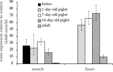
RT-qPCR of ACTC1 mRNA. Using RT-qPCR, the level of ACTC1 expression was estimated in foetal muscle, as well as in skeletal and heart muscle of piglets and adult pigs (Figure 1). It can be seen that the expression of ACTC1 in muscle of foetus and piglets (1–14 days of age) was much higher compared to that of adult pig skeletal muscle. In heart muscle, the ACTC1 expression in adult pigs was much higher than that in adult skeletal muscle, and the expressions in the piglet heart was clearly higher than that in adult pig heart.
[image:6.595.64.291.600.744.2]When studying expression of α-cardiac actin mRNA in hearts of neonatal and young rats, Carrier et al. (1992) found that in young rats the expres-sion was higher when compared with more aged hearts. The expression of α-cardiac actin during human muscle development was studied by Il-kovski et al. (2005). They used Western blotting with the specific α-cardiac actin antibodies. The protein was expressed throughout embryonic and Table 2. Allele frequencies at single nucleotide
polymor-phism FM212567.1:g.901C>G in eight pig breeds
Breed n C G HWE
χ2 P
Czech Large White 20 0.68 0.32 4.72 < 0.05 Czech Landrace 26 0.96 0.04 0.04 > 0.80 Czech Meat Pig 15 0.87 0.13 2.86 > 0.05 Piétrain 26 0.85 0.15 0.37 > 0.50 Prestice Black Pied 11 0.95 0.05 0.03 > 0.80 Hampshire 9 0.94 0.06 0.03 > 0.80
Duroc 15 1.00 0.00 – –
Meishan 15 1.00 0.00 – –
foetal development until birth, after which expres-sion was markedly downregulated. The α-cardiac actin was also detected in skeletal muscle about 1 month after birth, while it was not detected in the muscle at 6 months and in adults. In the mouse skeletal muscle, Ilkovski et al. (2005) observed low expression of cardiac actin at postnatal day 7, but no expression at 6 months. McHugh et al. (1991) studied the epression of α-cardiac actin in rat muscle and heart. In the skeletal muscle, α-cardiac actin was expressed at low levels during embryonic de-velopment and during first postnatal days. This expression then decreased to almost undetectable level in the adult. In the heart, α-cardiac actin was significantly expressed during embryonic and foetal development, as well as during 8 days postnatal, and decreased slightly in adults. Moreover, α-cardiac actin mRNA ratios in the skeletal muscle and heart appeared to be similar to the ratios of the respective proteins in the same tissues.
Although different methodological approaches have been used, it appears that in skeletal muscles of mammals, ACTC1 (both mRNA and protein) is highly expressed during embryonic and foetal development, and then is gradualy downregulated. Appreciable expression continues in the early period of postnatal development but expression is low in adult human and animals. Earlier reported absence of ACTC1 expression in adult humans and mouse may be due to the use of less-sensitive methodologies.
While there is no doubt as to the structural and functional role of ACTC1 in heart and skeletal
muscle (e.g. cardiac muscle tissue morphogenesis, cardiac myofibril assembly, heart contraction, cardiac muscle contraction, actomyosin structure organization, and actin-myosin filament sliding),
ACTC1 mRNA is expressed also in several other tissues. In humans, the protein has been found in other tissues, too (Ensembl) wherein the ACTC1 canonical role cannot be expected. It should have other functions, as indicated in the list of gene ontologies (e.g. ATP binding, ATPase activity, myosin binding, positive regulation of gene expres-sion, and several others) (genecards.org/cgi-bin/ carddisp.pl?id_type=hgnc&id=143).
CONCLUSION
In this study we report on the porcine mRNA and genomic sequences of the ACTC1 gene. The gene is composed of 7 exons and coding sequence contains 1134 bp. The gene was mapped by IMpRH mapping to chromosome 1 (the closest marker was SW65). Transcription profiling was studied in 28 tissues. The expression was high in some tissues ( m. lon-gissimus dorsi, m. biceps femoris, heart, tongue, diaphragm, jejunum, liver, and spleen), low in others (back fat, brain, lymph node, lung, kidney, ileum, colon ascendens (centripetal coil), bladder, pancreas, ureter, aorta, and adrenal gland), and in 8 tissues (oesophagus, stomach, duodenum, appendix, colon ascendens (centrifugal coil), rectum, gall bladder, and mesenterium) there was no expression of ACTC1. By using RT-qPCR, in foetal and piglet muscle (1, 7, and 14 days of age) the expression was higher compared to that in adult muscle; ACTC1 expression in adult porcine heart was higher than that in the skeletal muscle, and the expression in piglets’ heart was much higher than that in the heart of adult pigs.
[image:7.595.65.292.528.672.2]Acknowledgement. We thank Marie Datlová and RNDr. Martina Pinková for technical assistance. We are indebted to Drs. Martine Yerle and Denis Milan (INRA, Castanet-Tolosan, France) for provid-ing IMpRH panel, and Drs. Patrick Chardon and Karine Hugot (INRA, Jouy-en-Josas, France) for the BAC library screening. Porcine DNA samples were provided by Prof. Hermann Geldermann (University of Hohenheim, Stuttgart, Germany; Meishan and Pietrain pigs) and Prof. Alan L. Ar-chibald (Roslin Institute, Midlothian, Scotland, UK; Meishan pigs).
Figure 1. Relative quantification by RT-qPCR of ACTC1
mRNA expression in hind limb muscle of porcine foetus,
in m. biceps femoris and heart of 1-, 7-, and 14-day-old
piglets, and adult pigs. Expression values are rescaled against the value of the muscle of adult pig (= 1). Bars represent standard error
muscle heart
G
ene
ex
pr
ession
re
la
tive
to
con
tr
ol
(adult
m
us
cle)
30 40 50 60 70 80 90
ssi
on
r
el
at
iv
e
to
c
on
tr
ol
adu
lt m
us
cle
)
foetus 1-day-old piglet 7-day-old piglet 14-day-old piglet adult
0 10 20
muscle heart
G
en
e e
xp
re (a
30 40 50 60 70 80 90
ssi
on
re
la
tiv
e
to
c
on
tr
ol
adu
lt m
us
cle
)
foetus 1-day-old piglet 7-day-old piglet 14-day-old piglet adult
0 10 20
muscle heart
G
en
e e
xp
REFERENCES
Carrier L., Boheler K.R., Chassagne C., de la Bastie D., Wisnewski C., Lakatta E.G., Schwartz K. (1992): Expres-sion of the sarcomeric actin isogenes in the rat heart with development and senescence. Circulation Research, 70, 999–1005.
Chalupova P., Dvorakova V., Knoll A., Stratil A., Barten-schlager H., Stupka R., Citek J., Sprysl M., Palanova A., Horak P., Geldermann H. (2014): Polymorphism, linkage mapping, and association analysis with carcass traits of four porcine candidate genes selected from gene-expression profiles of Czech Large White and Wild Boar muscles. Czech Journal of Animal Science, 59, 116–127. Erkens T., Van Poucke M., Vandesompele J., Goossens K.,
Van Zeveren A., Peelman L.J. (2006): Development of a new set of reference genes for normalization of real-time RT-PCR data of porcine backfat and longissimus dorsi muscle, and evaluation with PPARGC1A. BMC Biotechnology, 6: 41.
Freeman T.C., Ivens A., Baillie J.K., Beraldi D., Barnett M.W., Dorward D., Downing A., Fairbairn L., Kapetanovic R., Raza S., Tomoiu A., Alberio R.,Wu C., Su A.I., Summers K.M., Tuggle C.K., Archibald A.L., Hume D.A. (2012): A gene expression atlas of the domestic pig. BMC Biology, 10: 90.
Groenen M.A.M., Archibald A.L., Uenishi H., Tuggle C.K., Takeuchi Y., Rothschild M.F., Rogel-Gaillard C., Park C., Milan D., Megens H.-J., Li S., Larkin D.M., Kim H., Frantz L.A.F., Caccamo M., et al. (2012): Analyses of pig genomes provide insight into porcine demography and evolution. Nature, 491, 393–398.
Gunning P., Ponte P., Blau H., Kedes L. (1983): α-Skeletal and α-cardiac actin genes are coexpressed in adult hu-man skeletal muscle and heart. Molecular and Cellular Biology, 3, 1985–1995.
Hawken R.J., Murtaugh J., Flickinger J.H., Yerle M., Robic A., Milan D., Gellin J., Beattie C.W., Schook L.B., Alexander L.J. (1999): A first-generation porcine whole-genome radiation hybrid map. Mammalian Genome, 10, 824–830. Horak P., Stratil A., Knoll A., Bilek K., Van Poucke M., Peel-man L.J. (2008): The porcine ACTC1 gene – structure, polymorphism, mapping and expression. In: Proc. 31st Internat. Conference on Animal Genetics, Amsterdam, the Netherlands, Abstract No. 2203.
Ilkovski B., Clement S., Sewry C., North K.N., Cooper S.T. (2005): Defining α-skeletal and α-cardiac actin expression in human heart and skeletal muscle explains the absence of cardiac involvement in ACTA1 nemaline myopathy. Neuromuscular Disorders, 15, 829–835.
McHugh K.M., Crawford K., Lessard J.L. (1991): A compre-hensive analysis of the developmental and tissue-specific expression of the isoactin multigene family in the rat. Developmental Biology, 148, 442–458.
Murani E., Muraniova M., Ponsuksili S., Schellander K., Wimmers K. (2007): Identification of genes differentially expressed during prenatal development of skeletal muscle in two pig breeds differing in muscularity. BMC Devel-opmental Biology, 7: 109.
Rogel-Gaillard C., Bourgeaux N., Billault A., Vaiman M., Chardon P. (1999): Construction of a swine BAC library: application to the characterization and mapping of por-cine type C endoviral elements. Cytogenetics and Cell Genetics, 85, 205–211.
Stratil A., Knoll A., Horak P., Bilek K., Bechynova R., Barten-schlager H., Van Poucke M., Peelman L.J., Svobodova K., Geldermann H. (2008): Mapping of the porcine FBN2, YWHAQ, CNN3, DCN, POSTN, SPARC, RBM39 and GNAS genes, expressed in foetal skeletal muscles. Animal Genetics, 39, 204–205.
Svobodova K., Horak P., Stratil A., Bartenschlager H., Van Poucke M., Chalupova P., Dvorakova V., Knorr C., Stup-ka R., Citek J., Sprysl M., Palanova A., Peelman L.J., Gel-dermann H., Knoll A. (2015): Porcine EEF1A1 and EEF1A2 genes: genomic structure, polymorphism, mapping and expression. Molecular Biology Reports, 42, 1257–1264. Te Pas M.F.W., de Wit A.A.W., Priem J., Cagnazzo M., Davoli
R., Russo V., Pool M.H. (2005): Transcriptome expression profiles in prenatal pigs in relation to myogenesis. Journal of Muscle Research and Cell Motility, 26, 157–165. Van Poucke M., Melkebeek V., Erkens T., Van Zeveren A.,
Cox E., Peelman L.J. (2009): Molecular cloning and char-acterization of the porcine prostaglandin transporter (SLCO2A1): evaluation of its role in F4 mediated neonatal diarrhoea. BMC Genetics, 10: 64.
Wimmers K., Murani E., Te Pas M.F.W., Chang K.C., Davo-li R., Merks J.W.M., Henne H., Muraniova M., da Costa N., Harlizius B., Schellander K., Boll I., Braglia S., de Wit A.A.C., Cagnazzo M., Fontanesi L., Prins D., Ponsuksili S. (2007): Association of functional candidate genes derived from gene-expression profiles of prenatal porcine muscle tissue with meat quality and muscle deposition. Animal Genetics, 38, 474–484.
Yerle M., Pinton P., Robic A., Alfonso A., Palvadeau Y., Del-cros C., Hawken R., Alexander L., Beattie C., Schook L., Milan D., Gellin J. (1998): Construction of a whole-genome radiation hybrid panel for high-resolution gene mapping in pigs. Cytogenetics and Cell Genetics, 82, 182–188.