Original Article
A herbal extract treats type 2 diabetes mellitus
effectively by down-regulating expression of CD14
Lifang Li1,2, Dewen Yan2, Yan Zou2, Tingji Zhang2, Guangfeng Lin2, Jianhua Xiao1
1Institute of Pathogenic Biology, Hengyang Medical College, University of South China, Hengyang 421001, P. R.
China; 2The Second People’s Hospital of Shenzhen, Shenzhen 518037, P. R. China
Received July 5, 2018; Accepted September 12, 2018; Epub February 15, 2019; Published February 28, 2019
Abstract: The incidence of Type 2 diabetes mellitus (T2DM) has increased worldwide. This study investigated the effect of a herbal extract, named as Baihu decoction (BHD), in treating T2DM by using mouse adipose cells and a mouse T2DM model. Mouse 3T3-L1 preadipocytes were cultured in DMEM culture medium and used for experi-ments when about 90% fat-differentiated cells were mature. The KM male mouse diabetes model was established by injecting streptozotocin. Thirty two KM T2DM mice were randomly divided into 4 groups, with 8 mice in each group. Enzyme-linked immunosorbent assays (ELISA) and Reverse Transcription-quantitative PCR (RT-real time-PCR)
were performed to investigate the levels of inflammatory factors and adipokines related to diabetes and their mRNA levels. Western blot experiments were carried out to determine levels of a series of proteins. BHD reduced inflam -matory responses induced by lipopolysaccharide in mouse adipose cells and repressed the activity of the CD14/
TLR4-NF-κB signal pathway. Our siRNA experimental results demonstrated that decreased CD14 gene expression
reduced the levels of inflammation significantly. The animal experimental results indicated that BHD has an effec -tive effect on reduction of blood sugar level of T2DM mice. Furthermore, this anti-diabetic effect was due to
inhibi-tion of the activity of the CD14/TLR4-NF-κB signal pathway, especially CD14. BHD has anti-diabetic effect both in vitro and in vivo by down-regulating the activity of the CD14/TLR4-NF-κB signaling pathway, especially CD14.
Keywords: Type 2 diabetes mellitus, Baihu decoction, adipose tissue, chronic inflammation, CD14
Introduction
Diabetes is a complex chronic disease associ-ated with high blood sugar levels, induced by deficiencies in insulin secretion, activation, or both. If patients cannot receive high quality care and frequent hospitalization, complicati- ons will be resulted [1]. Type 2 diabetes melli-tus (T2DM) is a common and increasingly prev-alent disease and thus a major public health concern worldwide [2]. Patients with T2DM may develop morbid complications of diabetic keto-acidosis [3]. T2DM is characterized by relative insulin deficiency caused by pancreatic β-cell dysfunction and insulin resistance in target or- gans. As the sixth leading cause of disability in 2015, diabetes places considerable socio-eco-nomic burden, estimated at $825 billion in the world [4, 5]. T2DM patients need intensive managements of sugar and lipid concentra-tions as well as blood pressure to minimize risk of complications occurring and progression [6].
The incidence of T2DM is affected by genetic and environmental factors. Genetic factors ex- ert their effects following exposure to an obeso-genic environment characterized by sedentary behavior and excessive sugar and fat consump-tion [7, 8]. In patients who are younger than 55 years of age and have a glycated haemoglobin concentration of 6.9% or less, it is twice-fold higher risk of mortality compared with people without diabetes [9]. Previous study have sh- own that diabetes is associated with increased risk of coronary heart disease, ischemic stroke, and other deaths related to vascular disease [10]. Patients with T2DM can present with established complications such as retinopathy [11].
nd that one of the mechanisms underlying dia-betes and insulin resistance is chronic inflam-mation [13]. It also reported that dysbiosis could be linked between chronic inflammation, inflammatory bowel disease, colorectal cancer, and T2DM through crosstalk between molecu-lar signal pathways, in particumolecu-lar, NF-κB and TNF-α [14]. The intestinal epithelium has an important function as a protective barrier ag- ainst luminal antigens. However, the barrier can be breached, therefore disruption of the intesti-nal homeostasis can promote chronic inflam-mation [15]. Normally, inflaminflam-mation is a benefi-cial activity as it rejuvenates injured tissues and removes the foreign agents disturbing homeostasis. Inflammation is achieved through a complex inflammatory response that may involve a balance between a huge panel of bio-active molecules and autoimmunity. However, chronic inflammation may increase the risk of several pathologic responses such as inflam-matory bowel disease, colorectal cancer, and T2DM [14].
The CD14/TLR4-NF-κB pathway are found to be associated with various biological functions im- plicated in development of inflammation. CD14 is a lipopolysaccharide-binding protein, which functions as an endotoxin receptor. It is anch- ored to the cell surface by linkage to glyco-sylphosphatidylinositol. CD14 is strongly posi-tive in monocytes and most tissue macroph- ages, but is weakly expressed or negative in monoblasts and promonocytes. Myeloblasts and other granulocytic precursors do not ex- press CD14, but neutrophils and a small pro-portion of B lymphocytes may weakly express. The CD14 receptor is a pattern recognition mol-ecule in the innate immune response against other exogenous and endogenous stress fac-tors [16]. It has been demonstrated that chron-ic infection by Gram negative mchron-icroorganisms may contribute to the inflammatory component of atherosclerosis.
Lipopolysaccharide (LPS) or endotoxin generat-ed by Gram negative bacteria exerts proathero-genic effects by contributing to low density lipo-protein oxidation, foam cell formation, and th- rombogenesis [17]. LPS binds to the LPS-bin- ding protein, which in turn is coupled to CD14 on the cell surface of monocytes. Then, LPS-CD14 interacts with toll-like receptor 4 (TLR4) and forms a complex with another accessory protein MD-2. The TLR4 signaling cascade is
initiated after binding with the adaptor protein MyD88, which leads to a series of events, and subsequently triggers activation and transloca-tion of NF-κB and causes transcriptransloca-tion of cyto-kines, such as TNF-α. The CD14/TLR4-NF-κB pathway can be inhibited by pyrrolidine dithio-carbamate (PDTC, an NF-κB inhibitor), thus reducing inflammatory reaction.
Many compounds such as insulin secretago- gue, α-glucosidase inhibitor, intestinal lipase inhibitor, and insulin sensitizer have been used for treatment of T2DM [18-20]. These drugs have various adverse effects and induce com-plications such as gastrointestinal discomfort, hypoglycemia, and pulmonary edema, although they can decrease the level of blood glucose [21, 22]. Therefore, it is urgent to study new medicines those have high effectiveness, low adverse effects, and clear action mechanism. Baihu decoction (BHD), a traditional herb medi-cine curative for inflammation and high blood sugar, has be used for a long term in China and other Asian countries. BHD with addition of Ginseng has effective hypoglycemic and anti-oxidant effects [23]. Application of BHD com-bined with insulin to treat acute hyperglycemia in type 2 diabetes can significantly reduce the levels of TNF-α, IL-6, high-sensitivity C-reactive protein, Leptin, and adiponectin in patients [24, 25]. These findings implies that BHD might have functions to treat T2DM by decreasing blood sugar level.
Adipose cells, a kind of endocrine cells, are an important base for inflammation. Adipose tis-sue secretes a variety of affinity or anti-inflam-matory adipocytokines that can cause, medi-ate, or antagonize inflammatory responses as well as insulin resistance. In obese human and rodent models, it has been demonstrated that enhanced expression of proinflammatory adi-pocytokines can induce insulin resistance [26]. In the pathophysiology of inflammation-insulin resistance-type 2 diabetes, dysfunction of adi-pocyte endocrine regulation plays an important role.
The animal experimental results also demon-strated that BHD efficiently inhibits mouse blo- od sugar levels by inhibiting the CD14/TLR4-NF-κB signal pathway.
Materials and methods
Cell culture
Mouse 3T3-L1 preadipocytes were purchased from American Type Culture Collection (ATCC; Manassas, VA, USA) and cultured in DMEM cul-ture medium containing 10% fetal bovine se- rum (FBS; Gibco-BRL, Gaithersburg, MD, USA) at 37°C with 5% CO2 and 100% humidity. Two days after the cell contact inhibition was ob- served, cells were cultured in DMEM medium containing 10% fetal bovine serum, 0.5 mM IBMX, 0.25 μM 10 g/ml dexamethasone, and 10 μg/ml insulin for 3 days. When about 90% fat-differentiated cells were mature, cells can be used for the following experiments.
BHD preparation
An extract was prepared by decocting the dry herbs (Gypsum fibrosum, Anemarrhena aspho-deloides Bge, Oryza sativa L, and Glycyrrhiza uralensis Fisch) with a ratio of 9:3:1.5:1 in boil-ing water for 60 minutes to a concentration of 1 g/ml. Since Gypsum fibrosum is the main ingre-dient and it is also called “baihu”, the extract is named as Baihu decoction (BHD) for conveni- ece. BHD was filtrated with a 2 layer gauze and then membrane with pores of a 0.2 μm diame-ter, saved in -20°C for use. Before use, BHD was diluted into a series of concentrations, including 0.05 g/ml (5%) and 0.25 g/ml (25%). The 4 kinds of Chinese herbal medicines were purchased from Chinese herbal medicine Pie- ces Factory (Shenzhen, China).
siRNA experiments
Mouse 3T3-L1 cells were transfected with 80 pmol of siRNA against the mouse CD14 (cat #: sc-29962), TLR4 (cat #: sc-40261), and NF NF-κB p65 (cat #: sc-29411) messages, or a ne- gative control siRNA (cat #: sc-sc-44233 using X-tremeGENE (Roche, Shanghai, China). All si- RNAs were purchased from Santa Cruz Bio- technology, Santa Cruz, USA. At 48 hours post-transfection, total protein was harvested, sepa-rated on 10% SDS/PAGE gels, and subjected to immunoblot analyses.
Establishment of the mouse diabetes model
All animal experiments were performed in acc- ordance with the guidelines of the Animal Ethics Committee of the University of South China. Forty male KM mice, weighed 20-22 g, were pu- rchased from and maintained in the Animal Biosafety Laboratory of the University of South China. Mice were fed for 1 week before the fol-lowing experiments. Eight mice were fed con-tinuously as usually, but the left 32 mice were used to establish T2DM model. After 30 days of high fat and high sugar feeding, mice were fast-ed for 12 hours but water was providfast-ed and then mice were intraperitoneally injected with streptozotocin (STZ, 80 mg/kg/mouse). Three days later, the mice received the same treat-ment as described above one more time. After these treatments, free diets were provided for one week. Then, blood samples were obtained from the mouse tail vein after the mice was fasted for 5 hours. The mouse blood sugar con-centration was measured. All mouse grew nor-mally. If mouse blood sugar concentration was greater than 11.1 mM, the mouse diabetes mo- del was considered as successfully establi- shed. Blood glucose meter was purchased from Johnson & Johnson (Shanghai) Medical Equ- ipment Co, Ltd, China.
Animal grouping
Eight-week-old male KM T2DM mice were ran-domly divided into 4 groups, with 8 mice in each group and all mice being given with high fat diet. The 4 T2DM mouse groups were de- signed as follows: (i) in the phosphate buffered saline (PBS) control group, mice were intragas-trically administered with PBS for 8 weeks; (ii) in the 5% BHD group, mice were intragastrically administered with 5% BHD for 8 weeks; (iii) in the 25% BHD group, mice were intragastrically administered with 25% BHD for 8 weeks; and (iv) in the 25% BHD + PDTC group, mice were intragastrically administered with 25% BHD and injected with PDTC (200 mg/kg/d) subcu-taneously for 8 weeks.
Enzyme-linked immunosorbent assay (ELISA)
temperature. After plate was washed 4 times, enzyme binding solution (100 μl/well) was add- ed, followed by an incubation for 30 minutes at room temperature. After 4-time washes, color-ing solution was added. After 10-20 minutes, stop solution was added, and the OD value of 450 was determined.
Reverse Transcription-quantitative PCR (RT-real time-PCR)
Total RNAs were extracted using TRIzol reagent (Invitrogen, Grand Island, New York, USA) acc- ording to the manufacturer’s instructions. First-strand cDNA was synthesized from the RNA using Reverse Transcription kit (TaKaRa, Tokyo, Japan) with random primers. The levels of mRNAs were quantified by CFX96 RT-real time-PCR detection system using a SYBR Premix Ex Taq kit (Takara, Tokyo, Japan). The relative mRNA levels were normalized to that of the house-keeping gene GAPDH. All primers were given in Table 1.
Western blotting
Adipose cells were harvested in RIPA buffer (Beyotime Institute of Biotechnology, Shanghai, China), supplemented with 0.5 mM phenyl-methylsufonyl fluoride and 0.5% cocktail prote-ase inhibitor (Roche, Bprote-asel, Switzerland) using micro-scrapers. After sonication and centrifu-gation, protein concentrations of samples were determined by the bicinchoninic acid method. Equal amounts of proteins were mixed with 5X loading buffer (250 nM Tris-HCl (PH 6.8), 0.5% BPB, 10% SDS, 50% glycerol, 5% β-merca- ptoethanol). Proteins were subjected to 10% SDS-polyacrylamide gel electrophoresis, and then transferred onto a polyvinylidene difluride
Leptin (cat. no ab3583, 1:500; Abcam), CD14 (cat. no ab133335, 1:500; Abcam), TLR4 (cat. no ab22048, 1:1000; Abcam), NF-κBp65 (cat. no ab16502, 1:1000; Cell Signaling Tech- nology), β-actin (cat. no sc-47778, 1:10,000; Santz Cruz), followed by incubation with horse-radish peroxidase-labelled secondary antibod-ies for 2 hours. Immuno-reactivity was detect-ed using the ECL system (Bio-Rad Laboratories, California, USA). Band gray values were mea-sured by Image J software (National Institutes of Health, Bethesda, Maryland, USA).
Statistical analysis
Data are presented as the mean ± standard deviation (mean ± SD) of 3-6 independent experiments. The statistical significance of the difference was determined by the Students’ t test. p < 0.05 was considered to be significant-ly different. p < 0.01 was considered to be very significantly different.
Results
BHD reduces inflammatory responses induced by LPS in mouse adipose cells.
Since T2DM is tightly related to inflammation and BHD has been used to treat inflammation for a long term in China and other Asian coun-tries, we first examined whether BHD could inhibit inflammatory activity. Mouse adipose 3T3-L1 cells were stimulated by LPS to induce the inflammatory responses. The unstimulated cells served as the control group. The LPS-stimulated cells were treated with LPS, LPS + 5% (0.05 g/ml) BHD, LPS + 25% (0.25 g/ml) BHD, LPS + PDTC, or LPS + 25% BHD + PDTC, respectively. After 48 hours, cells were exam-Table 1. Primers used in Real-time PCR
Primers Forward Backward
Universal primers F27 R1492
AGAGTTTGATCMTGGCTCAG TAGGYTACCTTGTTACGACT IL-6 GAGGATACCACTCCCAACAGACC AAGTGCATCATCGTTGTTCATACA TNF-α GGATCTCAAAGACAACCAAC ACAGAGCAATGACTCCAAAG CD14 TTGGCTTGTTGCTGTTGCTTC GCGGAGGTTCAAGATGTTGAGAT TLR-4 GAAACTCAGCAAAGTCCCTG GAAAGGCTTGGTCTTGAATG NF-κBp65 ATGTGCATCGGCAAGTGG CAGAAGTTGAGTTTCGGGTAG Apelin TGCTCTGGCTCTCCTTGACT ATGGGTCCCTTATGGGAGAG Leptin GGACCAGACATTGGCGATCTAC CCGGAGGTTCTCCAGGTCA GAPDH ATGGGTVAGAAGGACTCCTATG ATCTCCTGCTCGAAGTCTAGAG
[image:4.612.91.403.84.231.2]ined by microscope. The shape of cells in all 6 groups was observed to be similar, without de- tectable difference, showing that BHD has no visibly toxic effect on cells. The culture super-natants and cell lysates were harvested sepa-rately. The collected culture supernatants were used to examine levels of factors by ELISA. As shown in Figure 1A, the levels of IL-6, TNF-α, Apelin, and Leptin in the LPS group were elevat-ed to 8.61, 8.23, 11.69, and 9.25 fold (p < 0.01), respectively, in comparison with the unstimulated control group, indicating the inflammatory responses were indeed stimulat-ed successfully. When comparstimulat-ed with the LPS
19.25%, 22.78%, 18.01%, and 13.34%, respec-tively, relative to those in the LPS groups (p < 0.01). However, the addition of PDTC even decreased the levels to 10.51%, 11.02%, 8.98%, and 7.11% (p < 0.01), respectively. Since PDTC is a CD14/TLR4-NF-κB signal path-way inhibitor, the function of BHD may be relat-ed to the CD14/TLR4-NF-κB signal pathway.
BHD inhibits activity of the CD14/TLR4-NF-κB
signal pathway in adipose cells
To further confirm whether the inhibitory effe- ct of BHD on inflammation was related to the group, levels of the 4 kinds of factors in the LPS + 25% BHD group were all decreased sig-nificantly (p < 0.01), indicating that BHD with a concentration ratio of 25% can effectively inhibit inflammation. The pres-ence of PDTC enhanced the inhibitory effect of BHD by about 50%, when comparing LPS + 25% BHD + PDTC group with LPS + 25% BHD group, suggesting that the function of BHD is related to CD14/TLR4-NF-κB signal pathway since PDTC is an inhibitor of the sig-nal pathway, and PDTC may have an additive effect. But lev-els of the 4 kinds of factors in the LPS + 5% BHD group were only slightly reduced, indicating 5% BHD is not an effective con-centration. When compared wi- th the LPS group, levels of the 4 kinds of factors in the LPS + PDTC group were not changed obviously, showing that the presence of PDTC did not de- tectably affect the factor lev-els. Therefore, in the following experiments, the unstimulated control, LPS + 5% BHD, and LPS + PDTC groups were not included.
[image:5.612.93.366.73.406.2]Cell lysates were used to iso-lated the total RNAs. As shown in Figure 1B, RT-real time-PCR results showed that the mRNA levels of IL-6, TNF-α, Apelin, and Leptin in the LPS + 25% BHD group were reduced to Figure 1. BHD affects secretion of inflammatory factors and adipocytokines
of LPS-stimulated 3T3-L1 cells. 3T3-L1 cells were treated with medium (as the control), LPS (1 μg/ml), LPS (1 μg/ml) + 5% BHD, LPS (1 μg/ml) + 25% BHD, LPS (1 μg/ml) + PDTC (10 μM), or LPS (1 μg/ml) + 25% BHD + PDTC (10 μM), respectively. The culture supernatants and cells were harvested for ELISA and RT-real time-PCR experiments, respectively. (A) The levels of
IL-6, TNF-α, Apelin, and Leptin were determined by ELISA. Values shown
are mean ± SD of 6 independent experiments. **p < 0.01, compared to the
control group. ##p < 0.01, compared to the LPS group. &&p < 0.01, compared
to the LPS + 25% BHD group. (B) mRNA levels of IL-6, TNF-α, Apelin, and
Leptin were determined by RT-real time-PCR. Values shown are mean ± SD of 3 independent experiments. **p < 0.01, compared to the LPS group. ##p
CD14/TLR4-NF-κB signal pathway, 3T3-L1 cells were treated with LPS, LPS + 25% BHD, or LPS + 25% BHD + PDTC, respectively. After 48 ho- urs, microscopic examination showed that cell growth was the same among all groups, con-firming both BHD and PDTC have no observable toxic effect on cells. The cell lysates were har-vested for Western blot and RT-real time-PCR analyses. As shown in Figure 2, protein (Figure 2A) and mRNA (Figure 2B) levels of CD14, TLR4, and NF-κB p65 were all decreased in the
ed that the protein levels of CD14, TLR4, and p65 were all significantly reduced (p < 0.05) as expected.
[image:6.612.91.371.71.180.2]The culture supernatants were harvested for determining levels of IL-6 and Apelin. In these experiments, only IL-6 and Apelin were deter-mined by ELISA since TNF-α and Leptin were found to be affected at similar degrees with IL-6 and Apelin, respectively in Figure 1A. As shown in Figure 3B, results showed that IL-6 Figure 2. BHD inhibits the activity of CD14/TLR4-NFκB signal pathway in
3T3-L1 cells. Mouse 3T3-L1 cells were treated with LPS, LPS + 25% BHD, or LPS + 25% BHD + PDTC, respectively. Cells were harvested for isolation of the total protein and RNAs. (A) Western blotting was performed to determine the expression of CD14, TLR4, and p65. β-actin served as a loading control. All experiments were performed for 3 times independently and one of them was shown. (B) RT-real time-PCR was conducted to examine the mRNA ex-pression. Values shown are mean ± SD of 3 independent experiments. **p
< 0.01, compared to the LPS group. ##p < 0.01, compared to the LPS + 25%
BHD group. #p < 0.05, compared to the LPS + 25% BHD group.
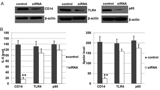
Figure 3. CD14 mRNA expression affects the levels of inflammatory fac -tor IL-6 and Adipokine Apelin. Negative control siRNA and siRNA targeting CD14, TLR4, or p65 were transfected into LPS-stimulated 3T3-L1 cells. Af-ter 48 hours, the cells and culture supernatants were harvested for west-ern blotting and ELISA experiments, respectively. (A) The total proteins were extracted for determining protein expression. β-actin served as a loading control. All experiments were performed 3 times independently and one of them was shown. (B) The levels of IL-6 and Apelin were determined by ELISA. Values shown are mean ± SD of 6 independent experiments. **p <
0.01, compared to the Negative control siRNA group.
25% BHD group when com-pared to the LPS group (p < 0.01). Those in the 25% BHD + PDTC were even reduced rela-tive to those in the 25% BHD group (p < 0.01 or p < 0.05). Importantly, among the 3 teins, the levels of CD14 pro-tein and mRNA were changed as the highest degree, sug-gesting that CD14 is possibly the most sensitive one among the 3 proteins in the signal pathway. The results show that the anti-inflammation effect of BHD is due to inhibition of the activity of CD14/TLR4-NF-κB signal pathway, especially CD14.
Reduced CD14 gene expres-sion decreases the levels of
inflammatory factor and adi -pokine
[image:6.612.91.372.313.472.2]level in the siRNA-against CD14 group was decreased to 11.22% (p < 0.01) of the negative control siRNA group. However, the levels of TLR4 and p65 in the siRNA groups were de- creased to only 81.92% and 85.05% of the neg-ative control siRNA group. A similar situation was found for Apelin. These results suggest that CD14 expression is the most important factor for inhibiting inflammation of LPS-sti- mulated 3T3-L1 cells. Together with the results in Figure 2, it is suggested that BHD decreases inflammation mainly by altering CD14 gene expression.
BHD has an anti-diabetic effect on T2DM km
mice
T2DM, as a complicated metabolic disorder, is associated with inflammation. It has also been found that reduced CD14 gene expression decreases the levels of inflammatory factor and
adipokine in above results, therefore we further investigated whether BHD has an anti-diabetic effect on T2DM. A T2DM animal model was established using KM mice. Blood sugar con-centration of every mouse, including 8 mice in the normal non-T2DM group and 32 mice in the T2DM group, was detected. As expected, blood sugar mean level (15.65 ± 0.92 mM) of mice in the T2DM group were increased to 3.39 fold in comparison with that (4.61 ± 0.26) of the nor-mal non-T2DM mice, indicating that the aninor-mal model is established successfully.
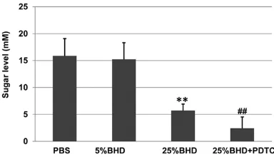
To determine whether BHD can inhibit the sugar level of mice, T2DM mice were divided into 4
groups, including PBS, 5% BHD, 25% BHD, and 25% BHD + PDTC. Since the presence of PDTC does not affect inflammation as shown above, the animal experiment does include the condi-tion of PDTC only in the following animal experi-ments. Mice in all groups grew normally, indi-cating that BHD and PDTC have no detectably toxic effects on mouse. As shown in Figure 4, the sugar levels of mice in the 5% BHD group were only slightly reduced, but those of mice in the 25% BHD group were decreased significant-ly relative to the PBS group (p < 0.01). The pres-ence of PDTC enhanced the inhibition of BHD when comparing those of the 25% BHD + PDTC group to the 25% BHD group (p < 0.01). These results suggest that BHD has an anti-diabetic effect on T2DM KM mice.
BHD inhibits activity of the CD14/TLR4-NF-κB signal pathway by down-regulating the level of
CD14 mRNA level in T2DM km mice, especially CD14
To further determine if the inhibitory effect of BHD on sugar level of T2DM mice was caused by changes in CD14, TLR4, and p65 expres-sion, the mice in the PBS, 25% BHD, and 25% BHD + PDTC groups used in the above experi-ments excepting the 5% BHD group, were euth-anized. Then adipose tissues were collected to isolate total proteins and RNAs. As shown in Figure 5A, Western blotting results showed that CD14 protein level in the 25% BHD group were decreased significantly different relative to the PBS group (p < 0.01), but TLR4 and p65 levels were decreased not so much (p < 0.05). The presence of PDTC enhanced inhibition of BHD when comparing those of the 25% BHD + PDTC group to the 25% BHD group (p < 0.05). Although the levels of CD14, TLR4, and p65 were all decreased, the CD14 levels were affected mostly.
[image:7.612.92.284.74.184.2]To examine the mechanism underlying the alt- ered protein expression, the mRNA levels were investigated by RT-real-time PCR. As shown in Figure 5B, the mRNA levels of CD14, TLR4, and p65 in the 25% BHD group were decreased sig-nificantly relative to the PBS group (p < 0.01). The presence of PDTC enhanced the inhibition of BHD when comparing those of the 25% BHD + PDTC group to the 25% BHD group (p < 0.01 or p < 0.05). Although the mRNA levels of CD14, TLR4, and p65 were also changed among the 3 groups, the CD14 levels were reduced mostly. These findings suggest that BHD reduces sugar Figure 4. BHD has an anti-diabetic effect on T2DM
KM mice. All groups of the mice were treated as
de-scribed in the METHOD section. Blood samples were
obtained from the mouse tail vein after the mice was fasted for 5 hours. The mouse blood sugar concen-tration was measured using blood glucose meter. Values shown are mean ± SD. **p < 0.01, compared
to the PBS group. ##p < 0.01, compared to the 25%
levels by decreasing the expression of CD14, possibly by affecting the promoter activity of CD14.
Discussion
Diabetes is increasingly occurring worldwide, which causes many complications, such as hea- rt and blood vessel disease, neuropathy, ne- phropathy, eye and foot damage, hearing im- pairment, etc. T2DM accounts for more than 90% of diabetes. Although patients can take drugs like insulin secretagogues, diterpenoids, glycosidase inhibitors, and so on, these drugs all have various side effects. Insulin injection might results in hypoglycemia, and even severe hypoglycemic induced coma. Thus new drugs with reduced side effects are urgently needed for patients. Drugs from the natural medicinal herbs are a good resource, since usually those kinds of drugs contains multiple compounds that has many different targets and few toxic effect. In this study, BHD, a traditional herb medicine, was found to have anti-diabetic eff- ects.
The complicated interactions among insulin resistance, adipose tissues, and inflammation were very important for explaining the underly-ing mechanisms of diseases, includunderly-ing chronic inflammation, T2DM, insulin resistance, athero-sclerosis, hyperlipidemia, and nonalcoholic fatty liver. Since inflammation is an important reason to result in T2DM, we first investigated whether BHD could inhibit inflammatory res-
T2DM mice. These results are essential as they implied that BHD may be an effective drug, without obvious side effects, for treating T2DM patients.
The molecular mechanism of BHD inhibiting the sugar level of T2DM mice was also investigated at molecular, cellular, and animal levels. An anti-inflammation effect of BHD was found to be due to inhibition of the activity of CD14/ TLR4-NF-κB signal pathway, especially CD14. Furthermore, reduced CD14 gene expression by siRNA decreases the levels of inflammatory factor and adipokine. It was showed that IL-6 level in the siRNA-against CD14 group was decreased to 11.22% of the negative siRNA group. However, the levels of TLR4 and p65 in the negative control group was decreased to only 81.92% and 85.05%. A similar situation was found for Apelin. These results indicate that CD14 is key factor since CD14 is on the upstream of the CD14/TLR4-NF-κB signal pathway.
[image:8.612.88.373.74.180.2]A mouse model was established to research the BHD effect on T2DM. As found in the ani-mal experiments, the sugar levels of mice in the 25% BHD group were decreased significantly relative to the PBS group (p < 0.01). The pres-ence of PDTC enhanced the inhibition of BHD on sugar levels of mice when comparing those of the 25% BHD + PDTC group to the 25% BHD group (p < 0.01). These results suggest that BHD has an anti-diabetic effect on T2DM KM mice. PDTC facilitates the inhibitory effect of Figure 5. BHD inhibits the activity of CD14/TLR4-NFκB signal pathway in
T2DM KM mice. Mice in the PBS, 25% BHD, and 25% BHD + PDTC groups were euthanized, followed by collecting adipose tissues from the mice and isolating of total proteins and RNAs. (A) Western blotting was performed to determine the expression of CD14, TLR4, and p65. β-actin served as a loa- ding control. All experiments were performed for 3 times independently and one of them was shown. (B) RT-real time-PCR was conducted to examine the mRNA expression of CD14, TLR4, and p65. Values shown are mean ± SD of 3 independent experiments. **p < 0.01, compared to the PBS group. ##p < 0.01, compared to the 25% BHD group. #p < 0.05, compared to the
25% BHD group.
BHD on sugar level of the T2DM mice. PDTC is an NF-κB inhibitor and our results suggest that BHD is a CD14 inhibitor. It is reasonable to imagine that both PDTC and BHD target the CD14/TLR4-NF-κB signal pathway.
BHD reduces the activity of the CD14/TLR4-NF-κB signal pathway by repressing the level of CD14 mRNA level in T2DM KM mice, especially CD14. These results are consistent with those in the adipose cells, suggesting that CD14 gene expression is essential for BHD to inhibit the T2DM. Although the underlying molecular mechanisms are emerging, it is noted that the interaction among signal pathways are com-plex. One or multiple components of BHD might inhibit the activity of CD14 promoter. Further study of the effects of the single component of BHD on T2DM are required in the future. The extract isolated from 2 kinds of herbs, Huangbai and Zhimu, can reduce the levels of d-glucose, hexadecanoic acid, octadecanoic ac- id, propanoic acid, 3-hydroxybutyric acid, and 2,3-dihydroxybutanoic acid in urine of T2DM mice [27]. A herbal extract isolated from Hawthorn decreased blood glucose level and increased plasma insulin release from pancre-as, representing another agent for prevention or treatment of T2DM found by using a rat model [28]. It was recently reported that Xiexin Tang, an extract prepared by decocting 3 kinds of herbs, can modify of gut microbiota and improve the symptom of type 2 diabetic rats [29]. Although Xiexin Tang can improve the symptom of type 2 diabetic rats, but it is acting by modification of inflammation due to gut microbiota, an indirect mechanism on diabe-tes. Furthermore, it was not found the clear molecular mechanisms underlying effects of Xiexin Tang on blood glucose levels, like we did in this study.
In this study, BHD would found to repress chronic inflammation as illustrated using mo- use cells and hyperglycemia using a mouse model. The effects on inflammation and hyper-glycemia were due to the inhibition of activity of the CD14/TLR4-NF-κB signal pathway, espe-cially CD14. Next, human cells will be used to research the influence of BHD on T2DM patients. To furthermore define the mecha-nism, knock-out mice lacking CD14, TLR4, and
NF-κB p65 genes will be generated to study activity of their promoters before applying clinical application.
Acknowledgments
The work was supported by the Science and technology project of Shenzhen city (grant No. JCYJ20150330102720160), Hunan Provincial Key Laboratory for Special Pathogens Pre- vention and Control Foundation (Grant No. 2014-5), and Hunan Province Cooperative in- novation Center for Molecular Target New Drug Study (No. 2015-8).
Disclosure of conflict of interest
None.
Address correspondence to: Jianhua Xiao, Institute of Pathogenic Biology, Hengyang Medical College, University of South China, #28 Changsheng Road, Hengyang 421001, P. R. China. Tel: +86-734-828- 2913; E-mail: jhxiao223@163.com
References
[1] Meece J. The role of the pharmacist in manag-ing type 2 diabetes with glucagon-like pep-tide-1 receptor agonists as add-on therapy. Adv Ther 2017; 34: 638-57.
[2] Vangoitsenhoven R, Maris M, Overbergh L, Van
Loco J, Mathieu C and Van der Schueren B. Ce-reulide food toxin, beta cell function and diabe-tes: facts and hypotheses. Diabetes Res Clin Pract 2015; 109: 1-5.
[3] Stratton IM, Adler AI, Neil HA, Matthews DR, Manley SE, Cull CA, Hadden D, Turner RC and Holman RR. Association of glycaemia with macrovascular and microvascular complica-tions of type 2 diabetes (UKPDS 35): prospec-tive observational study. BMJ 2000; 321: 405-12.
[4] GBD 2015 Disease and Injury Incidence and Prevalence Collaborators. Global, regional, and national incidence, prevalence, and years lived with disability for 310 diseases and inju-ries, 1990-2015: a systematic analysis for the Global Burden of Disease Study 2015. Lancet 2016; 388: 1545-1602.
[5] Wang Y, Yeo QQ and Ko Y. Economic evalua-tions of pharmacist-managed services in peo-ple with diabetes mellitus: a systematic review. Diabet Med 2016; 33: 421-7.
[6] Gaede P, Vedel P, Larsen N, Jensen GV, Parving
HH and Pedersen O. Multifactorial intervention
and cardiovascular disease in patients with type 2 diabetes. N Engl J Med 2003; 348: 383-93.
[8] Grarup N, Sandholt CH, Hansen T and
Peders-en O. GPeders-enetic susceptibility to type 2 diabetes
and obesity: from genome-wide association studies to rare variants and beyond. Diabetolo-gia 2014; 57: 1528-41.
[9] Tancredi M, Rosengren A, Svensson AM, Kosi-borod M, Pivodic A, Gudbjornsdottir S, Wedel H, Clements M, Dahlqvist S and Lind M. Excess Mortality among Persons with Type 2 Diabetes. N Engl J Med 2015; 373: 1720-32.
[10] Sarwar N, Gao P, Seshasai SR, Gobin R, Kap-toge S, Di Angelantonio E, Ingelsson E, Lawlor DA, Selvin E, Stampfer M, Stehouwer CD, Lew-ington S, Pennells L, Thompson A, Sattar N, White IR, Ray KK and Danesh J. Diabetes mel-litus, fasting blood glucose concentration, and risk of vascular disease: a collaborative meta-analysis of 102 prospective studies. Lancet 2010; 375: 2215-22.
[11] Agouridis AP, Rizos CV, Elisaf MS and Filippa-tos TD. Does combination therapy with statins
and fibrates prevent cardiovascular disease in
diabetic patients with atherogenic mixed dys-lipidemia? Rev Diabet Stud 2013; 10: 171-90. [12] Pouvreau C, Dayre A, Butkowski EG, de Jong B
and Jelinek HF. Inflammation and oxidative
stress markers in diabetes and hypertension. J
Inflamm Res 2018; 11: 61-68.
[13] Morris G, Berk M, Carvalho AF, Caso JR, Sanz Y and Maes M. The role of microbiota and intes-tinal permeability in the pathophysiology of au-toimmune and neuroimmune processes with
an emphasis on inflammatory bowel disease
type 1 diabetes and chronic fatigue syndr- ome. Curr Pharm Des 2016; 22: 6058-75. [14] Jurjus A, Eid A, Al Kattar S, Zeenny MN,
Gerges-Geagea A, Haydar H, Hilal A, Oueidat D, Matar
M, Tawilah J, Hussein IH, Schembri-Wismayer P, Cappello F, Tomasello G, Leone A and Jurj-
us RA. Inflammatory bowel disease,
colorect-al cancer and type 2 diabetes mellitus: the links. BBA Clin 2016; 5: 16-24.
[15] Bondarenko VM and Riabichenko EV. [Role of intestinal barrier dysfunction in maintenance
of chronic inflammatory process of different
localization]. Zh Mikrobiol Epidemiol Immuno-biol 2010; 92-100.
[16] Arroyo-Espliguero R, Avanzas P, Jeffery S and Kaski JC. CD14 and toll-like receptor 4: a link between infection and acute coronary events? Heart 2004; 90: 983-8.
[17] Halmos T and Suba I. [Physiological patterns of intestinal microbiota. The role of dysbacterio-sis in obesity, insulin redysbacterio-sistance, diabetes and
metabolic syndrome]. Orv Hetil 2016; 157:
13-22.
[18] Refaat R, Sakr A, Salama M and El Sarha A. Combination of vildagliptin and pioglitazone in experimental type 2 diabetes in male rats. Drug Dev Res 2016; 77: 300-9.
[19] Ferrario MG, Lizan L, Montagnoli R and Ram- irez de Arellano A. Liraglutide vs. sitagliptin add-on to metformin treatment for type 2 dia-betes mellitus: short-term cost-per-controlled patient in Italy. Prim Care Diabetes 2016; 10: 220-6.
[20] Ngubane PS, Hadebe SI, Serumula MR and Musabayane CT. The effects of transdermal in-sulin treatment of streptozotocin-induced dia-betic rats on kidney function and renal expres-sion of glucose transporters. Ren Fail 2015; 37: 151-159.
[21] Cheng AY and Fantus IG. Oral antihyperglyce -mic therapy for type 2 diabetes mellitus. CMAJ 2005; 172: 213-26.
[22] Shah PK, Mudaliar S, Chang AR, Aroda V, An-dre M, Burke P and Henry RR. Effects of inten-sive insulin therapy alone and in combination with pioglitazone on body weight, composition, distribution and liver fat content in patients
with type 2 diabetes. Diabetes Obes Metab
2011; 13: 505-10.
[23] Baosheng Zhao WZ, Xiuli Wang. Study on ef-fect and mechanism of renshen baihu decoc-tion in treating diabetes. New Drugs Andclini-cal Pharmacology of Traditional Chinese Med- icine 2010; 21: 493-5.
[24] Li P. Baihu Decoction combined with insulin treating acute hyperglycemia in type 2 diabe-tes 26 cases. Henan Traditional Chinese Medi-cine 2014; 2: 25-27.
[25] Wang Y. Clinical efficacy of baihu decoction
combined with insulin in the treatment of type 2 diabetes mellitus with acute hyperglycemia. Diabetes New World 2016; 7: 38-42.
[26] Kwon H and Pessin JE. Adipokines mediate
in-flammation and insulin resistance. Front Endo -crinol (Lausanne) 2013; 4: 71.
[27] Song L, Liu H, Wang Y, Wang Y, Liu J, Zhou Z, Chu H, Zhuang P and Zhang Y. Application of
GC/MS-based metabonomic profiling in study -ing the therapeutic effects of Huangbai-Zhimu herb-pair (HZ) extract on streptozotocin-indu- ced type 2 diabetes in mice. J Chromatogr B Analyt Technol Biomed Life Sci 2015; 997: 96-104.
[28] Aierken A, Buchholz T, Chen C, Zhang X and Melzig MF. Hypoglycemic effect of hawthorn in type II diabetes mellitus rat model. J Sci Food Agric 2017; 97: 4557-61.