Original Article
Analysis of risk factors for axillary metastasis in sentinel
lymph node positive breast cancer patients
Li Huang
1,2, Jun Zhang
1, Zhicheng Ge
1, Xiang Qu
11Department of General Surgery, Beijing Friendship Hospital, Capital Medical University, Beijing City, China; 2Department of Breast Surgery Ward No.3, The Affiliated Tumor Hospital of Shanxi Medical University, Shanxi
Tumor Hospital, Taiyuan, Shanxi Province, China
Received October 24, 2018; Accepted November 27, 2018; Epub February 15, 2019; Published February 28, 2019
Abstract: Objective: To investigate the risk factors for axillary metastasis in patients with sentinel lymph nodes (SLN) positive breast cancer (BC). Methods: Pathological data of 192 BC patients with positive results of sentinel lymph node biopsy who underwent axillary lymph node dissection (ALND) were analyzed. The predictive model of the Memorial Sloan-Kettering Cancer Center (MSKCC) was used to predict the risk of axillary non-SLN metastasis in patients, and the accuracy of the model was evaluated by the receiver operating characteristic (ROC) curves. Results: The results showed no statistical differences in age, tumor type, estrogen receptor expression, tissue dif-ferentiation, and the number of negative SLNs (all P > 0.05), but statistical differences in tumor size, tumor number,
the number of positive SLNs, and SLN metastasis rates were significant (all P < 0.05). Multivariate logistic analysis
showed that the tumor size, the number of tumors, and positive SLN rates were independent risk factors for non-SLN metastasis. The area under the ROC curve of the MSKCC model was 0.771. Conclusion: Tumor size, the num-ber of tumors, and positive SLN rates are independent risk factors for axillary non-SLN metastasis in SLN-positive patients. If the SLN is positive associated with the presence of these clinical symptoms, ALND is therefore advised. The MSKCC model has a high predictive accuracy for breast cancer patients with non-SLN metastasis.
Keywords: Breast cancer, sentinel lymph node, axillary metastasis, risk factors
Introduction
Breast cancer (BC) is the most common
malig-nant tumor in women currently [1]. More than
249,000 people were diagnosed with BC in the
United States in 2016, and over 40,000 people
die from BC or its complications annually [2]. In
recent years, the age of onset is younger with
an increasing incidence year by year, which
imposes a great threat to both quality of life
and life expectancy of patients [3]. Due to the
advances in medical technologies, early
screen-ing of BC has promoted; therapies for BC are
improving constantly. Studies now suggest
that, the number of BC patients is increasing on
a yearly basis, so does the 5-year survival rate
[4].
Axillary lymph node dissection (ALND) is the
standard treatment regimen for metastatic
tumors of BC patients [5]. Axillary lymph node
(ALN) is an important indicator that is used to
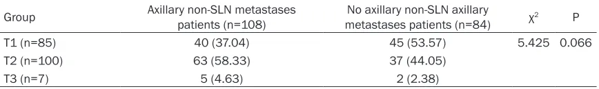
Table 1.
Lymph node metastasis
Group Axillary non-SLN metastases patients (n=108) metastases patients (n=84)No axillary non-SLN axillary χ2 P
T1 (n=85) 40 (37.04) 45 (53.57) 5.425 0.066
T2 (n=100) 63 (58.33) 37 (44.05)
T3 (n=7) 5 (4.63) 2 (2.38)
Note: SLN, sentinel lymph node.
pitals and medical centers [9]. Study has also
reported that the accuracy of non-SLN
predic-tions can be improved using the MSKCC model
[10].
In this study, we retrospectively analyzed the
pathological data of patients in the Thyroid and
Breast Surgery Clinic who underwent SLNB to
verify the MSKCC model for its predictive
capabilities.
Materials and methods
Patients
The study was approved by the Medical Ethics
Committee of the Beijing Friendship Hospital,
and all patients signed written informed con-
sent.
The pathological data of 192 BC patients with
positive SLNB who underwent ALND in Beijing
Friendship Hospital from January 2013 to
December 2016 were analyzed. In this study
cohort, all patients were female. The age of the
patients was 35-71 years, with an average age
of 50.4±12.8 years. The seventh edition of the
American Joint Committee on Cancer was
applied to perform tumor, node and metastasis
staging in BC patients [11]. Specific clinical
data of the patients was collected.
Inclusion criteria: a) Patient older than 18 years
old; b) Diagnosed with BC by cancer pathologic
biopsy prior to surgery, with a tumor size < 50
mm; c) No chemotherapy or radiotherapy prior
to surgery; d) At least 1-2 SLN metastases
con-firmed by ALND; e) Suspicious ALN positive
indi-cated by ultrasound.
Exclusion criteria: Patients with congenital
dis-eases, family genetic history, defective limbs
and immunodeficiency associated disease;
with incomplete clinical data; with other
malig-nant tumors, or distal metastases confirmed by
preoperative imaging examination.
Identification of SLN and case examination
In this study, all BC patients were injected with
methylene blue of 2 mL around the breast
mass using the 4-point method, followed by
massaging for 5 minutes. An arcuate incision
was made in the outer edge of the pectoralis
major, which was cut layer by layer; blue-stained
lymphatic vessels were identified and tracked.
Blue-stained lymph node tissue was removed
for frozen biopsy, HE staining and biopsy along
with other tumor specimens.
Statistical analysis
All collected data were assessed for statistical
analyses by using SPSS 20.0 software, and the
desired images were drawn using Graphpad
Prism 7.0. Enumeration data are expressed as
a percentage (%) and assessed using the Chi
square test. Taking the single factor P < 0.05
index as the independent variable, and the BC
non-SLN metastasis as the dependent
vari-able; stepwise logistic regression was used for
multivariate analysis. P < 0.05 indicates a
sta-tistically significant difference.
Results
Lymph node metastasis
Amongst the SLN-positive patients, 108 had
axillary non-SLN metastases and 84 patients
had no axillary non-SLN axillary metastases.
This included 85 patients of T1, 100 patients of
T2, and 7 patients of T3. No differences be-
tween the two groups were observed (P > 0.05;
Table 1
).
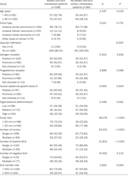
Univariate analysis of axillary non-SLN
metas-tases and clinical pathological data
and the number of negative SLNss were ob-
[image:3.612.94.521.90.677.2]served between the two groups (all P > 0.05).
However, significant differences in tumor size,
the number of tumors, the number of positive
Table 2.
Univariate analysis of axillary non-SLN metastases and clinical pathological data of patients
Group metastases patients Axillary non-SLN (n=108)
No axillary non-SLN axillary metastases
patients (n=84) χ
2 P
Age (year) 2.337 0.126
> 45 (n=92) 57 (52.78) 35 (41.67)
≤ 45 (n=100) 51 (47.22) 49 (58.33)
Tumor type 0.101 0.751
Invasive ductal carcinoma (n=150) 85 (78.71) 65 (77.38) Invasive lobular carcinoma (n=20) 12 (11.11) 8 (9.52) Invasive mixed carcinoma (n=13) 7 (6.48) 6 (7.15) Other types of cancer (n=9) 4 (3.70) 5 (5.95)
Vascular infiltration 0.505*
Yes (n=2) 2 (1.85) 0 (0.00)
No (n=190) 106 (98.15) 84 (100.00)
Estrogen receptor 0.252 0.616
Positive (n=100) 55 (50.93) 45 (53.57)
Feminine (n=80) 45 (41.67) 35 (41.67)
Not checked (n=12) 8 (7.40) 4 (4.76)
Progesterone receptor 2.896 0.089
Positive (n=95) 60 (55.56) 35 (41.67)
Feminine (n=85) 41 (37.96) 44 (52.38)
Not checked (n=12) 7 (6.48) 5 (5.95)
Human epidermal growth factor-2 0.005 0.944
Positive (n=97) 55 (50.93) 43 (51.19)
Feminine (n=83) 47 (43.52) 35 (41.67)
Not checked (n=12) 6 (5.55) 6 (7.14)
Organizational differentiation 0.338 0.561
Low (n=58) 37 (34.26) 21 (25.00)
Medium (n=72) 35 (32.41) 37 (44.05)
High (n=62) 36 (33.33) 26 (30.95)
Tumor size 48.276 < 0.001
≥ 20 mm (n=98) 79 (73.15) 19 (22.62)
< 20 mm (n=94) 29 (26.85) 65 (77.38)
Number of tumors 19.231 < 0.001
Single (n=108) 46 (42.59) 62 (73.81)
Multiple (n=84) 62 (57.41) 22 (26.19)
Number of positive SLN 21.815 < 0.001
Single (n=133) 60 (55.56) 73 (86.90)
Multiple (n=59) 48 (44.44) 11 (13.10)
Number of negative SLN 47.492 0.115
Single (n=115) 70 (64.81) 45 (53.57)
Multiple (n=77) 38 (35.19) 39 (46.43)
SLN transfer rate 2.924 0.004
> 50% (n=129) 82 (75.93) 47 (55.95)
SLNs, and SLN metastasis rates were observed
(all
P < 0.05;
Table 2
).
Multivariate analysis of non-SLN metastases
Following univariate analysis, multivariate
logis-tic analysis was performed. This analysis show-
ed that tumor size, the number of tumors, and
the positive SLN rates were independent risk
factors for non-SLN metastasis (
Table 3
).
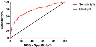
MSKCC predictive model verification
Calculation of the non-SLN metastasis rate
using the MSKCC model and the receiver
oper-ating characteristic curve in the current stu-
dy showed that the area under the curve
(AUC)=0.771 and the 95% CI was 0.724, 0.818
(
Figure 1
).
Discussion
As a common malignant tumor in women, BC
often presents as a painless breast mass, and
in the advanced stages, skin depression and
shape changes may occur [12]. Study has
shown that a lack of exercise, obesity, and
fam-ily history (amongst other factors) are all BC risk
factors [13]. The 5-year survival rate of BC
patients in developed countries can be up to
towards younger people, which has a bad
impact on life expectancy and future quality of
life in the population. In the present day, the
major treatments for BC include surgical
resec-tion, radiotherapy, chemotherapy and
endo-crine therapy [16, 17]. For many years, the
sur-gery was widely employed in clinical practice.
However, with advanced treatment protocols,
more care is taken to preserve general health
without compromising therapeutic benefits.
This requires the support of more advanced
medical knowledge and technology [18].
ALND is an important part of the surgical
proce-dure for invasive BC. However, ALND can easily
lead to a series of adverse reactions, including
lymphedema and a loss of consciousness,
which not only prolongs hospitalization time of
patients with increasing economic burden of
the family, but also seriously impacts quality of
life of the patients [19]. SLNB is an important
operative procedure for early ALN staging in BC
patients. SLNB can avoid dissection in SLN
negative patients; thus, it reduces the
occur-rence of postoperative complications [20].
ALND is required only when SLNB is positive.
However, ALND is generally performed in all
SLN-positive patients, but non-SLN metastasis
are not present in up to 50% of cases, resulting
in over-treatment [21]. Thus, when SLN is
posi-tive, early BC patients can be prevented from
overtreatment by means of prediction of
non-SLN metastasis.
[image:4.612.92.378.85.139.2]In this study, we retrospectively analyzed 192
SLN-positive to identify the risk factors of
axil-lary non-SLN metastasis in patients with SLN
metastasis. Univariate analysis of the two
groups showed that age, tumor type, estrogen
receptor expression, tissue differentiation, and
the number of negative SLNs were not
associ-ated with axillary non-SLN metastasis, whilst
tumor size, tumor number, the number of
posi-tive SLNs, and SLN metastasis rate were
asso-ciated with axillary non-SLN metastasis. In the
Table 3.
Multivariate analysis of non-SLN metastases
Factor β SD χ2 OR 95% CI P
Tumor size 0.782 0.425 5.941 0.472 0.225, 0.836 0.013 Number of tumors 0.584 0.148 8.684 0.638 0.387, 0.805 0.008 Positive SLN rate 0.841 0.465 6.251 0.428 0.201, 0.825 0.019
Note: SLN, sentinel lymph node; SD, standard deviation; OR, odds ratio; CI, confi -dence interval.
Figure 1. MSKCC predictive model verification. The
analysis revealed that the AUC=0.771 and 95% CI was 0.724, 0.818. MSKCC, Memorial Sloan-Ketter-ing Cancer Center; AUC, area under the curve; CI,
confidence interval.
[image:4.612.91.288.184.281.2]study by Mittendorf et al., univariate analysis
also showed that tumor size, tumor number,
the number of positive SLNs and SLN
metasta-sis rates were correlated with axillary non-SLN
metastasis, which was consistent with the
results of this study [22].
Subsequently, multivariate logistic analysis
showed that tumor size, tumor number and
positive SLN rates were independent risk
fac-tors for axillary non-SLN metastasis. In the
study by Viale et al., multivariate analysis also
showed that tumor size, the number of tumors
and SLN positive rates were independent risk
factors for axillary non-SLN metastasis, again
consistent with our data [23]. The MSKCC
axil-lary non-SLN metastasis prediction model
obtained an AUC of 0.76 by verifying the clinical
data of 373 SLN-positive BC patients, which
was in accordance with the range of 0.58-0.86
obtained by many national medical centers and
hospitals [24, 25]. In this study, the AUC was
0.771, indicating that the model could well
pre-dict the non-SLN metastasis.
There are however, some limitations of this
study. The small sample size impacted the
accuracy of the data and we did not perform
follow-ups after surgery. Thus, whether BC
recurrence occurred in the post-surgery patient
was not clear. In future studies, we hope to
increase the sample size and verify all findings
with long-term follow-ups.
In conclusion, this study confirmed that tumor
size, the number of tumors and SLN positive
rates were independent risk factors for axillary
non-SLN metastasis in SLN-positive patients.
When patients were positive for SLN and
pre-sented these clinical symptoms, they were
rec-ommended to undergo ALND. The MSKCC
model displayed a high accuracy for predicting
non-SLN BC in patients.
Disclosure of conflict of interest
None.
Address correspondence to: Jun Zhang, Department of General Surgery, Beijing Friendship Hospital, Capital Medical University, No.95 Yong’an Road, Xicheng District, Beijing City 100050, China. Tel: +86-13811055986; Fax: +86-010-63138754; E-mail: zhangjun5ac@163.com
References
[1] Burstein HJ, Lacchetti C, Griggs JJ. Adjuvant endocrine therapy for women with hormone receptor-positive breast cancer: american so-ciety of clinical oncology clinical practice guide-line update on ovarian suppression summary. J Oncol Pract 2016; 12: 390-393.
[2] Siegel RL, Miller KD, Jemal A. Cancer statistics, 2016. CA Cancer J Clin 2016; 66: 7-30. [3] Francis PA, Regan MM, Fleming GF. Adjuvant
ovarian suppression in premenopausal breast cancer. N Engl J Med 2015; 372: 1673. [4] Desantis CE, Ma J, Goding SA, Newman LA,
Je-mal A. Breast cancer statistics, 2017, racial disparity in mortality by state. CA Cancer J Clin 2017; 67: 439-448.
[5] Lyman GH, Somerfield MR, Giuliano AE. Senti -nel lymph node biopsy for patients with early-stage breast cancer: 2016 american society of clinical oncology clinical practice guideline up-date summary. J Oncol Pract 2017; 13: 196-198.
[6] Giuliano AE, Ballman K, Mccall L, Beitsch P, Whitworth PW. Locoregional recurrence after sentinel lymph node dissection with or without axillary dissection in patients with sentinel lymph node metastases: long-term follow-up from the American college of surgeons oncol-ogy group (Alliance) ACOSOG Z0011 random-ized trial. Ann Surg 2016; 264: 413-420. [7] Rossi EC, Kowalski LD, Scalici J, Cantrell L,
Schuler K. A comparison of sentinel lymph node biopsy to lymphadenectomy for endome-trial cancer staging (FIRES endome-trial): a multicentre, prospective, cohort study. Lancet Oncol 2017; 18: 384-392.
[8] Pilewskie M, Mautner SK, Stempel M, Eaton A, Morrow M. Does a positive axillary lymph node needle biopsy result predict the need for an axillary lymph node dissection in clinically node-negative breast cancer patients in the ACOSOG Z0011 Era? Ann Surg Oncol 2016; 23: 1123-1128.
[9] Bennett AV, Keenoy K, Shouery M, Basch E, Temple LK. Evaluation of mode equivalence of the mskcc bowel function instrument, lasa
quality of life, and subjective significance ques -tionnaire items administered by web, interac-tive voice response system (IVRS), and paper. Qual Life Res 2016; 25: 1123-1130.
[11] Sobin LH, Compton CC. TNM seventh edition: what's new, what's changed: communication from the international union against cancer and the American joint committee on cancer. Cancer 2010; 116: 5336-5339.
[12] Chlebowski RT, Rohan TE, Manson JE, Aragaki AK, Kaunitz A. Breast cancer after use of estro-gen plus progestin and estroestro-gen alone: analy-ses of data from 2 women's health initiative randomized clinical trials. JAMA Oncol 2015; 1: 296-305.
[13] Rock CL, Flatt SW, Byers TE, Colditz GA, Demark-wahnefried W. Results of the exercise and nutrition to enhance recovery and good health for you (ENERGY) trial: a behavioral weight loss intervention in overweight or obese breast cancer survivors. J Clin Oncol 2015; 33: 3169-3176.
[14] Chen W, Zheng R, Baade PD. Cancer statistics in China, 2015. CA Cancer J Clin 2016; 66: 115-132.
[15] WHO. Global status report on alcohol and health 2014. World Health Organization 2014; 18: 1-57.
[16] Taylor C, Correa C, Duane FK, Aznar MC, Anderson SJ. Estimating the risks of breast cancer radiotherapy: evidence from modern radiation doses to the lungs and heart and from previous randomized trials. J Clin Oncol 2017; 35: 1641-1649.
[17] Boughey JC, Ballman KV, Le-Petross HT, McCall LM, Mittendorf EA, Ahrendt GM, Wilke LG,
Taback B, Feliberti EC, Hunt KK. Identification
and resection of clipped node decreases the false-negative rate of sentinel lymph node sur-gery in patients presenting with node-positive breast cancer (T0-T4, N1-N2) who receive neo-adjuvant chemotherapy: results from ACOSOG Z1071 (Alliance). Ann Surg 2016; 263: 802-807.
[18] Sheikh IA, Jiffri EH, Kamal MA, Ashraf GM, Beg MA. Lactoperoxidase, an antimicrobial milk protein, as a potential activator of carcino- genic heterocyclic amines in breast cancer. Anticancer Res 2017; 37: 6415-6420.
[19] Li CZ, Zhang P, Li RW, Wu CT, Zhang XP. Axillary lymph node dissection versus sentinel lymph node biopsy alone for early breast cancer with sentinel node metastasis: a meta-analysis. Eur J Surg Oncol 2015; 41: 958-966.
[20] Leiter U, Stadler R, Mauch C, Hohenberger W, Brockmeyer N. Complete lymph node dissec-tion versus no dissecdissec-tion in patients with senti-nel lymph node biopsy positive melanoma (DeCOG-SLT): a multicentre, randomised, phase 3 trial. Lancet Oncol 2016; 17: 757-767. [21] Pilewskie M, Jochelson M, Gooch JC, Patil S,
Stempel M. Is preoperative axillary imaging
beneficial in identifying clinically node-nega -tive patients requiring axillary lymph node dis-section? J Am Coll Surg 2016; 222: 138-145. [22] Mittendorf EA, Hunt KK, Boughey JC, Bassett
R, Degnim AC. Incorporation of sentinel lymph node metastasis size into a nomogram predict-ing nonsentinel lymph node involvement in breast cancer patients with a positive sentinel lymph node. Ann Surg 2012; 255: 109-115. [23] Viale G, Maiorano E, Pruneri G, Mastropasqua
MG, Valentini S. Predicting the risk for addi-tional axillary metastases in patients with breast carcinoma and positive sentinel lymph node biopsy. Ann Surg 2005; 241: 319-325. [24] Padmanabhan N, Ayub MF, Hussain K, Kurien
A, Radhakrishna S. Factors influencing
non-sentinel node involvement in non-sentinel node positive patients and validation of MSKCC no-mogram in Indian breast cancer population. Indian J Surg Oncol 2015; 6: 337-345.