Voltage-Gated Proton Channels
Thomas E. DeCoursey*1ABSTRACT
Voltage-gated proton channels, HV1, have vaulted from the realm of the esoteric into the forefront of a central question facing ion channel biophysicists, namely, the mechanism by which voltage-dependent gating occurs. This transformation is the result of several factors. Identification of the gene in 2006 revealed that proton channels are homologues of the voltage-sensing domain of most other voltage-gated ion channels. Unique, or at least eccentric, properties of proton channels include dimeric architecture with dual conduction pathways, perfect proton selectivity, a single-channel conductance approximately 103times smaller than most ion channels, voltage-dependent gating that is strongly modulated by the pH gradient,pH, and potent inhibition by Zn2+(in many species) but an absence of other potent inhibitors. The recent identification of H
V1 in three unicellular marine plankton species has dramatically expanded the phylogenetic family tree. Interest in proton channels in their own right has increased as important physiological roles have been identified in many cells. Proton channels trigger the bioluminescent flash of dinoflagellates, facilitate calcification by coccolithophores, regulate pH-dependent processes in eggs and sperm during fertilization, secrete acid to control the pH of airway fluids, facilitate histamine secretion by basophils, and play a signaling role in facilitating B-cell receptor mediated responses in B-lymphocytes. The most elaborate and best-established functions occur in phagocytes, where proton channels optimize the activity of NADPH oxidase, an important producer of reactive oxygen species. Proton efflux mediated by HV1 balances the charge translocated across the membrane by electrons through NADPH oxidase, minimizes changes in cytoplasmic and phagosomal pH, limits osmotic swelling of the phagosome, and provides substrate H+for the production of H2O2 and HOCl, reactive oxygen species crucial to killing pathogens.C 2012 American Physiological Society.Compr Physiol2:1355-1385, 2012.
Introduction
Proton pathways through proteins are a stalwart element in nu-merous bioenergetic molecules, in which proton and electron movements are elaborately choreographed (27,177,269,272). In these molecules, protons generally cross partway through the membrane, perform some function, or await a catalytic step, and then return or continue across the membrane. In con-trast, the voltage-gated proton channel behaves like a simple, passive conduction pathway that spans the entire membrane. Like all biological channels, proton channels are gated; that is, the channel opens to conduct protons when required, and then closes. They are called “voltage-gated” because mem-brane potential regulates the probability that the channel will be open, with depolarization promoting opening.
Voltage-gated proton channels are attractive subjects of research for three broad reasons. First, there is a rapidly expanding list of important physiological roles played by voltage-gated proton channels in a variety of cells and species. Second, by virtue of their homology to the voltage-sensing domains (VSDs) of other voltage-gated ion channels, they provide a unique device for evaluating gating mechanisms. Third, they violate so many rules adhered to by other ion channels that they require a new vision and a whole new set of questions. Their characterization as ion channels has oc-casionally been questioned on several grounds, but by most
criteria, they are unquestionably ion channels. This review will summarize current knowledge in these three areas, with an emphasis on their physiological roles.
History
The first explicit proposal of a voltage-gated proton channel by Woody Hastings and colleagues in 1972 (96), postulated a role for the proton channel in triggering a bioluminescent light flash in a dinoflagellate, Gonyaulax polyedra, when the cell is mechanically disturbed (Fig. 1). Further information on pro-ton channels in dinoflagellates is presented later (see Section “Bioluminescence and other functions in dinoflagellates”).
Proton channels were identified as bona fide ion chan-nels in 1982 by Roger Thomas and Bob Meech (260) who performed voltage-clamp studies of Helix aspersa snail neu-rons that had been impaled by a battery of microelectrodes. Protons in the form of HCl were injected into the cell bod-ies and outward membrane current carried by protons could
*Correspondence to tdecours@rush.edu
1Department of Molecular Biophysics and Physiology, Rush University
Medical Center, Chicago, Illinois
Published online, April 2012 (comprehensivephysiology.com) DOI: 10.1002/cphy.c100071
H+
H+
LH2
H+
LBP L’ASE LBP
H+ H+
[image:2.684.40.279.56.180.2]LH2
Figure 1 Voltage-gated proton channels trigger the bioluminescent light flash in dinoflagellates. The proton concentration is high in the flotation vacuole (below the membrane in the diagram). An action po-tential depolarizes the membrane, opening proton channels, allowing protons to flow down their electrochemical gradient into the interior of the scintillon (upper compartment). The sudden drop in pH in the scintillon triggers the flash by two concerted mechanisms. Upon pro-tonation, the light-emitting pigment, luciferin (LH2) is released from
luciferin-binding protein (LBP) making it available as a substrate for luciferase, which is itself activated by acidification. [Adapted, with per-mission, from reference (109). Originally published inBioluminescence in Action, edited by P. Herring, Academic Press, London. pp. 129-170, 1978. Copyright Elsevier.]
be detected when the membrane potential was clamped to positive voltages. During sustained depolarization, the out-ward current declined as the intracellular pH (pHi) increased. Despite the small single-channel conductance (see Section “Minuscule single channel conductance”), proton channels were sufficiently abundant in the membrane that their activity restored pHi on a time scale of tens of minutes. These neu-rons were 100 to 200μm in diameter (56); in small cells like leukocytes (6-10μm diameter) proton current can restore pHi after an acid load in tens of seconds (62, 76, 134), because of the more favorable surface/volume ratio. A thorough voltage-clamp characterization carried out by Lou Byerly, Bob Meech, and Bill Moody demonstrated a critical shift of the position of the gH-V relationship along the voltage axis when either pHo or pHi was changed (29). Also at UCLA, Mike Bar-ish and Christiane Baud identified and characterized similar proton currents in Ambystoma salamander oocytes (19). Sub-stantial effort was expended in these early studies to establish convincingly that proton channels were genuine ion chan-nels and that protons did not simply permeate other chanchan-nels adventitiously (29, 170). Polyvalent cations were found to in-hibit proton currents (260), and Martyn Mahaut-Smith (166) showed that Zn2+inhibited proton currents 80 times more po-tently than Ca2+currents in Helix neurons. Byerly and Suen (30) studied current fluctuations in membrane patches from
Lymnaea stagnalis, and concluded that the single-channel
proton conductance was extremely small (<50 fS). Based on measurements of pH changes and membrane potential, Lydia Henderson, Brian Chappell, and Owen Jones deduced the probable existence of proton channels in human neu-trophils, where they were proposed to compensate for the electrogenic activity of NADPH oxidase (Nox) (115, 116).
Nearly a decade after the first voltage-clamp study, pro-ton currents were discovered in mammalian cells, rat alveolar epithelial cells (54). These cells exhibit high levels of proton current, enabling a series of studies in which the main bio-physical properties of proton channels were explored. The H+ current was not much larger with 100 mmol/L buffer than with 10 mmol/L or even 1 mmol/L buffer, suggesting that the rate-determining step in permeation was not proton transfer from buffer to the channel, but occurred within the conduction path-way (64). Buffer concentration on the intracellular side of the membrane was, reasonably enough, shown to be important, with 100 mmol/L buffer providing markedly better control of pHithan 10 mmol/L (64). Several studies explored the phe-nomenon of proton depletion as an inevitable consequence of the passage of large proton currents across cell membranes. It became clear that the proton channel does not inactivate, but that when proton depletion is pronounced, the currents decay with time as pHiincreases (54, 76, 105, 134, 184, 186). In the classical sense, “inactivation” means that a channel enters an altered conformational state from which it cannot open until recovery to a resting state has occurred (121). That proton cur-rent decay reflects changes in pH, and consequently in both the driving force and the pH-dependent gating of the channel is clear from studies showing that—whether assessed by pH electrode, pH sensing fluorescent dyes, or by measurement of the proton current reversal potential—pHi changes roughly in proportion to the integral of the H+ flux during pulses (54,62,76,105,134,144,170,186,260). Separating the effects of proton depletion from “genuine” underlying proton cur-rent kinetics continues to challenge researchers in this field. However, because no proton channel has yet been shown to inactivate, it remains true that visible decay of proton currents constitutes prima facie evidence that significant pH changes occurred during the pulse.
In 1993, proton currents were recorded in three human cell types (24, 76), including human neutrophils (60), confirming the hypothesis of Henderson and colleagues. The number of cell types reported to express proton channels continued to expand. A breakthrough in physiological function was the discovery that in phagocytes with active Nox, the proper-ties of the proton conductance differed markedly from those in resting cells (16). The change was so profound (see Sec-tion “Enhanced gating mode”) that it was at first thought to reflect the appearance of a novel type of proton channel, which was hypothesized to comprise the gp91phoxcomponent
acetate), was shown to increase proton currents identically in PLB-985 cells with or without gp91phox(69). COS-7 cells that
lacked endogenous proton currents still lacked detectable pro-ton current when gp91phoxwas transfected into them together
with other components to produce a functional NADPH ox-idase complex (181). The coup de grˆace for the hypothesis was the demonstration that neutrophils from proton channel (HVCN1) knockout mice exhibited no detectable proton cur-rents, yet had normal electron currents when Nox was acti-vated, unambiguously demonstrating the presence of func-tioning gp91phox in their membranes (88, 178). Ironically,
in their first landmark paper that proposed the existence of voltage-gated proton channels in human neutrophils (114), Henderson and colleagues made six extremely accurate pre-dictions that remain true today:
“H+ions—movement is via a channel in the membrane, the
opening of which lags behind activation of the [NADPH] oxi-dase. The mechanism for initiating the opening of this channel is unknown at present, but it could be a voltage decrease (i.e., depolarization), an increase in the internal concentration of protons or a phosphorylation/conformational change.”
If this had been the last word on the subject, a decade of controversy could have been avoided.
The discovery of proton channel genes in 2006 (224, 234) inevitably fomented research into several areas previously not possible, namely, structure-function studies, as well as knockout and knockdown studies, and the development of antibodies. Completely unexpected was that the proton
chan-nel molecule would so closely resemble the voltage-sensing domain of most voltage-gated ion channels. Discovery of this feature immediately transformed the proton channel molecule into a new playground for the study of voltage-gating mecha-nisms. In the resulting flurry of activity, discoveries have been made at an astonishing pace.
Properties
Gene and molecular features
The proton channel protein, H
V1
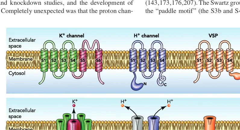
[image:3.684.90.488.410.626.2]Identification of proton channel genes in human (224), mouse, and Ciona intestinalis (sea squirt) (234) in 2006 revealed sur-prising properties, the most remarkable of which is the sim-ilarity of the HV1 protein to the VSD of most voltage-gated ion channels (Fig. 2). The first VSD to be identified that was not part of a voltage-gated ion channel was a voltage-sensing phosphatase (VSP) (191). The VSP senses membrane po-tential and exhibits “gating currents” like those associated with voltage-gated ion channels, but the response to depo-larization is modulation (an increase) of phosphatase activ-ity. The subsequent identification of the voltage-gated proton channel gene added impetus to the idea that the VSD might be viewed as a modular unit that could be attached to an-other molecule and provide voltage sensitivity to its function (143,173,176,207). The Swartz group have further shown that the “paddle motif” (the S3b and S4 transmembrane helices)
Figure 2 Membrane topology of voltage-gated K+channels (left), voltage-gated proton channels (center), and voltage-sensitive phosphatases (right). The K+channel (lower panel) is a tetramer of the six transmembrane domain monomer shown in the top; the proton channel is a homodimer of the four transmembrane domain monomer in the top panel, and the phosphatase is a monomer. The assembled K+channel has a single conduction pathway, but the proton channel has two, one in each protomer. The voltage-sensing phosphatase (VSP) senses voltage, but does not conduct. The proton channel cartoon illustrates schematically that the dimer is held together by C-terminal coiled-coil interactions, and that the channel can be phosphorylated at Thr29in the intracellular N terminus to produce the
is indeed modular, and continues to function when swapped among K+channels, HV1, and VSP (7).
Perhaps, the most distinctive feature of the proton channel is the absence of S5-S6 domains that form the conduction pathway in ordinary ion channels. Because conduction can be demonstrated when purified HV1 protein is reconstituted in synthetic liposomes, no accessory protein is required (151). The conduction pathway must exist within the HV1 molecule itself. Homology models have been proposed, based on the similarity of the sequence to that of the VSD of K+channels (199,223,270). The channel resembles an hourglass filled with water, with a constriction near the center. Proton selectivity is established by an essential aspartate residue in the S1 domain, Asp112 in hH
V1 (200) and Asp51 in kHV1 (251), which is thought to be located at or near the constriction when the channel is in the open state.
Three new proton channel genes were discovered in 2010, all in unicellular marine organisms; two coccolithophores,
Emiliania huxleyi and Coccolithus pelagicus (259) and a
dinoflagellate, Karlodinium veneficum (251). These chan-nels greatly expand the phylogenetic representation of HV1 (Fig. 3). One putative plant HV1 is shown. Only seven of the 37 putative HV1 genes have been confirmed by
heterolo-gous expression and electrophysiological recording; in addi-tion to the six just discussed, Strongylocentrotus purpuratus was confirmed by Y. Okamura (personal communication). The confirmation that HV1s in several unicellular species are genuine proton channels is an important validation of the cri-teria used to identify putative proton channels from sequence analysis alone. We proposed recently that a protein with four transmembrane domains that includes (a) an appropriately po-sitioned Asp residue in S1 to act as the selectivity filter and (b) two or preferably three appropriately spaced Arg residues in
C. sa vign
y
100
Chl orell
a P
olysphondylium
Kar lodinium
E. huxle
yi
C. pelagi
cus
Ph
ae
oda
ctylu
m
Thalass iosir a
Nem ato
stel la
Trichop lax
Bra nchiostoma
Strongylocentrotus
99
82
86 69
96 76
88
001
84
90
76
67 97
Lottia
C. intestinalis Single celled
organisms
Invertebrates
Amphibians
Fish
Birds Gallus
Taeniop ygia
Equu s
Lo xodonta Or
ycto lagus
Macaca Homo Sore
x
My otis
M on odelph
is Canis1
Canis2 Sus
Echino ps
Sper mophi
lus Tupa
ia
Bos Ratt
us Mus
Danio
Gasterosteus
Oryzias
T. laevis
X. tropicalis
[image:4.684.137.442.278.647.2]Mammals
Figure 3 A phylogenetic analysis showing evolutionary relationships among 37 puta-tive HV1 voltage-sensing domain (VSD) sequences. This maximum likelihood phylogenetic
tree with 100 boostraps was constructed from a multiple sequence alignment of the VSD portion of 37 HV1s; N- and C-termini were truncated. Branch lengths are displayed to
S4, that include the WRxxR motif, can be considered highly likely to be a voltage-gated proton channel (251).
The phylogenetic tree in Figure 3 is drawn to scale, so that the radial length of each branch reflects the extent of sequence differences. It is tempting to overinterpret this phy-logram. Mammalian HV1s are very similar to each other, but as one ventures toward more primordial species, variation in-creases. Invertebrate HV1s exhibit a high degree of individu-ality, and HV1s in unicellular organisms are riotously diverse. The most deviant among this group is kHV1, a dinoflagellate proton channel. One might predict that in general, sequence diversity may presage functional diversity. As we will discuss later (see Section “Bioluminescence and other functions in di-noflagellates”), the Karlodinium proton channel violates the hitherto sacred rule of conducting exclusively outward current (251), and thus its unique behavior matches its idiosyncratic sequence.
Nomenclature
The discoverers of the first proton channel genes adopted different systems for nomenclature. Ramsey and colleagues (224) christened the human channel HV1: H for H+ selec-tive, V for voltage-gated, and 1 for its being the first example identified in humans. Sasaki and colleagues adopted more evocative abbreviations, mVSOP and CiVSOP: m for mouse, Ci for C. intestinalis, and VSOP for voltage-sensor-only pro-tein (234), thus incorporating the most surprising feature of the gene product into its designation. The official gene name for all species is HVCN1. Here, I will adopt the more aus-tere convention and refer to the protein as HV1, which will be qualified by a prefix that indicates species (hHV1 for hu-man, mHV1 for mouse, etc.). Although this appears to limit the spectrum to 26 species (and then only if they have differ-ent initials), the list can be expanded by including genus and species, for example, CiHV1, EhHV1, or CpHV1. To date, only one proton channel gene has been identified in any species, so for now, the suffix is 1 for all species.
The proton channel is a dimer, exhibiting
cooperative gating
Several types of evidence led to the conclusion that the pro-ton channel molecule exists mainly as a dimer, both in het-erologous expression systems (139, 150, 261) and in native cells (217), with a conduction pathway in each protomer (139, 261). It is puzzling why the channel goes to the trou-ble of assembling as a dimer, in light of evidence that the monomer exhibits the main features of native proton cur-rent: activation upon depolarization, extreme proton selectiv-ity, and exquisite sensitivity of gating to both pHo and pHi (103, 139, 199, 261). A precedent exists for a dimeric channel with conduction pathways in each monomer, in the ClC fam-ily of Cl−channels and Cl−/H+antiporters (1, 108, 159, 163, 171, 172, 174). Like ClC-0 (108), the two protomers com-prising the proton channel dimer appear to interact during
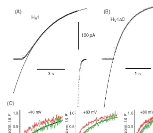
gating (103, 198, 199, 262). This interaction has been inter-preted either as positive cooperativity in which the opening of a single protomer greatly increases the probability of the second protomer opening (262), or a strict requirement that both protomers undergo a conformational change before ei-ther can conduct (103, 198, 199). Coiled coil interactions at the C terminus appear to provide the main glue that holds the dimer together (103, 139, 151, 156, 198, 199, 261). Trun-cating (or replacing) the C terminus results in expression of monomers (103,139, 198, 199, 261) that open 5-6 times more rapidly than the wild-type (WT) dimer (139, 198, 261). As illustrated in Figure 4, the proton conductance of the dimer activates with a sigmoid time course, consistent with classi-cal Hodgkin-Huxley n2kinetics (122), whereas the monomer activates exponentially (103, 199). Figure 4C shows that dur-ing depolarizdur-ing pulses a fluorescent probe attached to the S4 domain moves with an exponential time course, whereas the current turns on after a small delay and with a sigmoid time course. Reflecting the cooperativity of gating, the limiting slope of the gH-V relationship of the dimer is twice as steep as that of the monomer (103). These properties of dimeric gat-ing suggest an explanation for the utility of proton channels existing as dimers, at least in phagocytes (199). As will be seen (see Section “Charge compensation”), proton channels open in phagocytes to prevent excessive depolarization caused by the electrogenic activity of Nox. Consequently, it is critically important that proton channels activate with a steep voltage dependence, to prevent self-inhibition of Nox (73). If steep voltage dependence occurs at the expense of slower opening, little is lost. Depending on the type of stimulus, the time course of the respiratory burst is relatively slow, usually preceded by a delay of seconds to minutes, and persisting for minutes to hours (72).
The dimeric arrangement may not dictate an invariant in-terface. Two possible interfaces have been proposed, shown in Figure 5. On the basis of cross-linking studies for mu-tants in which Cys was introduced at various locations in the hHV1 molecule, several interactions at the external end of S1 (shown in red in Fig. 5A) were detected (150). However, based on Zn2+effects on hH
V1 mutants in which the Zn2+ -binding residues His140and His193were mutated, the interface shown in Figure 5B was proposed to occur at least occasion-ally (199). In the Cys scanning study, weak cross-links were observed at position 194, right next to His193, suggesting that this orientation can occur at least occasionally (150). If both kinds of interfaces are possible, then multimers could also oc-cur (Fig. 5C). Given substantial evidence that the protomers interact during gating, one imagines that channel opening might occur only from a particular orientation. Because Zn2+ appears to prevent channel opening (36), it is reasonable to suppose that Zn2+—which is thought to bind at the interface between protomers (199)—might act by preventing the dimer from adopting its preferred orientation.
HV1 H
V1ΔC
100 pA 30 pA
1 s 3 s
+40 mV +60 mV +80 mV
1,000 2,000
Time (ms)
1,000 2,000
Time (ms)
1,000 Time (ms)
Nor
m.
I & F
1.0
0.5
0
Nor
m.
I & F
1.0
0.5
0
Nor
m.
I & F
1.0
0.5
0
(B) (A)
[image:6.684.138.445.52.319.2](C)
Figure 4 Comparison of gating kinetics of the human proton channel dimer (A) and monomer (B), expressed in HEK-293 cells. Note the slower, sigmoid activation of the WT channel (A), and the faster, exponential turn-on of the monomer (C terminus truncated, HV1C) both during pulses to+50 mV at 23◦C at pHo 7.5, pHi 7.5. [Adapted from
reference (199). Originally published in The Journal of Physiology.] (C) The time course of movement of a fluorescent probe attached to the S4 domain of theCionaproton channel is exponential (red). This time course raised to the second power (green) matches the proton current recorded at the same time (black). [Adapted, with permission, from reference (103). Adapted, with permission, from Macmillan Publishers Ltd: Nature Structural & Molecular Biology17: 51-56, 2010.]
short linker to the N terminus of the second protomer func-tioned almost identically to the WT dimer (198, 199). The tandem dimer activated with a sigmoid time course (198), consistent with similar cooperative interactions between pro-tomers observed in WT dimers (103). Its gH-V relationship was also indistinguishable from WT, and the only difference detected was slower activation kinetics (198). Evidently, C terminal coiled-coil interactions tether the two protomers to-gether, but appear not to contribute critically to the inter-actions between protomers that occur during channel gating (198). However, a role of the C terminus in modulating gating by direct mechanical linkage has been suggested (99).
In contrast to the structural role played by the C termi-nus, the N terminus appears to interact closely with the gat-ing mechanism of the channel. The enhanced gatgat-ing mode (see Section “Enhanced gating mode”) is mainly the result of phosphorylation of Thr29 in the N terminus of hH
V1 (194). In addition, a mutation that results in M91T substitution (Met replaces Thr at position 91) in the N terminus has been iden-tified in the human population (128) (see Section “Acid se-cretion in epithelia of the respiratory tract”). This mutation shifts the gH-V relationship positively by 20 mV, reducing the likelihood of channel opening. It remains unclear how
these two residues scattered among the 100-residue N termi-nus of hHV1 are able to modulate gating, which as discussed previously presumably involves movement of the S4 domain relative to the other transmembrane domains.
Selectivity and permeation by protons
Perfect selectivity
(A) (B)
[image:7.684.298.537.52.222.2](C)
Figure 5 Possible dimer interfaces, with the transmembrane do-mains color coded as: S1 red, S2 yellow, S3 green, and S4 blue. The dimer in A was proposed on the basis of cysteine cross-linking studies (150); the dimer in B was proposed to explain Zn2+ binding
studies (199). External His residues that bind Zn2+(224) are shown in aqua (cf. Figure 14). [Adapted, with permission, from reference (198). Originally published in Channels (Austin) 4: 260-265, 2010.]
Proton conduction through proteins is widespread, ap-pears to occur fairly readily, and is presumed to be possi-ble whenever water molecules are identified inside proteins within hydrogen bonding distance of other waters or titratable groups. Many membrane proteins have been shown to be ca-pable of conducting protons, several of which were discussed previously in this context (56). However, despite the apparent facility of proton conduction, proton selective conduction is more demanding, and is discussed later. It is arguably more difficult to prevent protons from carrying current through an aqueous pathway, than to allow proton permeation. This chal-lenge is faced by aquaporins, whose job is to facilitate rapid water permeation (4), but to prevent permeation by ions, espe-cially protons. Precisely how protons are excluded remains de-batable (28, 33, 51, 127, 175, 190, 256). Intriguingly, mutation of just three amino acids turns AQP1 into a cation-conducting channel with a strong preference for protons (273).
Protons are chemically quite different from other cations. In aqueous solutions, protons exist as H3O+at least 99% of the time (47). By hopping from one water molecule to the next by the Grotthuss mechanism (3, 49, 52, 53), proton conduc-tion through water occurs five times more rapidly than other cations the same size as H3O+ (23, 86). Proton conduction through proteins occurs by a hydrogen-bonded chain (HBC) mechanism (202) in which protons hop from one molecule or group to the next (Fig. 6). The HBC may comprise titratable amino acid side chains or water molecules (202). Because protons are ubiquitous (the total concentration of hydrogen
C
C C
C
C
C
H H
H H
H H (A)
(B) H+
H
H
H H H H
O
O O
O
O
O
C
C C
C C
C
O
O O
O O
[image:7.684.39.278.53.289.2]O
Figure 6 Hydrogen-bonded chain (HBC) conduction. In this schematic example, hydroxyl groups (e.g., from Ser residues) form a HBC that spans the membrane. (A) Proton conduction occurs when a proton enters the chain from the left to form a positive ion, which then moves to the right by hopping of successive protons to effect a reversal of the direction of the hydrogen bonds between each pair of oxygen atoms. Proton conduction would also occur by loss of a proton on the right followed by movement of a negative ion (or fault) to the left. (B) To complete the process, reorientation of each hydrogen bond in the chain must occur, so that another proton can enter from the same side. [Redrawn, with permission, from reference (202).]
in water is 110 mol/L) and interchangeable, multiple protons contribute to the net flux of a single proton across the mem-brane. Other ions diffuse around water and other molecules; protons use water and other molecules as stepping stones. The HBC mechanism potentially enables proton selective conduc-tion, by virtue of the ability of protons to translocate along the chain without displacing the elements of the chain. Thus, protons are conducted through the gramicidin channel, which contains a linear row of a dozen water molecules (154), two orders of magnitude faster than any other ion (201), because other ions must wait for the waters to diffuse through the pore. The protons hop across, leaving the water wire intact.
permeation occurred in the pore (64), the general opinion was that the permeation pathway through voltage-gated pro-ton channels likely contained at least one titratable group (56, 61, 62, 66, 67, 77, 117, 169). Recently, this hypothesis was tested by mutating every likely HBC element in the human HV1 proton channel (223). No single residue was found to be indispensable for conduction. By default, the mechanism of proton selectivity was suggested instead to involve non-diffusible waters “frozen” at a narrow constriction in the channel. These waters could transfer H+ but block perme-ation of ordinary ions. Even more recently, Asp112 located in the S1 TM domain was identified as the proton selectiv-ity filter of the human voltage-gated proton channel (200). Neutralizing mutations not only eliminated proton specific conduction, they resulted in permeability to anions. The con-servative replacement of Asp with Glu did not alter pro-ton selectivity, but the D112H mutant was anion permeable. The corresponding residue in a dinoflagellate proton channel, Asp51, also in the S1 domain, was found to exhibit identi-cal phenomenology; neutral substituents produced a strongly Cl− permeable channel, Glu acted like Asp, and the His mutant conducted Cl− (251). Evidently, when the Asp at the selectivity filter is neutralized, the rest of the channel molecule excludes cations. How proton-specific conduction is enforced is not yet understood in atomic detail, but an acid at this key location in the first transmembrane domain (S1) is essential.
A similar problem has been confronted by the M2 pro-ton channel of influenza A virus. This channel shares many biophysical properties with the voltage-gated proton chan-nel, including high selectivity (43, 158, 189), small conduc-tance (158, 189), and a large deuterium isotope effect (189). However, surprising new studies indicate that the M2 chan-nel is not exclusively proton selective, but that Na+ and K+ can occasionally permeate in the opposite direction, com-pensating electrically for H+influx (152, 215). If M2is less selective than the voltage-gated proton channel, then its di-rect relevance as a model for the selectivity mechanism of the voltage-gated proton channel may be questioned, but it remains heuristically useful. The M2 channel is a homote-tramer with a ring of four His37 residues that comprise an acid-activated gate. One conceptual model postulates that protonation of these His37, through electrostatic repulsion, expands the channel allowing waters to transfer protons down the center of the pore while still preventing other cations from permeating by a frozen water mechanism (136,142,233,252). Alternatively, His37 might be protonated and deprotonated with each conduction event, shuttling protons through the pore (124, 137, 148, 218, 241, 245, 248). Recently, a hybrid mechanism was suggested, in which the permeating proton is delocalized among the four His37 and associated water molecules (2). Whether a proton delocalization mechanism might also be applicable to the voltage-gated proton chan-nel remains to be determined. The idea seems seductively vague, but it may actually reflect reality. Surprisingly, mu-tation of Asp112 in hH
V1 to His (D112H) failed to
pro-duce proton-selective conductance; instead, the D112H mu-tant was anion permeable (200). Precisely, the same result was seen in the D51H mutant of the dinoflagellate pro-ton channel kHV1 (251). Evidently, His can shuttle pro-tons, but even at a constriction, does not guarantee proton specificity.
Minuscule single channel conductance
One of the most exciting possibilities enabled by the discovery of the patch-clamp technique was the direct measurement of current flowing through a single molecule, an open ion chan-nel (106). Most ion chanchan-nels have a conductance of 2 to 400 pS (119), and conduct currents of a few picoamperes, that are resolvable by direct recording from a patch of membrane, or even from whole cells that are electrically tight [such as human lymphocytes (59) or eosinophils (90)]. However, the voltage-gated proton channel has a conductance approximately 3 or-ders of magnitude smaller than most other channels, and for many years single channel currents were thought to be too small to resolve by direct measurement. Due to the difficulty in resolving single-channel currents, a preferred approach is to evaluate current fluctuations, which provides similar infor-mation, as illustrated in Figure 7. At subthreshold voltages, the current exhibits very little noise. Above threshold, the random gating of proton channels introduces noise into the current, from which the single-channel current amplitude, iH, can be derived: iH =σ2/[IH × (1-Popen)], where σ2 is the variance and Popen is the open probability (119). These pa-rameters are all directly measurable, excepting Popen, which can be accounted for by selecting conditions that minimize its effect; by recording just above Vthresholdwhere Popen≈0 (60), or by analyzing data from multiple voltages simultaneously (39, 244). Determined in this way, the unitary conductance increased as pHidecreased, from 38 fS at pHi6.5 to 140 fS at pHi5.5 at room temperature (39). The unitary conductance was independent of pHo, as might be expected for exclusively outward currents. Incidentally, this behavior is consistent with H+ being the conducted species, rather than OH−, which would be expected to have the opposite dependence on pH changes. In some patches, Cherny and colleagues observed step-like events that resembled single-channel currents, with amplitudes 7-16 fA fluctuations (39). These were just on the boundary of being resolvable, and required seal resistances in the T(1015) range; because at the low frequencies of proton channel gating, the resolution is limited by extraneous noise contributed by the seal resistance (153). The ampli-tude of these apparent unitary currents was about double that calculated in the same patches from fluctuations (39). This result is intriguing in light of the proton channel existing as a dimer (139, 150, 261), whose two subunits each have separate conduction pathways and function with strong cooperativity (103, 198, 199, 262).
2 pA
2.5 s
20
–20
–60
pHi 5.0
2 pA
2 pA
2.5 s
40
40 20 0
–20 –60, –40 2.5 s
60
50
40
30
20
10
0
–100 –80 –60 –40 –20
Voltage (mV) gH
(pS)
Pmax Pmax
pHi 5.0
5.5
6.5 pHi 5.0
1.0 0.95
0.9 0.8
0.5 0.1
1.0 0.95
0.9 0.8
0.5 0.1
V1/2 V1/2 V1/2
pHi 5.5
pHi 6.5
0 20
–80 10–28 10–27
Model v
ar
iance (A
2)
–60 –40 –20
Voltage (mV)
Model variance: 200 channels @ 50 fS Model Popen –V Relationships
(A) (D)
(B) (C)
V1/2 = –40 mV
Popen
Vrev = –120 mV k = 8 mV
0 20
–80 1.0
0.8
0.6
0.4
0.2
0.0
–60 –40 –20
Voltage (mV)
0 20
40 –100
1000
100
10
1
0.1
–80 –60 –40 –20 Voltage (mV)
V
ar
iance (x10
–28
A
2 )
0 20 40
0
–40
pHi 5.5
[image:9.684.64.514.54.612.2]pHi 6.5
Figure 7 Determination of the single channel conductance from proton current fluctuations that reflect the stochastic opening and closing of proton channels. “A” shows families of proton currents in an excised, inside-out patch from a human eosinophil at three pHivalues. At subthreshold voltages, the current is quiet, but just aboveVthreshold, proton channels begin to open, and the current
becomes distinctly noisy. It is noteworthy that (at low pHi) the noise first increases with depolarization, but then decreases for large
depolarizations. “B” showsgH-Vrelationships from this patch. The variance of the current fluctuation, measured at quasi-steady-state,
is plotted in “C.” The variance increases more than 100-fold at voltages where the proton conductance is active, and is maximal near the midpoint of thegH-Vrelationship at each pHi(indicated asV1/2). “D” shows that the expected variance given the simplest possible
two-state model of gating (closed↔open) coincides with the observed voltage dependence. The nonmonotonic increase in variance with depolarization is consistent with the maximumPopenlimiting to approximately 0.95 at pHi5.5. [Adapted, with permission, from
be 0.75 at pHi 6.5, increasing to 0.95 at pHi 5.5 or lower (39). Thus, lowering pHi increases proton currents both by increasing the unitary conductance and by increasing Pmax.
Regulation of gating by the pH Gradient,
pH
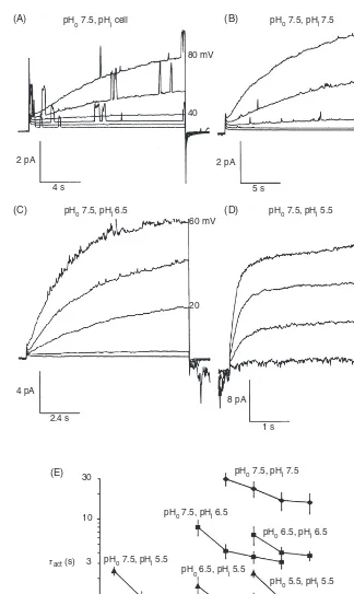
A feature displayed universally by voltage-gated proton chan-nels is that the voltage dependence of their gating shifts dras-tically when pH is changed on either side of the membrane. Lowering pHo shifts the gH-V relationship positively and tends to slow channel opening (larger activation time con-stant,τact) (19, 29). Byerly and colleagues (29) observed that the slowing of activation at lower pHowas greater than could be accounted for by the size of the gH-V relationship shift (i.e., on the assumption that all gating parameters shifted equally), and concluded that external protons directly inhibited chan-nel opening in snail neurons. Conversely, lowering pHishifts the gH-V relationship negatively (29,167). Figure 8 illustrates the effects of changes in pHion proton currents in inside-out patches of membrane from rat alveolar epithelial cells (63). Qualitatively, the effects are like those described in snail neu-rons, but there are subtle differences. Effects of pHo onτact appear to be accounted for by the 40 mV/unit pH shift of thegH-V relationship, in contrast with snail. However, lowering pHiin rat increased the current amplitude and greatly accel-erated activation. In rat epithelium,τact decreased 5-fold per unit decrease in pHi, which cannot be ascribed to a voltage shift. These minor differences probably reflect species differ-ences, but some subtle discrepancies seem to exist between properties observed in excised membrane patches compared with those in whole-cell configuration. Nevertheless, the qual-itative effects of pHoand pHiare quite consistent among all species studied to date. The physiological implication of this intricate regulation by pH is that the proton channel normally conducts only outward current, and consequently, its primary function must be acid extrusion from cells.
In stark contrast to the effects of pHo on channel ac-tivation, deactivation was completely insensitive to pHo in human THP-1 cells (65). A weak sensitivity to pHowas seen in rat alveolar epithelial cells if the tail currents were force-fit by a single exponential, but when a double exponential fit was used, the rapid τtail component was pHo independent. Near Vthreshold, a slower component of tail current decay ap-peared that was kinetically closer toτactand was more steeply voltage dependent, as well as being shifted∼approximately 40 mV/unit change in pHo(38).
In a systematic study of the regulation of the voltage-activation curve by pH, it was discovered that increasing pHo or decreasing pHiby one unit shifted the gH-V relationship by −40 mV (38), as illustrated in Figure 9A and B. This relationship was quantified as the Rule of Forty:
Vthreshold= −40 mV×(pH)+20 mV (1) where Vthreshold is the “threshold” voltage at which proton currents are first evident andpH=pHo −pHi. Although
Vthresholdis an artificial construct, when used rationally and consistently, it is an extremely useful tool for evaluating
changes in the position of the gH-V relationship. A metric more in vogue for other voltage-gated channels is to fit their
g-V relationship to a Boltzmann function, to obtain a slope
factor and a midpoint voltage. However, for proton channels, this approach provides only the illusion of quantification. Un-der most experimental conditions, proton depletion combined with slow activation kinetics precludes straightforward mea-surement of the gH-V relationship, so that Boltzmann param-eters become meaningless (196). An alternative form of Eq. (1) that does not require precise knowledge of pHi (which often deviates from its nominal value, even when attempts are made to control it) is:
Vthreshold=0.76×Vrev+18 mV (2) in which Vrev is the measured reversal potential (66). Both Eqs. (1) and (2) were determined in rat alveolar epithelial cells. When data from all studies published by 2003 were included, encompassing 15 cell types, the result was quite similar, as illustrated in Figure 10:
Vthreshold=0.79×Vrev+23 mV (3) The physiological meaning of this relationship, as alluded to previously, is that the proton channel evidently evolved into an ideal acid extrusion mechanism. In fact, the regu-lation of its gating by pH suggests that the voltage-gated proton channel was engineered to solve “the central problem of pHiregulation: the neutralization of intracellular acid de-rived from a variety of sources” (228). Over a very wide range of pHo and pHi encompassing most or all physio-logical conditions, proton channels open only when there is an outward electrochemical gradient. They open only when doing so will result in outward H+ current, that is, acid extrusion (56).
Now that we have enshrined an absolute rule, we will immediately identify an exception. A spectacular deviation from the universality of Eq. (3) was identifed recently in dinoflagellates (251). The proton channel from K. veneficum, kHV1, is regulated by pH with an offest of −37 mV, so that its Vthresholdis always 60 mV more negative than in other proton channels:
Vthreshold=0.79×Vrev−37 mV (4) Figure 10 illustrates that Vthresholddata from kHV1 (green dots) always fall below the red dashed line of equality between
Vrev and Vthreshold. The consequence is that kHV1 conducts
pHo 7.5, pHi cell
(A) (B)
(C)
(E)
(D)
pH
o 7.5, pHi 7.5
pHo 7.5, pHi 6.5 pHo 7.5, pHi 5.5
pHo 7.5, pHi 7.5
pHo 7.5, pHi 6.5
pHo 6.5, pHi 6.5
pH
o 5.5, pHi 5.5
pHo 6.5, pHi 5.5 pHo 7.5, pHi 5.5
80 mV
40
80 mV
40
20 mV 60 mV
20
–20 2 pA
4 s
4 pA
30
10
3 τact (s)
1
0.3
–40 0 40
Voltage (mV)
80 2.4 s
8 pA
1 s 2 pA
[image:11.684.125.449.48.591.2]5 s
Figure 8 (A-D) Effects of pHi on voltage-gated proton currents in an inside-out membrane
patch from a rat alveolar epithelial cell. The pipette pH (i.e., pHo) was 7.5. “A” shows proton
currents in a cell-attached patch that increase gradually with time during each pulse because the single channel proton currents are too small to resolve. Superimposed are large single channel currents most likely due to Kv1.3 delayed rectifier channels that dominate macroscopic currents in these cells (71). After this patch was excised into K+-free solutions, the same population of proton channels generated the currents shown in “B-D” at the indicated pHi. As pHi was
decreased, the currents became larger and activated much more rapidly (note the changing calibrations). The graph inEshows average activation time constants (τact) obtained by single
exponential fits in many patches. Changing pHoshifts the kinetics along the voltage axis with
little change in the range ofτactvalues. In contrast, changes in pHiprofoundly affectτact, with an
approximately 5-fold slowing per unit increase in pHi. [Adapted, with permission, from reference
(A)
20
20
–20 60
pH 8.0/ /7.5
60 100
60 100
60
20 20
–20
1.5
2.5 20
10
0.5
–0.5
–1.5
100 50
–100 –50 0
Voltage (mV)
Δ pH
–20
–60
100 pH 7.0/ /7.5
100 140
pH 6.0/ /7.5
pH 8.0/ /6.5
60
20
pH 7.0/ /6.5 pH 6.0/ /6.5
1 s
10 pA/pF
pH 8.0/ /5.5 pH 7.0/ /5.5
I (pA/pF)
pH 6.0/ /5.5
[image:12.684.135.441.51.517.2](B)
Figure 9 (A) Families of proton currents at different pHo//pHi in three rat alveolar
epithelial cells (one in each row). The most obvious effect of pH is to vary the voltage range in which proton channels open. (B) Average current-voltage relationships at the indicated
pH, wherepH=pHo−pHi, illustrate that the position of thegH-Vrelationship is
completely determined bypH. The “Rule of Forty” (Eqs. 1-3) expresses the 40-mV shift in the position of thegH-Vrelationship that occurs when pH changes by one unit,
regardless of whether pHoor pHiis changed. [Adapted, with permission, from reference
(38)©Cherny et al., 1995. Originally published in The Journal of General Physiology 105:861-896.]
from humans, it is noteworthy that kHV1 still obeys the Rule of Forty (251). It is difficult to escape the conclusion that the gating of all voltage-gated proton channels is regulated bypH by a similar mechanism. The details of this mecha-nism remain obscure. The possibility that a single titratable residue might govern the pH dependence was eliminated by a comprehensive study in which most such groups were
–100 –50 EH P J
AJ AG
G B
N G
GG
G
H N
KET H P
G
K A
A P E E K
N PP P JP M
G
S T K AN
T
A
PP S O
–100 –50 0 50 100
0
Vrev (mV)
Vthreshold =0.79 Vrev – 37 mV
Vthreshold =0.79 Vrev + 23 mV
Vthreshold =Vrev
Vthreshold
(mV)
50 100
[image:13.684.132.444.52.280.2]–150
Figure 10 Regulation of the position of thegH-Vrelationship bypH occurs in all known
voltage-gated proton channels. In most species, this regulation results inVthresholdalways
being positive to the reversal potential, Vrev, thus ensuring that when proton channels
open, acid will be extruded. The blue line shows the result of linear regression on data reported in 15 cell types that are listed in reference (56); the dashed red line shows equality betweenVthresholdandVrevfor comparison. In the recently identified kHv1 channel in the
dinoflagellate,Karlodiniun veneficum,Vthresholdis shifted by−60 mV relative to all other
species (green data points and line), with the result that depolarization aboveVthresholdwill
produce inward H+current in kHV1 at allpH (251).
Gating kinetics
Voltage-gated proton currents appear to electrophysiologists very similar to many other voltage-gated channel currents. In fact, to someone familiar with delayed rectifier K+currents, proton currents evoke a sense of d´ej`a vu, with the excep-tion of the time scale being orders of magnitude slower. Both currents activate sigmoidally; this appears to reflect n2 ki-netics for dimeric proton channels (103), but n4 kinetics for tetrameric K+channels (122). Both H+and K+channels ex-hibit the Cole-Moore effect, in which the delay before the cur-rent turns on during depolarization increases with preceding hyperpolarization (46, 62). Both H+and K+currents deacti-vate exponentially upon repolarization, andτtail is generally faster thanτact, presumably because a closing conformational change in just one subunit suffices to abolish conduction. Both H+ and K+ currents are inhibited by extracellular di-valent cations like Zn2+in most species by a mechanism in
whichτactis slowed drastically, the g-V relationship is shifted positively, andτtailis affected only weakly (36, 98, 101).
Proton currents in different species differ most obviously in gating kinetics. Mammalian proton currents activate with
τact in the range of seconds at room temperature, whereas in snail neurons, τact is a few milliseconds. These kinetic differences are consistent with the proposed functions of ton channels in these cells. In most mammalian cells, pro-ton currents extrude acid, which is not a job that needs to be rushed. Even when proton currents compensate charge in
phagocytes (see Section “Charge compensation”), the kinet-ics of Nox activation is sufficiently slow that a rapid com-pensatory response is not necessary. However, in snail neu-rons, the proposed function is to open during action potentials (29, 167, 170, 242, 260), and consequently, speedy activation is essential.
–40
20.5ºC 36.5ºC
20.5ºC
36.5ºC
20 pA 1 s 50 pA 0.1 s
–80
–120
–40
–80
–120
–140 –120 1000
(A) (B) (C)
100
τtail
(ms)
10
–100 –80 Voltage (mV)
[image:14.684.49.536.52.215.2]–60 –40 –20
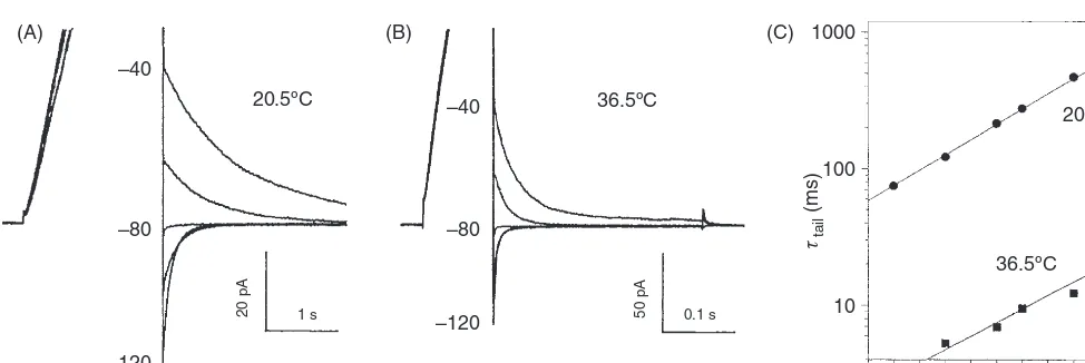
Figure 11 Proton channel gating is strongly temperature dependent. (A, B) In a human neutrophil at pHo 7.0, pHi5.5 at the indicated
temperatures, a depolarizing prepulse activated thegH, then the voltage was stepped to test voltages, illustrated in 20-mV increments. Note
the change in time base. The “tail current” (deactivation or closing) time constant,τtail, was obtained by fitting a single decaying exponential
to each current. TheQ10was identical at all voltages (C) and was 8.5 in this cell, corresponding to an activation energy of approximately
38 kcal/mol. [Adapted, with permission, from reference (67)©DeCoursey & Cherny, 1998. Originally published in The Journal of General Physiology.]
gating transition during opening, with a reverse conforma-tional change in only one subunit being sufficient to close the channel (67). This class of mechanism again resembles the classical Hodgkin-Huxley model for Na+and K+channel gating. The revelation that in many species, the proton channel functions as a dimer in which both protomers must undergo an opening conformational change before either can conduct (103, 199, 262) is in excellent agreement with the predictions that were based on temperature effects on gating kinetics.
Another clue to the gating mechanism comes from deu-terium isotope effects. In most respects, proton channels func-tioned similarly in normal and heavy water. However, chan-nel opening was 3-fold slower in deuterium, whereas chanchan-nel closing was at most weakly affected (66). Because deuterons bind with higher affinity to most titratable groups, the slowing of activation by deuterium is consistent with deprotonation being a rate-determining step in channel opening. A 3-fold slowing is consistent with a 0.5 unit increase in the pKaof the group in D2O (66), which in turn is more consistent with a car-boxylic or ammonium group being involved than a sulfhydryl acid (66, 238). Intriguingly, and perhaps coincidentally, both pHo and D2O have large effects onτact and small effects on
τtail. Deprotonation of hypothetical groups accessible to the internal solutions was proposed to be a prerequisite step in channel opening in a model proposed to explain the depen-dence of the gH-V relationship on both voltage and the pH gradient,pH (38). In this model, the protonation state of titratable groups accessible to one side of the membrane or the other regulates proton channel gating (38).
Given their structural parallels (Fig. 2), it is tempting to assume that gating of HV1 will entail the same outward move-ment of the S4 domain that is thought to occur in the VSD of K+and Na+channels (131, 147, 161, 253, 275, 276). Indeed, Cys scanning supports the general similarity of S4 movement in CiHV1 to that of other VSDs (103), as do paddle motif
swaps (7). However, a remarkable study of mHV1 revealed that mutants in which the entire C terminus was truncated along with S4 as far as between the second and third Arg residues in the S4 domain still retained proton channel func-tion (230). Although each of the three S4 Arg residues in HV1 contributes to voltage sensing (104), their movement must not be as extensive as in K+channels, unless the movement in the C truncated mutant was reduced compared to the WT channel. The charged residues in S4 of the Shaker K+channel VSD are proposed to move through a “gating charge transfer center” (258); the first (R1) is in this center in closed channels, but the fifth (K5) resides there in the open state (157).
At present, most fundamental questions regarding gating remain unanswered or incompletely answered. What is the structural difference between open and closed proton chan-nels? How does the molecule move during gating? How do the two protomers interact during gating in the dimer? How does pH regulate the voltage-dependence of gating? Although recent discoveries resulting from structure-function studies seem to have complicated rather than clarified these ques-tions, the voltage-gated proton channel continues to be an ex-ceptional system in which to explore fundamental questions about voltage gating mechanisms.
Inhibition of proton currents by Zn
2+is
surprisingly revealing
Essentially every study that has reported the existence of pro-ton currents in a new cell type has used inhibition by Zn2+ as a defining feature. Occasionally, Cd2+or other polyvalent metals are included; a large number have generally similar ef-fects, including Cu2+, Ni2+, Co2+, Mn2+, Be2+, Pb2+, Hg2+, La3+, Gd3+, and Al3+, with Zn2+, Cu2+, and La3+
pHo 7.0
200 pA
1 s
200 pA
1 s
20 pA
2 s
pHo 6.0
pHo 5.0
1 μmol/L ZnCl2 10 μmol/L ZnCl2
10 μmol/L ZnCl2 100 μmol/L ZnCl2
[image:15.684.40.278.48.365.2]100 μmol/L ZnCl2 1000 μmol/L ZnCl2
Figure 12 Modulation of Zn2+ effects on proton currents by pHo
reveals strong competition between Zn2+ and H+for external
bind-ing sites. Identical families of pulses were applied in each row at the indicated pHo and Zn2+concentrations. Zn2+slows activation of
the proton current at a given voltage and shifts thegH-Vrelationship
positively. [Adapted, with permission, from reference (36)©Cherny & DeCoursey, 1999. Originally published in The Journal of General Physiology.]
physiological effects of proton channel function in cells. The advent of the HVCN1 knockout mouse has provided an alter-native to this classical pharmacological lesion experiment. As more homologs of proton channels are identified and charac-terized, some have been discovered to be relatively insensitive to inhibition by Zn2+. The proton channel of the sea squirt, C.
intestinalis, can be inhibited by Zn2+, but only at concentra-tions 27-fold higher than the mouse channel (234). A proton channel in E. huxleyi, a marine phytoplankton, is also only weakly inhibited by Zn2+(259), as is kH
V1, a dinoflagellate channel (unpublished data with S.M.E. Smith, D. Morgan, B. Musset, V.V. Cherny, A.R. Place, and J.W. Hastings). Thus, there is a hint of a pattern of weaker metal sensitivity for proton channels in marine than terrestrial life. However, pro-ton currents in the nudibranch mollusc, Tripro-tonia diomedea, appear to exhibit high Zn2+ sensitivity (271), as do proton currents in C. pelagicus, a marine coccolithophore (259). In any case, the idea that lower Zn2+sensitivity in marine species
might be a protective adaptation to the heavy metal content of seawater seems unlikely, because the metal concentrations
are too low to produce much inhibition of even the more sen-sitive human proton channel: 76 nmol/L Zn2+, 112 nmol/L Ni2+, 14 nmol/L Cu2+, 1 nmol/L Cd2+, and 21 pmol/L La3+ (263). Low sensitivity to metal will become more important as the seas become polluted, but at present there seems to be a reasonable safety factor.
The effects of polyvalent cations on voltage-gated pro-ton channels resemble the effects of these metals on most voltage-gated ion channels. Metal ions slow channel opening, shift the g-V relationship positively, and may reduce the max-imum conductance (36, 89, 119). As illustrated in Figure 12, although Zn2+appears to make the current smaller, most of this is the result of slower activation kinetics (largerτact) and a positive shift of the gH-V relationship. Both of these effects were quantified over a range of pHoand the pattern was evalu-ated using several possible models of competition. Ironically, despite the widespread use of Zn2+to characterize proton cur-rents, one of the most remarkable features of the inhibition of proton currents by Zn2+, namely, the strong modulation of Zn2+effects by pH, was overlooked until 17 years after the first report of voltage-gated proton channels (36). To a first approximation, the inhibition produced by 1μmol/L Zn2+at pHo 7.0 requires 10μmol/L Zn2+ at pHo 6.0, and approxi-mately 1000μmol/L Zn2+at pHo5.0 (Fig. 12). The antago-nism between low pH and Zn2+suggests that H+and Zn2+
complete for a binding site on the proton channel molecule. Figure 13 illustrates the predictions of the preferred model in which Zn2+ binds competitively with H+ at three sites with pKa6.3, a metal-binding constant pKM 6.5, and a
pKa 6.3
pHo 8 10
9 8 7 6 5
4
3
2
1 0.9 0.8
0.01 0.1 1 10
[ZnCl2] corrected for buffer binding (μmol/L)
τact
(ZnCl
2
)/
τact
100 1000 10000
7 6 5.5 5
pKM 6.5 a 0.03
Figure 13 The pHo dependence of the slowing of proton current
activation can be explained if Zn2+binds to a site consisting of three
groups withpKa6.3, with affinity 316 nmol/L, and cooperativity factor
a=0.03 (see text). All curves were drawn with these values and no other adjustable parameters. Adequate fits were also obtained by assuming two titratable groups, but not with only one. At high pHocompetition
with protons disappears and limits to simple metal binding. See text for further details. [Adapted, with permission, from reference (36)©
[image:15.684.298.538.424.609.2]cooperativity factor of 0.03, meaning that Zn2+can still bind if one of the three groups is protonated, but with approxi-mately 30-fold lower affinity. There were no other adjustable parameters in the model, which simply assumed that the proton channel cannot open when Zn2+ is bound (36). Two other, even simpler models also provided good fits to the data; these had two or three titratable groups that coordinated Zn2+ strictly competitively with protons. In contrast, simple 1:1 competition could not explain the data, because the effectiveness of Zn2+ decreases by a maximum
of 10-fold/unit pH in such a model, and the data clearly indicated a 100-fold shift between pHo6 and pHo5 (Figures 12 and 13). The outcome of this exercise was a prediction that Zn2+inhibits proton currents by binding simultaneously to at least two groups with pKa6-7, in the range of His (36). This prediction appeared to be borne out nicely when the human proton channel was identified 7 years later. Ramsey and colleagues (224) showed that two His residues, His140 and His193, that were expected to be accessible to the external solution, both contributed to Zn2+inhibition.
To our consternation, a homology model of the human proton channel structure, based on the crystal structure of the VSD from K+channels, indicated that the two Zn2+-binding His residues, His140 and His193, were approximately 14 ˚A apart, too far to plausibly coordinate a single Zn2+atom (199).
Another homology model predicted a comparable separation between the two His residues (223). However, the discovery that the proton channel was a dimer (139,150,261), presented a possible explanation. As shown in Figure 5B, if the dimer adopts a position with the complementary His residues fac-ing each other, Zn2+ could bind at the interface between the protomers. This hypothesis was explored by creation of a se-ries of channels with mutations to His140and His193as well as tandem dimers. Figure 14A and B illustrates the generally similar pattern observed for Zn2+shifting the gH-V relation-ship and slowing activation, respectively. Mutation of either His140 (H140A) or His193 (H193A) individually attenuated Zn2+ effects, indicating that both residues contribute to the effects observed in WT channels. H140A, H193A and the tandem dimer H193A-H140A all exhibited distinct Zn2+
ef-fects that were weaker than in WT. Remarkably, the tandem dimer WT-H140A/H193A was as insensitive to Zn2+ as the double mutant that lacked both His (H140A/H193A). In par-ticular, the slowing effect of Zn2+occurs only when there is at least one His in each protomer, and is absent when both His are present in the same protomer. This pattern suggests that Zn2+slows proton channel opening when it binds at the interface between protomers. Presumably, the Zn2+-bound channel dimer cannot undergo a conformational change re-quired for opening.
Functions
As discussed previously (see Section “Regulation of gating by the pH gradient,pH”), the properties of the voltage-gated
proton channel suggest that in most species, this molecule is ideally suited to solve “the central problem of pHiregulation: the neutralization of intracellular acid derived from a variety of sources” (228). Proton channels have been shown to con-tribute to pHi recovery following an acid load in a variety of cells: snail neurons (260), rabbit osteoclasts (211), human neutrophils (75), mouse mast cells (145), rat microglia (187), a human bronchial epithelial cell line (16HBE14o-) (264), rat alveolar epithelial cells (192), rat hippocampal neurons (35, 246), murine osteoclasts (183), TsA201 cells transfected with the mouse proton channel gene mVSOP (234), and in a rat microglial cell line GMI-R1 (185). In many other cells, proton channels extrude acid under specific conditions that occur physiologically. Hypotonic shock leads to depolariza-tion and alkalinizadepolariza-tion in bovine articular chondrocytes that may be mediated by proton channels (231, 232). Roles in reg-ulating both pHiand membrane potential have been proposed in human cardiac fibroblasts (87). Although acid extrusion remains the main general function of proton channels, several quite different functions have been identified in a number of cells, which are described in the following Sections.
Bioluminescence and other functions
in dinoflagellates
As mentioned previously (Fig. 1), the first explicit proposal of a voltage-gated proton channel was in 1972 by Fogel and Hast-ings in the bioluminescent dinoflagellate, G. polyedra (96). It had been shown previously that current injection into the large central vacuole of another bioluminescent dinoflagellate,
Noc-tiluca scintillans (N. miliaris), elicited an all-or-nothing action
(A) WT H140A H193A H140A/H193A WT-WT tandem H193A-H140A tandem WT-H140A/H193A tandem
WT H140A H193A H140A/H193A WT-WT tandem H193A-H140A tandem WT-H140A/H193A tandem No effect
WT dimer
WT-WT tandem
H140A
H193A-H140A tandem
H193A
WT-H140A/H193A tandem
H140A/H193A
WT dimer
WT-WT tandem
H140A
H193A-H140A tandem H193A
WT-H140A/H193A tandem H140A/H193A
80 100
60
Vthreshold
shift (mV)
τact
(Zn
2+
)/
τact
40
20
0
20
15
10
5
0
1 10
[Zn2+] (μmol/L)
100 1000
1 10
[Zn2+] (μmol/L)
[image:17.684.109.464.52.509.2]100 1000
Figure 14 Effects of Zn2+ on Vthreshold (A) and on the slowing of proton current activation (B) in
constructs with several His mutations. Dimeric proton channels are illustrated with diagrams in which solid symbols indicate His and open symbols indicate Ala substituted for His. Three tandem dimers are shown with their C- and N-termini linked to constrain the His positions in the dimer.Vthreshold
shifts were estimated fromgH-Vrelationships plotted semilogarithmically. Statistical comparisons are
to WT channel parameters ( P<0.05, *P<0.01). [Adapted, with permission, from reference (199). Originally published in The Journal of Physiology 588:1435-1449, 2010.]
(25, 95, 141). Second, luciferase has a pH optimum at pH 6.3, and is inactive at pH 7.5 (141, 237). It seems very likely that voltage-gated proton channels play a crucial role in trigger-ing bioluminescence in dinoflagellates, by orchestrattrigger-ing the concerted activation of both mechanisms.
A further, more spectacular role has been speculated to exist, namely, mediating the action potential itself (209). The flash-triggering action potential coincides with decreased impedance, indicating that a conductance increase is respon-sible (83). The peak of the action potential is not affected
Cytoplasmic membrane
Schematic scintillons pH triggering of flash
Cytoplasm neutral pH Theca
Action Potential triggers proton flux
H+
Vacuole low pH
[image:18.684.88.231.53.339.2]Vacuole membrane
Figure 15 Proposed mechanism by which proton flux through voltage-gated proton channels triggers light flashes in dinoflagellates. Mechanical stimulation elicits an action potential conducted along the tonoplast, the membrane separating the central vacuole and a thin layer of cytoplasm (gray). When the action potential invades the scintil-lons (grape-like structures formed by evagination of tonoplast), proton flux from the vacuole at low pH into the scintillon activates luciferase, triggering the flash. [Adapted, with permission, from reference (110). Originally published in Hastings JW. Bioluminescence. In:Cell Physiol-ogy Sourcebook: A Molecular Approach(3rd ed.), Sperelakis N, editor. San Diego: Academic Press, 2001, p. 1115-1131.]
channels in other species, including obeying the Rule of Forty [Eq. (4)], it differs in the absolute position of the gH-V rela-tionship. As illustrated in Figure 10, at any givenpH, the kHV1 channel opens at voltages 60 mV more negative than in all other species (251). Consequently, there is a substantial voltage range within which inward H+current occurs. These properties are reminiscent of voltage-gated Na+channels and strongly suggest the ability of these channels to mediate an action potential. In any case, the negative voltage range of activation of kHV1 indicates a stark difference in function, compared with all other known proton channels. Whereas other proton channels are designed to allow only proton ex-trusion; kHV1 activation will produce H+ influx. H+ influx could initiate or propagate action potentials, or produce a drop in pHithat might have signaling functions. A role of H+flux into the cytoplasm was proposed to trigger tentacle flexion in
N. miliaris (212). As originally proposed (96), HV1-mediated H+flux from the vacuole into the scintillon triggers the flash (251) (Fig. 15).
A variety of functions might exist for proton channels in nonbioluminescent dinoflagellate species. For example, in mixotrophic species like K. veneficum, H+ flux might be in-volved in capturing prey by extrusion of trichocysts or prey di-gestion. A phylogenetic analysis of VSD regions from several dozen HV1 sequences revealed high sequence diversity among the single-celled species and among invertebrates (Fig. 3). Although this observation may partially reflect the genetic liability of dinoflagellates and other unicellular species (11), diverse sequences could well produce other novel functions of HV1. As in multicellular organisms, ion channels in di-noflagellates undoubtedly play a variety of roles in regulating basic life functions, which makes them excellent targets for controlling dinoflagellate populations and behavior.
Calcification by coccolithophores
New proton channel genes were recently identified in two ma-rine unicellular algae, the coccolithophores E. huxleyi and C.
pelagicus ssp braarudii (259). The properties of these
chan-nels in heterologous expression systems were quite similar to those of other species, with the exception that the E. huxleyi channel (EhHV1) was only weakly inhibited by Zn2+. Coc-colithophores are abundant phytoplankton that play a major role in calcification; that is, the conversion of HCO3−(derived from CO2 dissolved in ocean water) into calcium carbonate, CaCO3 via the intermediate reaction HCO3− → CO32− + H+. Each Ca2+ that is converted into CaCO
3 generates a proton, which must be eliminated from the cytosol of the coc-colithophore. Inhibiting the Zn2+-sensitive CpH
V1 with 30
μmol/L Zn2+produced an immediate drop in pH
iand a sus-tained decrease in calcite production (259). Decreasing pHo lowers pHi, suggesting that ocean acidification due to disso-lution of atmospheric CO2 derived from human activity (or, for those who harbor doubts as to its source, by magic) could inhibit calcification directly. However, a stronger correlation was found between coccolith mass and [CO32−] than with pH or pCO2, indicating complex control (21). Nevertheless, since the Last Glacial Maximum (ice age), ocean CO2levels have increased by approximately 50% (from∼190 to∼280 parts per million), and coccolith mass has decreased by almost 50% (from 13.6 to 7.5 pg) (21).