DESIGN AND DEVELOPMENT OF LIPOSOME BY
MICROENCAPSULATION VESICLE METHOD AND VAGINAL
DELIVERY SYSTEM FOR VORICONAZOLE GEL
P. Ravindra*, Dr. P. Arun, P. Shailendra, P. Bhaveshand D. Neelesh
Department of Pharmacy, Shri Ram Group of Institution, ITI Madhotal Jabalpur-482002.
1. ABSTRACT
The present investigation constitutes the design and development of
liposome by microencapsulation vesicle method and vaginal delivery
system for voriconazole and its in-vitro diffusion & ex-vivo membrane
permeation study. Amount of polymer and the number of sonication
cycle was varied in different batches of formulations. Size, size
distribution, surface charge, entrapment efficiency and drug content
were studied for each formulation batch. Optimization of formulation
and process parameter resulted in the production of Voriconazole
loaded liposomal suspension with particle size distribution ranging
between 130 to 240 nm and entrapment efficiency around 55%. In-
vitro diffusion characterization using dialysis membrane was carried out to evaluate the
release characteristics of the drug from optimized liposomal gel formulations with respect to
the pure drug in gel. An increase in the cumulative release of the drug (1.160 mg) is observed
in comparison to the pure drug (0.743 mg) at specified time. In case of ex-vivo permeation
study on vaginal membrane of the goat, supported the in vitro results.
2. KEYWORDS: Liposome, microencapsulation, voriconazole, in-vitro diffusion, polymer.
3. INTRODUCTION
Drugs, in the form of vegetation and minerals. Have existed longer than man himself. Human
disease and man sinstict to survive have, through the ages led to their discovery.[1]
Pharmaceuticals knowledge has grown exponentially over the years. We now have a much
clearer understanding of how drugs are absorbed into, distributed within, and cleared from
the body.
Volume 8, Issue 9, 1358-1367. Research Article ISSN 2277– 7105
Article Received on 13 June 2019,
Revised on 03 July 2019, Accepted on 24 July 2019
DOI: 10.20959/wjpr20199-15552
*Corresponding Author
P. Ravindra
Department of Pharmacy,
Shri Ram Group of
Institution, ITI Madhotal
Development of new drug requires much of research a long development time and also
coordinated team effort of a large group of researcher in various fields. Instead of searching
for new drugs using random hits or miss approach the development of superior drug delivery
system which enhances the therapeutic efficacy of conventional drugs by controlling the
release rate or targeting to the tissue site may be an effective approach to improve the
efficacy of chemotherapeutic agents. Now a day, much of the research work is focused on
development of controlled sustained release medications for enhancement of clinical
efficacy.[7]
A more advanced version of controlled delivery is the targeted delivery. Targeted delivery
exhibit all the advantages of the controlled delivery and at the same time facilitates delivery
of the drug to the target site.[2]
In the era of the novel drug delivery system (NDDS) emphasis is given on spatial placement
of the drug for chronic condition. Targeted delivery of anti-fungal appears to be a challenging
but achievable task with the use of novel drug delivery system. Different novel approaches
used for delivering these drugs include liposome, microsphere, nanotechnology,
micro-emulsion, antibody-loaded drug delivery, magnetic microcapsule, implantable pumps,
noisome.[8]
1. Although opportunities to develop liposome-based efficient drug delivery system extend
into all therapeutic classes of pharmaceuticals, the development of effective treatment
modalities for the anti-fungal infection remains a financially and therapeutically
significantly need
4. MATERIAL SPECIFICATIONS
MATERIAL MANUFACTURER
Voriconazole : Jubilant Organosys, Noida, UP
Soya lecithin : Himedia laboratories pvt.ltd., Vadhani, Mumbai
Chloroform : Qualigens fine chemicals, GSK Pharmaceutical,
Mumbai
Double Distilled Water : Double Distillation Unit, Borosil
Cholesterol : Burgoyne urbidges & co. Mumbai
Carbopol 934 : Himedia laboratories pvt. ltd., Vadhani, Mumbai
Potassium dihydrogen phosphate
:
Merck Specialties pvt ltd. Worli, Mumbai.
Propylene glycol : Qualigens fine chemicals, GSK Pharmaceutical,
5. Method Development for Preparation of Voriconazole loaded Liposomal suspension
The micro encapsulation vesicle (MCV) method is a liposome preparation technique that
reproducibly produces liposomes with homogeneous particle sizes with high encapsulation
efficiency. Liposomes encapsulating water-soluble drugs, lipophilic drugs and an amphiphilic
drug were prepared by the MCV method and the encapsulation efficiency of the drugs was
examined.[3]
With the MCV method, theoretically the initial drug-containing water phase was always
separated from the dispersion medium by the lecithin-containing oil phase, which was
advantageous to maintaining a higher encapsulation efficiency of the water-soluble drug.
The encapsulation efficiency of these drugs strongly correlated to the log P octanol/water and
also tended to correlate to the log P chloroform/water for the order of the log P
chloroform/water was almost the same as the order of the log P octanol/water in the drugs
examined. As far as the results of this study, the log P octanol/water was considered to be a
better indicator of the encapsulation efficiency of a drug in the MCV method.[4]
Factors affecting the encapsulation efficiency of the drug in the liposomes are various and
come from the properties of both the liposomes and encapsulated drugs. Concerning the
encapsulated drugs, the encapsulation efficiency is affected by hydrophilic or lipophilic
properties and tended to interact with the membrane bilayer.[6] As for the liposome properties,
aqueous volume, membrane rigidity, surface area and preparation methods are reported to
have influenced the encapsulation efficiency.
The micro encapsulation vesicle (MCV) method is a preparation technique in which
liposomes are formed through a two-step emulsification and dispersion with mechanical
agitation. We have previously reported that the technique reproducibly produced liposomes
with homogeneous particle sizes, a high encapsulation efficiency and good stability in
preparations with purified soya lecithin.[5] The MCV method is expected to have advantages
in preparing drug-loaded liposomes, for a drug can theoretically be encapsulated easily if it
dissolves either in a water phase or an oil phase. A drug solution, regardless of the water
phase or the oil phase, forms a w/o emulsion first, which then forms a w/o/w emulsion and
finally the drug-loaded liposomes are generated. The encapsulation efficiency of drugs with
this study, water-in soluble drugs, one being a lipophilic compound was studied to examine
the encapsulation efficiency of the liposomes prepared by the MCV method.
5.1 METHOD OF PREPARATION OF LIPOSOMAL SUSPENSION
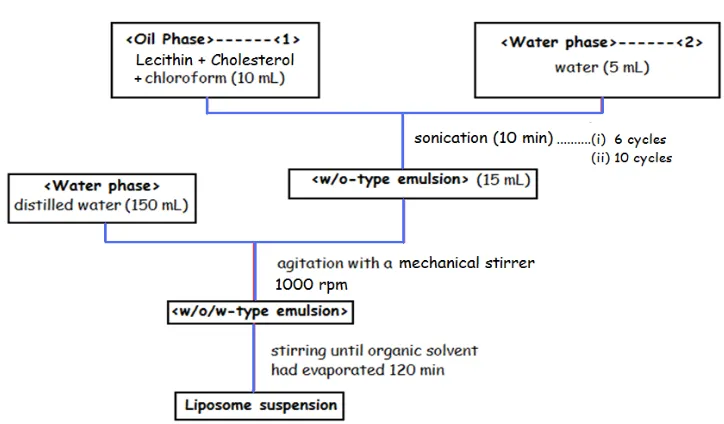
Fig.1: Schematic Representation of Preparation of Voriconazole loaded Liposomal
suspension by MCV method.
Liposomal suspension containing anti-fungal drug, voriconazole can be prepared by
micro-encapsulation vesicle method. In this method, drug, lecithin and cholesterol was dissolved in
10 ml of chloroform. Then to this oil phase 5 ml of water was added and sonicated for 10
minutes to form 15 ml of water-in-oil (w/o) emulsion which is white milky in colour, it
shows MLV. To this w/o emulsion 150 ml of distilled water was added and agitated using
mechanical stirrer at 1000 rpm in room temperature. This results into formation of
water-in-oil-in-water (w/o/w) emulsion which was transparent blue – grey colour, it shows SUV.
Agitation by mechanical stirrer at 1000 rpm for 120 min results in evaporation of the organic
solvent, chloroform thus, forming liposome suspension.
Table No.1:Batch Specification of VZ Liposomal Suspension.
Formulation code
PC:CH ratio
Amount of PC (mg)
Amount of PC (mg)
Amount of Drug (mg)
Sonication cycles
LP 1 9:1 180 20 20 6
LP 2 10:1 200 20 20 6
LP 3 12:1 220 20 20 6
LP 4 9:1 180 20 20 10
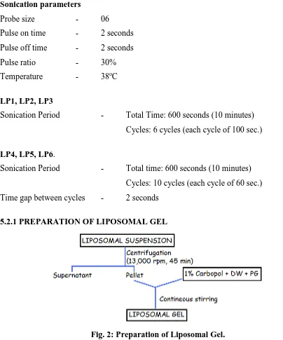
[image:4.595.116.478.144.356.2]Sonication parameters
Probe size - 06
Pulse on time - 2 seconds
Pulse off time - 2 seconds
Pulse ratio - 30%
Temperature - 38oC
LP1, LP2, LP3
Sonication Period - Total Time: 600 seconds (10 minutes)
Cycles: 6 cycles (each cycle of 100 sec.)
LP4, LP5, LP6.
Sonication Period - Total time: 600 seconds (10 minutes)
Cycles: 10 cycles (each cycle of 60 sec.)
Time gap between cycles - 2 seconds
[image:5.595.72.476.73.558.2]5.2.1 PREPARATION OF LIPOSOMAL GEL
Fig. 2: Preparation of Liposomal Gel.
5.2 Characterization Process of Voriconazole Loaded Liposomal suspension.
Parameters Characterization Methods
Vesicle size determination Malvern Zetasizer Surface and Internal Morphology SEM, TEM
Charge determination Zeta Potentiometer
Polydispersity index Malvern Zetasizer
Vesicle size and surface charge determination
The average diameter of the vesicles and their Zeta potential were determined using a
Zetamaster apparatus (Malvern Instruments, Malvern, UK) at a temperature of 25 ± 0.1o C.
liposome suspensions were suitably diluted with distilled water in order to avoid
multi-scattering phenomena. Microscopic observations allowed exclusion of drug crystallization
phenomena as a consequence of water dilution of the samples. The intensity of the laser light
scattered by the samples was detected at an angle of 900 with a photomultiplier. For each kind
of liposomal suspension, five independent samples were taken, each of which was measured
at least twice, up to four times. For each specimen, 10 autocorrelation functions were
analyzed using a cumulative analysis. From this analysis, the z-average value was obtained,
which is an approximation of the diameter of the liposomes. The polydispersity index was
used as a measurement of the width of the size distribution.
For liposome surface charge determinations, about 2 mL of each liposomal suspension,
suitably diluted with distilled water, was dropped into the Zetamaster electrophoretic cell and
the Zeta-potential was determined by Electrophoretic Mobility (l) measurements. The
mobility l was converted into a Zeta-potential by the Smoluchowski equation Z =µη/ε, where
6. RESULTS AND DISCUSSION
Table No. 2: Zeta potential, Average size, PDI and % Entrapment of all liposomal suspensions. Formulation code Zeta potential (mV)
Z – avg.
size (nm) PDI
%Entrapment efficiency (mean ± SD, n = 3)
LP 1 -48.0 183.6 0.551 53.303 ± 1.709
LP 2 -53.2 206.3 0.539 47.666 ± 1.900
LP 3 -44.3 134.5 0.469 36.233 ± 0.802
LP 4 -48 228 0.645 53.433 ± 3.023
LP 5 -44.7 238.3 0.655 52.533 ± 1.096
LP 6 -38 175.8 0.505 31.533 ± 1.331
6.2 DRUG CONTENT
Table No. 3: Drug content of optimized formulations.
FORMULATION DRUG CONTENT
LP 1- 9:1 (6) 3.4 ± 0.213 LP 2- 10:1 (6) 6.2 ± 0.405 LP 4- 9:1 (10) 1.5 ± 0.435 LP 5- 10:1 (10) 4.4 ± 0.374
It is clearly seen from the above Table No. 7 that the formulations containing 10:1 polymer:
drug ratio gives higher drug content than their corresponding 9:1, polymer: drug ratio
formulation. This may be because of the higher average sizes showed by the formulation LP
2 and LP 5.
7. CONCLUSION
In conclusion, Voriconazole loaded liposomal gel for vaginitis was prepared by
microencapsulation vesicle method varying the polymer amount and the number of
sonication cycle. Influence of both the formulation and process parameters in formulation of
Voriconazole loaded liposomal gel was characterized with respect to the size, size
distribution, surface charge, entrapment efficiency and drug content. In-vitro and ex-vivo
characterization was carried out to evaluate the release characteristics of the drug from
liposomal gel with respect to the pure drug. Increase in lipid concentration in liposomal gel
was able to control the release of the active for longer period of time, which shows the
sustained release behavior of formulation.
Future scope
Further, thixotropic behavior of the liposomal gel and in-vivo studies in animal models and
stability studies are needed to prove the enhanced bioavailability of Voriconazole loaded
8. REFERENCES
1. Howard & Ansel, Nicholas G Popovich [edn], In Pharmaceutical dosage form & drug
delivery, 5th Edn, 1990, Philadelphia.
2. Chien Y W in Transdermal controlled release drug administration in Novel Drug Delivery
System., fundamentals, Developmental concepts & Biomedical application, 1982 Marcel
Dekker, New York, 5.
3. Scheuplin. Rj, Mechanism of percutaneous absorption route of penetration & influence of
solubility J. Invest dermatol, 1965; 45: 334.
4. Li, V.H.K., Robinson, J.R and Lee, V.H.L., In; Controlled Drug Delivery: Fundamentals
and Applications, 2nd Edn., Vol29, Marcel Dekker, Inc., NY, 1987; 7.
5. Goldberg, E. P. Eds., In; Targeted Drugs, 2nd Edn., Wiley, New York, 1983; 312.
6. Riaz.M review article on Liposomes Preparation Methods, Pakistan Journal of
Pharmaceutical Sciences, January 1996; 19(1): 65-77.
7. Khar RK, Vyas SP. Targeted and Controlled drug delivery novel carrier systems. 1st ed.
New Delhi; CBS Publishers and Distributors, 2002; 384.
8. Vemuri. S Rhodes. C.T., Preparation and Characterisation of Liposomes as therapeutic