Acta Cryst.(2001). E57, o719±o720 DOI: 10.1107/S1600536801011424 George and Nangia C6H7N3O
o719
organic papers
Acta Crystallographica Section E Structure Reports Online
ISSN 1600-5368
N
-(3-Pyridyl)urea
Sumod George and Ashwini Nangia*
School of Chemistry, University of Hyderabad, Hyderabad 500 046, India
Correspondence e-mail: [email protected]
Key indicators Single-crystal X-ray study T= 293 K
Mean(C±C) = 0.002 AÊ Rfactor = 0.045 wRfactor = 0.134
Data-to-parameter ratio = 15.4
For details of how these key indicators were automatically derived from the article, see http://journals.iucr.org/e.
#2001 International Union of Crystallography Printed in Great Britain ± all rights reserved
The crystal structure of the title compound, C6H7N3O, exhibits
packing typical of amides, with NÐH O hydrogen-bond dimers forming a corrugated tape and NÐH N bonds connecting the tapes.
Comment
In a recent monograph on the role of amides in non-covalent syntheses (Palmore & MacDonald, 2000), the urea functional group is considered as part of the amide family. In connection with an ongoing crystallographic study of aromatic ureas (Georgeet al., 2001), we have determined the crystal structure ofN-(3-pyridyl) urea, (I).
The pyridyl ring in (I) is tilted to the urea plane by 163.94 (14)(C8/N7/C6/C5, Fig. 1). The crystal structure of (I)
contains a corrugated tape of (syn) N10ÐH8 O9 and (anti) N10ÐH9 O9 hydrogen-bond dimers along the aaxis, with an (anti) N7ÐH7 N4 bond connecting screw-axis-related molecules in theb direction (synandantirefer to ureido H atoms) (Fig. 2). Theaaxis of 4.8558 (10) AÊ in (I) is smaller than the characteristic 5.1 AÊ packing in carboxylic amides because of the zigzag corrugated pattern. A crystal structure assembled by NÐH N hydrogen bonding interaction was published recently in this journal (Lynch & McClenaghan, 2001). In contrast to the structure of (I), the molecular conformation and crystal packing inN-(2-pyridyl)urea
(Veli-kova et al., 1997) are quite different. This latter structure
contains parallel zigzag ribbons of molecules hydrogen bonded through (syn) NÐH O dimers along the ab diag-onal. The molecule adopts a planar conformation because of an intramolecular (anti) NÐH N interaction. Thus, isomeric 2- and 3-pyridylurea have very different crystal structures.
Experimental
Compound (I) was synthesized by slowly adding NaOCN (130 mg, 2 mmol) dissolved in hot water (1 ml) to a solution of 3-amino-pyridine (188 mg, 2 mmol) in glacial AcOH (0.2 ml) with stirring. The aqueous solution was neutralized and extracted with chloroform to remove the unreacted 3-aminopyridine. Column chromatography and recrystallization from EtOAc afforded crystals of (I) (m.p. 472± 474 K).
Crystal data C6H7N3O
Mr= 137.15
Monoclinic,P21/n
a= 4.8558 (10) AÊ b= 8.1621 (16) AÊ c= 15.919 (3) AÊ
= 92.16 (3)
V= 630.5 (2) AÊ3
Z= 4
Dx= 1.445 Mg mÿ3
MoKradiation Cell parameters from 25
re¯ections
= 9.0±10.6
= 0.10 mmÿ1
T= 293 (2) K Cube, colourless 0.120.110.10 mm Data collection
Enraf±Nonius CAD-4 diffractometer
!scans
2021 measured re¯ections 1835 independent re¯ections 1362 re¯ections withI> 2(I) Rint= 0.010
max= 30.0
h= 0!6 k= 0!11 l=ÿ22!22 3 standard re¯ections
every 150 re¯ections frequency: 90 min intensity decay: none
Re®nement Re®nement onF2
R[F2> 2(F2)] = 0.045
wR(F2) = 0.134
S= 1.08 1835 re¯ections 119 parameters
All H-atom parameters re®ned
w= 1/[2(F
o2) + (0.0665P)2
+ 0.1436P]
whereP= (Fo2+ 2Fc2)/3
(/)max= 0.019
max= 0.25 e AÊÿ3
min=ÿ0.25 e AÊÿ3
Table 1
Hydrogen-bonding geometry (AÊ,).
DÐH A DÐH H A D A DÐH A
N7ÐH7 N4i 0.888 (19) 2.156 (18) 2.984 (2) 155.0 (16)
N10ÐH8 O9ii 0.92 (2) 2.05 (2) 2.9657 (19) 173.0 (18)
N10ÐH9 O9iii 0.842 (19) 2.193 (19) 2.9734 (19) 154.2 (18)
C2ÐH2 O9iv 0.97 (2) 2.67 (2) 3.629 (2) 168.4 (16)
Symmetry codes: (i) ÿ1
2ÿx;yÿ12;32ÿz; (ii) ÿx;1ÿy;2ÿz; (iii) xÿ1;y;z; (iv) 1ÿx;2ÿy;2ÿz.
Data collection: CAD-4 Software (Enraf±Nonius, 1989); cell re®nement: CAD-4 Software; data reduction: Xtal3.5 (Hall et al., 1995); program(s) used to solve structure: SHELXS97 (Sheldrick, 1990); program(s) used to re®ne structure:SHELXL97 (Sheldrick, 1997); molecular graphics: PLUTON-(C) (Spek, 1979±1997); soft-ware used to prepare material for publication:SHELXL97.
The authors thank the DST for funding the X-ray diffractometer facility.
References
Enraf±Nonius (1989).CAD-4Software. Version 5.0. Enraf±Nonius, Delft, The Netherlands.
George, S., Nangia, A. & Lynch, V. M. (2001).Acta Cryst.C57, 777±778. Hall, S. R., Flack, H. D. & Stewart, J. M. (1995). Editors.Xtal3.5Reference
Manual. Universities of Western Australia, Australia, Geneva, Switzerland, and Maryland, USA.
Johnson, C. K. (1976).ORTEPII. Report ORNL-5138. Oak Ridge National Laboratory, Tennessee, USA.
Lynch, D. E. & McClenaghan, I. (2001).Acta Cryst.E57, o4-o5.
Palmore, G. T. R. & MacDonald, J. C. (2000).The Amide Linkage: Selected Structural Aspects in Chemistry,Biochemistry and Material Science, edited by A. Greenberg, C. M. Breneman & J. F. Liebman, pp. 291±336. Chichester: John Wiley.
Sheldrick, G. M. (1990).Acta Cryst.A46, 467±473.
Sheldrick, G. M. (1997).SHELXL97. University of GoÈttingen, Germany. Spek, A. L. (1979±1997).PLUTON-(C). Utrecht University, The Netherlands. Velikova, V., Angelova, O. & Kossev, K. (1997).Acta Cryst.C53, 1273±1275. Figure 1
ORTEPII (Johnson, 1976) plot of (I) with 50% probability ellipsoids.
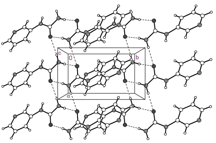
Figure 2
supporting information
sup-1
Acta Cryst. (2001). E57, o719–o720
supporting information
Acta Cryst. (2001). E57, o719–o720 [doi:10.1107/S1600536801011424]
N
-(3-Pyridyl)urea
Sumod George and Ashwini Nangia
S1. Comment
In a recent monograph on the role of amides in non-covalent syntheses (Palmore & MacDonald, 2000), the urea
functional group is considered as part of the amide family. In connection with an ongoing crystallographic study of
aromatic ureas (George et al., 2001), we have determined the crystal structure of N-(3-pyridyl) urea, (I).
The pyridyl ring in (I) is tilted to the urea plane by 163.94 (14)° (C8/N7/C6/C5, Fig.1). The crystal structure of (I)
contains a corrugated tape of (syn) N10—H8···O9 and (anti) N10—H9···O9 hydrogen-bond dimers along the a axis, with
an (anti) N7—H7···N4 bond connecting screw-axis-related molecules in the b direction (syn and anti refer to ureido H
atoms) (Fig. 2). The a axis of 4.8558 (10) Å in (I) is smaller than the characteristic 5.1 Å packing in carboxylic amides
because of the zigzag corrugated pattern. A crystal structure assembled by N—H···N hydrogen bonding interaction was
published recently in this journal (Lynch & McClenaghan, 2001). In contrast to the structure of (I), the molecular
conformation and crystal packing in N-(2-pyridyl)urea (Velikova et al., 1997) are quite different. This latter structure
contains parallel zigzag ribbons of molecules hydrogen bonded through (syn) N—H···O dimers along the ab diagonal.
The molecule adopts a planar conformation because of an intramolecular (anti) N—H···N interaction. Thus, isomeric 2-
and 3-pyridylurea have very different crystal structures.
S2. Experimental
Compound (I) was synthesized by slowly adding NaOCN (130 mg, 2 mmol) dissolved in hot water (1 ml) to a solution of
3-aminopyridine (188 mg, 2 mmol) in glacial AcOH (0.2 ml) with stirring. The aqueous solution was neutralized and
extracted with chloroform to remove the unreacted 3-aminopyridine. Column chromatography and recrystallization from
Figure 1
ORTEPII (Johnson, 1976) plot of (I) with 50% probability ellipsoids.
Figure 2
The corrugated tape of N—H.·O hydrogen bonds along [100]. For clarity, the N—H···N bond is not shown. Notice the
van der Waals packing of pyridyl rings between the hydrogen-bond tapes.
N-(3-Pyridyl)urea
Crystal data
C6H7N3O Mr = 137.15 Monoclinic, P21/n a = 4.8558 (10) Å b = 8.1621 (16) Å c = 15.919 (3) Å β = 92.16 (3)°
Z = 4 F(000) = 288 Dx = 1.445 Mg m−3
[image:4.610.127.487.287.526.2]supporting information
sup-3
Acta Cryst. (2001). E57, o719–o720 T = 293 K
Cube, colourless
0.12 × 0.11 × 0.10 mm
Data collection
Enraf-Nonius CAD-4 diffractometer
Radiation source: fine-focus sealed tube Graphite monochromator
ω scans
2021 measured reflections 1835 independent reflections 1362 reflections with I > 2σ(I)
Rint = 0.010
θmax = 30.0°, θmin = 2.6° h = 0→6
k = 0→11 l = −22→22
3 standard reflections every 150 reflections intensity decay: none
Refinement
Refinement on F2 Least-squares matrix: full R[F2 > 2σ(F2)] = 0.045 wR(F2) = 0.134 S = 1.08 1835 reflections 119 parameters 0 restraints
Primary atom site location: structure-invariant direct methods
Secondary atom site location: difference Fourier map
Hydrogen site location: inferred from neighbouring sites
All H-atom parameters refined w = 1/[σ2(F
o2) + (0.0665P)2 + 0.1436P] where P = (Fo2 + 2Fc2)/3
(Δ/σ)max = 0.019 Δρmax = 0.25 e Å−3 Δρmin = −0.25 e Å−3
Special details
Geometry. All e.s.d.'s (except the e.s.d. in the dihedral angle between two l.s. planes) are estimated using the full covariance matrix. The cell e.s.d.'s are taken into account individually in the estimation of e.s.d.'s in distances, angles and torsion angles; correlations between e.s.d.'s in cell parameters are only used when they are defined by crystal symmetry. An approximate (isotropic) treatment of cell e.s.d.'s is used for estimating e.s.d.'s involving l.s. planes.
Refinement. Refinement of F2 against ALL reflections. The weighted R-factor wR and goodness of fit S are based on F2, conventional R-factors R are based on F, with F set to zero for negative F2. The threshold expression of F2 > σ(F2) is used only for calculating R-factors(gt) etc. and is not relevant to the choice of reflections for refinement. R-factors based on F2 are statistically about twice as large as those based on F, and R- factors based on ALL data will be even larger.
Fractional atomic coordinates and isotropic or equivalent isotropic displacement parameters (Å2)
x y z Uiso*/Ueq
O9 0.1643 (2) 0.68331 (14) 0.95030 (7) 0.0415 (3)
N7 −0.1512 (3) 0.82764 (15) 0.86940 (8) 0.0349 (3)
N10 −0.2605 (3) 0.57747 (17) 0.92223 (9) 0.0378 (3)
C6 −0.0056 (3) 0.97288 (16) 0.85816 (8) 0.0287 (3)
N4 0.0190 (3) 1.21723 (17) 0.77396 (8) 0.0418 (3)
C8 −0.0680 (3) 0.69528 (16) 0.91682 (8) 0.0302 (3)
C5 −0.0936 (3) 1.07302 (18) 0.79136 (9) 0.0351 (3)
C1 0.2116 (3) 1.02820 (19) 0.90977 (9) 0.0358 (3)
C3 0.2266 (4) 1.2688 (2) 0.82488 (10) 0.0441 (4)
C2 0.3274 (3) 1.1782 (2) 0.89246 (10) 0.0404 (4)
H5 −0.245 (4) 1.037 (2) 0.7548 (12) 0.044 (5)*
H1 0.282 (4) 0.962 (2) 0.9579 (12) 0.050 (5)*
H7 −0.298 (4) 0.813 (2) 0.8353 (11) 0.042 (5)*
H3 0.299 (4) 1.374 (2) 0.8116 (12) 0.047 (5)*
H8 −0.218 (4) 0.494 (3) 0.9593 (13) 0.057 (6)*
H9 −0.426 (4) 0.605 (2) 0.9130 (12) 0.045 (5)*
Atomic displacement parameters (Å2)
U11 U22 U33 U12 U13 U23
O9 0.0292 (5) 0.0409 (6) 0.0535 (7) 0.0003 (4) −0.0105 (5) 0.0149 (5)
N7 0.0337 (6) 0.0307 (6) 0.0390 (6) −0.0028 (5) −0.0149 (5) 0.0050 (5)
N10 0.0314 (6) 0.0349 (7) 0.0464 (7) −0.0024 (5) −0.0083 (5) 0.0091 (5)
C6 0.0295 (6) 0.0274 (6) 0.0289 (6) 0.0025 (5) −0.0034 (5) −0.0005 (5)
N4 0.0480 (8) 0.0362 (7) 0.0405 (7) −0.0017 (6) −0.0066 (6) 0.0089 (5)
C8 0.0301 (6) 0.0289 (6) 0.0311 (6) 0.0026 (5) −0.0035 (5) 0.0009 (5)
C5 0.0382 (7) 0.0334 (7) 0.0329 (6) 0.0015 (6) −0.0087 (5) 0.0024 (5)
C1 0.0361 (7) 0.0372 (7) 0.0334 (7) −0.0026 (6) −0.0090 (5) 0.0028 (6)
C3 0.0506 (9) 0.0359 (8) 0.0454 (8) −0.0099 (7) −0.0039 (7) 0.0060 (7)
C2 0.0398 (8) 0.0398 (8) 0.0409 (8) −0.0080 (6) −0.0073 (6) −0.0021 (6)
Geometric parameters (Å, º)
O9—C8 1.2331 (17) N4—C5 1.331 (2)
N7—C8 1.3702 (17) N4—C3 1.337 (2)
N7—C6 1.3953 (18) C5—H5 0.97 (2)
N7—H7 0.886 (19) C1—C2 1.379 (2)
N10—C8 1.3459 (19) C1—H1 0.986 (19)
N10—H8 0.92 (2) C3—C2 1.380 (2)
N10—H9 0.84 (2) C3—H3 0.95 (2)
C6—C1 1.3879 (19) C2—H2 0.97 (2)
C6—C5 1.3954 (19)
C8—N7—C6 126.89 (12) N4—C5—C6 124.05 (14)
C8—N7—H7 116.3 (12) N4—C5—H5 117.0 (12)
C6—N7—H7 115.9 (12) C6—C5—H5 119.0 (12)
C8—N10—H8 115.6 (14) C2—C1—C6 118.48 (13)
C8—N10—H9 117.4 (14) C2—C1—H1 120.3 (12)
H8—N10—H9 120 (2) C6—C1—H1 121.2 (12)
C1—C6—N7 125.31 (12) N4—C3—C2 122.77 (15)
C1—C6—C5 117.69 (13) N4—C3—H3 115.1 (12)
N7—C6—C5 116.95 (12) C2—C3—H3 122.1 (12)
C5—N4—C3 117.27 (13) C1—C2—C3 119.74 (14)
O9—C8—N10 122.81 (13) C1—C2—H2 121.1 (12)
O9—C8—N7 123.13 (13) C3—C2—H2 119.2 (12)
N10—C8—N7 114.05 (13)
Hydrogen-bond geometry (Å, º)
D—H···A D—H H···A D···A D—H···A
supporting information
sup-5
Acta Cryst. (2001). E57, o719–o720
N10—H8···O9ii 0.92 (2) 2.05 (2) 2.9657 (19) 173.0 (18)
N10—H9···O9iii 0.842 (19) 2.193 (19) 2.9734 (19) 154.2 (18)
C2—H2···O9iv 0.97 (2) 2.67 (2) 3.629 (2) 168.4 (16)