CORRELATION OF LATERAL ATLANTO
*
Dr. Santanu
Department of Anatomy, Dr. D. Y. Patil Medical College, Navi Mumbai, India
ARTICLE INFO ABSTRACT
Introduction:
referred to the head from either bony structures or soft tissues of the neck. Since 1980, it was believed that there was an involvement of the cervical facet joints in the neck pain and Cervicogenic headache due to trauma (whiplash injury) or
the cause for cervicogenic headache. Case report:
neck, had an h/o car accident after which the pain started. When conservative treatments as well as cervical facet joint injection failed to give satisfactory pain relief, a diagnostic lateral atlanto joint block on the affected side was a great help to the patient.
Conclusion:
required to come to the diagnosis.
Diagnostic blocks are very useful to rule out the causes. Thorough anatomical knowledge is very important for every clinician.
Copyright © 2015 Dr. Santanu P Mallik and Dr. Sawant
permits unrestricted use, distribution, and reproduction in any medium, provided the original work is properly cited.
INTRODUCTION
Cervicogenic headache is a syndrome characterized by chronic unilateral headache that is referred to the head or scalp from either bony structures or soft tissues of the neck. Since 1980, it was believed that there was an involvement of the cervical facet joints in the neck pain and Cervicogenic headache due to trauma (whiplash injury) or arthritic changes
Marsland, 1988). But like other cervical facet joints, lateral atlanto-axial joint pathology can also be the cause for
cervicogenic headache (Bogduk, 1992).
Case report
A 30 year old car mechanic was having chronic headache for last three years affecting the left upper lateral part of the neck and scalp. He had an h/o car accident after which the pain started. He was treated with conservative treatmen Orthopedics, Neurologists as well as Psychiatrists and all the routine investigation including related X-ray / CT / MRI were within normal limits. Though C3, C4 and
joint injections achieved little relief but still he was
*Corresponding author: Dr. Santanu P Mallik ,
Department of Anatomy, Dr. D. Y. Patil Medical College, Navi Mumbai, India.
ISSN: 0975-833X
Vol.
Article History:
Received 19th August, 2015
Received in revised form 08th September, 2015
Accepted 09th October, 2015
Published online 30th November,2015
Citation: Dr. Santanu P Mallik and Dr. Sawant
report”, International Journal of Current Research
Key words:
Cervicogenic headache, Lateral Atlanto-axial joint, Cervical facet joint.
CASE ARTICLE
CORRELATION OF LATERAL ATLANTO-AXIAL JOINT WITH CERVICOGENIC HEADACHE
REPORT
Santanu P Mallik and Dr. Sawant, V. G.
f Anatomy, Dr. D. Y. Patil Medical College, Navi Mumbai, India
ABSTRACT
Introduction: Cervicogenic headache is a syndrome characterized by chronic hemicranial
referred to the head from either bony structures or soft tissues of the neck. Since 1980, it was believed that there was an involvement of the cervical facet joints in the neck pain and Cervicogenic headache due to trauma (whiplash injury) or arthritic changes. Lateral atlanto
the cause for cervicogenic headache.
Case report: A 30 year old car mechanic, having chronic headache affecting the left upper part of the neck, had an h/o car accident after which the pain started. When conservative treatments as well as cervical facet joint injection failed to give satisfactory pain relief, a diagnostic lateral atlanto
nt block on the affected side was a great help to the patient.
Conclusion: Headache is a complex phenomenon. Proper history, examination and investigation is required to come to the diagnosis. In refractory cases cervical spine should be assessed properly. Diagnostic blocks are very useful to rule out the causes. Thorough anatomical knowledge is very important for every clinician.
allik and Dr. Sawant. This is an open access article distributed under the Creative Commons use, distribution, and reproduction in any medium, provided the original work is properly cited.
headache is a syndrome characterized by chronic unilateral headache that is referred to the head or scalp from either bony structures or soft tissues of the neck. Since 1980, it was believed that there was an involvement of the cervical e neck pain and Cervicogenic headache due to
trauma (whiplash injury) or arthritic changes (Bogduk and
But like other cervical facet joints, lateral axial joint pathology can also be the cause for
A 30 year old car mechanic was having chronic headache for last three years affecting the left upper lateral part of the neck and scalp. He had an h/o car accident after which the pain started. He was treated with conservative treatments by Orthopedics, Neurologists as well as Psychiatrists and all the ray / CT / MRI were C5 cervical facet achieved little relief but still he was
Department of Anatomy, Dr. D. Y. Patil Medical College, Navi
complaining of pain on the upper left
lateral rotation on the same side. Then a diagnostic lateral Atlanto-Axial joint intra-articular steroid with local anesthetic injection under fluoroscopic guidance on the affected side was a great help to the patient.
DISCUSSION
Approximately 47% of the global population suffers from
headache (Stovner et al., 2007
headaches are cervicogenic and Female: Male = 4: 1. Leaving different intracranial causes and extra
from other structures from neck, it was believed
an involvement of the cervical facet joints in the neck pain and Cervicogenic headache due to trauma (whiplash injury) or arthritic changes. Since a huge portion of the spine's mechanical loading is supported by the facet joint, a variety of mechanical, physical, and chemical cascades are initiated in response to loading of the individual tissue components comprising the facet joint which may ranging from the macroscopic tissue-level, to cellular and molecular levels via many mechano-transduction mechanisms
The zygapophyseal or facet joints are complicated
biomechanical structures in the spine, with complex anatomy, mechanical performance and effects on overall spine behavior
International Journal of Current Research
Vol. 7, Issue, 11, pp.22728-22733, November, 2015
INTERNATIONAL
allik and Dr. Sawant, 2015. “Correlation of Lateral Atlanto-Axial Joint with Cervicogenic Headache
International Journal of Current Research, 7, (11), 22728-22733.
AXIAL JOINT WITH CERVICOGENIC HEADACHE – A CASE
f Anatomy, Dr. D. Y. Patil Medical College, Navi Mumbai, India
Cervicogenic headache is a syndrome characterized by chronic hemicranial pain that is referred to the head from either bony structures or soft tissues of the neck. Since 1980, it was believed that there was an involvement of the cervical facet joints in the neck pain and Cervicogenic headache Lateral atlanto-axial joints pathology can also be
headache affecting the left upper part of the neck, had an h/o car accident after which the pain started. When conservative treatments as well as cervical facet joint injection failed to give satisfactory pain relief, a diagnostic lateral atlanto-axial
Proper history, examination and investigation is In refractory cases cervical spine should be assessed properly. Diagnostic blocks are very useful to rule out the causes. Thorough anatomical knowledge is very
is an open access article distributed under the Creative Commons Attribution License, which
complaining of pain on the upper left-lower part of the scalp on lateral rotation on the same side. Then a diagnostic lateral articular steroid with local anesthetic injection under fluoroscopic guidance on the affected side was
Approximately 47% of the global population suffers from
., 2007); 15-20 percent of those headaches are cervicogenic and Female: Male = 4: 1. Leaving different intracranial causes and extra-cranial causes coming from other structures from neck, it was believed that there was an involvement of the cervical facet joints in the neck pain and Cervicogenic headache due to trauma (whiplash injury) or arthritic changes. Since a huge portion of the spine's mechanical loading is supported by the facet joint, a variety of mechanical, physical, and chemical cascades are initiated in response to loading of the individual tissue components comprising the facet joint which may ranging from the level, to cellular and molecular levels via
tion mechanisms (Adams et al., 2009).
The zygapophyseal or facet joints are complicated
biomechanical structures in the spine, with complex anatomy, mechanical performance and effects on overall spine behavior
INTERNATIONAL JOURNAL OF CURRENT RESEARCH
and health. At each spinal level, there is a pair of facet joints located on the postero-lateral aspects of each motion segment,
spanning from the cervical to the lumbar spine. These facet
joints are typical diarthrodial joints with cartilage surfaces (Figure 1) that provide a low-friction interface to facilitate motion during normal conditions in a healthy spine.
An a vascular layer of hyaline cartilage, with variable thickness in different spine region covers the articulating surfaces of each
facet5. The cartilage is thinner at the edges and gradually
increases to its thickest (∼1 mm) towards the center of the
articulating joint in cervical region. On the other hand the lumbar facet capsule has been reported to be 2.0 – 3.2 mm
thick6.The synovium of the facet joint is a thin and soft
peri-articular connective tissue with two main layers that secrete synovial fluid components involved in the maintenance of the synovial fluid used to lubricate and nourish the cartilaginous articular surfaces. However, subchondral bone can be exposed to mechanical compression during some loading scenarios. This not only presents the possibility for direct trauma to the bone but has also been hypothesized to lead to pain in some cases. The gap in cartilage coverage is greater in females, which may play a role in the greater susceptibility of women to
suffer neck traumas (Yoganandan et al., 2003).
The capsule, as well as the subchondral bone, synovium and
folds, are richly innervated with mechanoreceptive,
proprioceptive and nociceptive nerve endings. Therefore, mechanical loading of any of those innervated tissues in the facet joint could activate nerve endings and modulate the signals in the nervous system to initiate the development and maintenance of pain and/or cellular dysfunction (Womack et
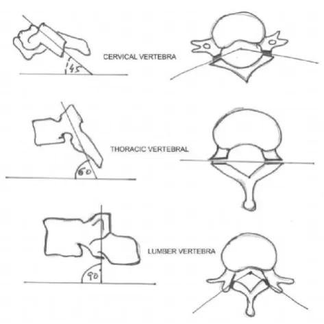
al., 2008). Facet joints are located at the junction between the
lamina and the lateral masses in the cervical region of the spine; whereas, in the thoracic and lumbar regions, they are joined to the vertebral body via the bony pedicles (Figure 2a, 2b, 2c). In general, the inclination angle (Figure 3) of the articular surfaces of the facet joint in the sagittal plane ranges from 20°–78° in the cervical region, 55°–80° in the thoracic region, and 82°–86° in the lumbar region. The angle between the articulating surfaces in the axial plane range from 70°–96°, 85°–120°, and 15°–70° off of the midline in the cervical, thoracic, and lumbar regions, respectively.
So, articular surfaces are more horizontally-oriented in the cervical and upper thoracic spinal regions than lumber region (White and Panjabi, 1990; Panjabi et al., 1993; Pal et al.,
2001). The Facet joints help in the motion of the vertebrae,
[image:2.595.81.534.140.310.2]while also facilitates the transmission of the loads applied to the spine.
Figure 2a. Cervical vertebra
Figure 2b. Thoracic vertebra
When the compressive load is 3–25% is carried by the Lumber facets (“facet force”), it is 22 - 24% in the cervical and upper thoracic spine (Yang and King, 1984). On the other hand, straining of the fibers in the facet capsule not only results from vertebral motion but also from the activation of the muscles that can occur during mechanical loading of the spine as the outer surface of the capsular ligament, covered by the surrounding paraspinal muscles.
[image:2.595.308.550.337.638.2]Figure 2c. Lumber vertebra
Figure 3. Inclination of Facet joints
When the capsule is stretched, the nerve afferents that innervate it are also stretched, which has been shown to trigger the generation of neuronal signaling to the central nervous system (CNS). Some studies found that the capsule contained afferents that responded with firing at both low- and high-thresholds of strain (10% and 47%, respectively) and also that afferents of both types exhibited persistent generation of after discharge for up to 5 mins after the release of the applied strain (39–57%)
that did not produce tissue rupture (Lu et al., 2005). So this
may be the possible mechanism for chronic pain because the after discharge was hypothesized as having long term effects in the CNS. The neural signals from the capsule travel via the primary afferents to the dorsal root ganglion (DRG) and spinal cord, and can induce several hallmarks of neuro-inflammation,
including glial activation (Winkelstein and Santos, 2008) and
cytokine upregulation (McMahon et al., 2005; Igarashi et al.,
2007)
The particular relationship between the mechanical, chemical, and cellular responses to compression in the cartilaginous matrix of the different zones remains largely unreported for the human spinal facet joint. Nevertheless, damage to the cartilage
structure elicits an inflammatory response (Pelletier et al.,
2001; Côté et al., 2001), which itself can also elicit not just osteoarthritis of the joint but can modulate pain signals from other regions of the joint. For example, one study showed that the inflammatory cytokines IL-6 and IL-1β were present in the facet cartilage retrieved from patients undergoing surgery for
lumbar spinal canal stenosis and disc herniation (Igarashi et al.,
2007). This result led to the conclusion that pain symptoms
might be due not only to mechanical tissue insults but also to chemical irritation of the tissue from the inflammatory agents leaking from the facet joint into the spinal space.
Facet joint injuries result most-often from motor vehicle and sports trauma, such as skiing, snowboarding, cycling, and
diving (Stemper et al., 2004), and include a wide range of bony
and ligamentous lesions depending on the extent and type of
tissue trauma (Hadley et al., 1992). In fact, even sub
catastrophic or “subfailure” loading of the facet capsule has
been shown to induce pain in a variety of scenarios21. Perhaps
the best example of sub catastrophic capsular distortions modulating physiological responses is highlighted by spinal
loading and whiplash-associated disorders (Stemper et al.,
2005; Quinn et al., 2007). Whiplash-associated disorders are particularly challenging in terms of understanding the biomechanics of the tissue injury and the physiological consequences because there is often no radiologic or other imaging evidence that reveals any obvious indicators of tissue
trauma (Chen et al., 2009; Barnsley et al., 1994).
Figure 4. Pain pattern from cervical facet joint disorders
[image:3.595.317.555.443.661.2]joints and intervertebral disc are inter-dependent, degeneration of the facet joint will also affect the mechanical behavior of the whole vertebral motion segment, and similarly, disc degeneration can impact the overall spinal degenerative cascades. Tissue degradation occurs at the structural and cellular levels during degeneration and that process can result
from and/or be associated with aging (Taylor and Twomey,
1986; Manchikanti et al., 2008), injury (Barnsley et al., 1994), and also infection or inflammation (septic arthritis, synovitis,
and rheumatoid arthritis) (Chan et al., 1987).
However, defining degeneration of the articular cartilage is more challenging and necessitates the use of MRI because this tissue is enclosed in the joint and it is “transparent” to X-rays (Ryu et al., 2010). The above discussion was needed to show that how cervical facet joint is responsible to generate neck pain and headache in our day-to-day activities. This pain pattern (Figure 4) showing that when the upper cervical facets are getting affected the pain radiates to the upper part of neck and scalp which calls ‘Cervicogenic Headache” whereas the lower cervical facet pain radiates to lower part of neck and shoulder. But many a times Lateral Atlanto-Axial joints or
Atlanto-Occipital joints also cause symptoms like
“Cervicogenic Headache” (Bogduk, 1992).
Atlanto-Axial joints (AA) are the joint of “negative expression”, as these joints allow the head to rotate laterally. On the contrary Atlanto-Occipital (AO) joint is a joint of “Yes”
or nodding (flexion or extension) (Edmeads, 1988). As there is
no disc between AA and AO level, the facet joints and the numerous ligaments provide the major stability between the two bones, which are thought to possess the articular nociceptors and sensory afferents necessary to mediate the pain
and facet syndrome (Clemente, 2010). Two lateral
Atlanto-axial joints compliment the median Atlanto-Atlanto-axial joint. The lateral AA joints help to transfer the weight of the skull from the AO joint. Both lateral and median AA joints are morphologically equivalent to the uncovertebral joints of the
lower cervical vertebrae (Bogduk and Marsland, 1986). The
obliques capitis inferior, splenius capitis and rectus capitis posterior major muscles of one side of the body, work in tandem with the sternocleidomastoid of the other side, to control the motion of the AA joint. That’s why a spasm in these muscles can also create symptoms of Cervicogenic headache (Dreyfuss et al., 1994)
The articular cartilage of Lateral AA joint is quiet thick (1.4mm to 3.2mm) and accommodates large, intra-articular menisci emerging from the flaccid roomy joint capsule. Sometimes degeneration of this menisci cause sharp local
catching pain (Dreyfuss et al., 1994). Vertebral artery is lying
lateral to the AA joints, which is protected from the sharp osseous surface of the lateral joint by a pericapsular soft tissue. In old age, with severe degenerative changes, its course may vary. Neck pain may be associated with vertebrao-basilar insufficiency with or without head turning which create symptoms like headache, lightheadedness, dizziness, vertigo, facial numbness, nausea, vomiting, blurred vision, diplopia,
dysphagia, gait abnormality and tongue symptoms (Bogduk
and Marsland, 1986; Dreyfuss et al., 1994). A joint has a wide range of motion on all articulation in the neck, which doesn’t
happen at any other vertebral levels. Rotation around the axis through the dens is limited to 40 - 45 degrees to each side, but it allows only 5 – 10 degrees of flexion or extension. That’s why the pain pattern (Figure 5) is different from other cervical facet pain. It is focal pain around the joint area during lateral rotation of the neck on the same side. This actually helped to diagnose the actual cause of pain of the above mentioned
[image:4.595.311.560.158.319.2]patient (Jofe et al., 1989; Worth and Selvic, 1987).
Figure 5. Pain pattern from Atlanto-axial and Atlanto-occipital joint disorders
On account of its proximity to the brain stem, importance in stabilization, fracture or injury at this level can be catastrophic.
The common trauma and pathologies are (Dreyfuss et al.,
1994):
Fracture of Dens
Rupture of transverse ligament of the Atlas
Down syndrome exhibits laxity or agenesis of the
ligaments
Rupture of the Alar ligaments
Compression of the C2 spinal ganglion, with severe lateral
rotation in the hyper-extended state, which may cause prolonged severe headaches and excruciating cervico-occipital pain.
Death by judicial hanging may be due to a rupture of the
transverse ligaments of the Atlas or fracture of the Dens. As a result, the Atlas is dislocated from the axis and compresses the brain stem with a fatal damage.
In spite of having problems in this area some patients may
be relatively asymptomatic because of the “Steele Rules of
Thirds” i.e. approximately one third of the atlas ring
occupied by the dens, one third by the spinal cord and the remaining third by the free fluid space and tissue surrounding the cord. This is the reason why some patients with anterior displacement of the Atlas may be relatively asymptomatic until a large degree of movement (> 1/3 of the diameter of the Atlas ring) occurs.
Treatment
1. Intra-articular Steroid with Local anesthetic injection under
fluoroscopy (posterior and lateral approach), which helped the above mentioned patient satisfactorily (Bogduk et al., 2009; Racz et al., 1996).
2. C2 selective nerve root block under fluoroscopy (Bogduk
3. Radiofrequency Ablation (Pulse mode) of AA joint is a
good option (Halim et al., 2010).
Conclusion
Headache is a complex phenomenon or disorder. Proper history, examination and investigations are required to diagnose properly. Many times diagnostic injections are helpful to come to a conclusion. Cervical spine has a great role in development of Cervicogenic headache. Cervical Facet joint disorders as well as the Lateral axial joint or Atlanto-occipital joints are one of the most important structures involved in the pathogenesis of Occipital headache or Cervicogenic headache. Thorough anatomical knowledge is very important for the clinicians for proper diagnosis through exclusion of other causes. On the other hand interventional pain therapy under skilled hands will support these kinds of patients satisfactorily.
REFERENCES
Adams, M. A., Dolan, P. and McNally, D. S. 2009. “The Internal Mechanical Functioning of Intervertebral Discs and Articular Cartilage, and its Relevance to Matrix Biology,” Matrix Biol., 28(7), pp. 384–38
Armstrong, C. G. and Mow, V. C. 1982. “Variations in the Intrinsic Mechanical Properties of Human Articular Cartilage with Age, Degeneration, and Water Content,” J.
Bone Jt. Surg. Am., 64(1), pp. 88–94
Barnsley, L., Lord, S. and Bogduk, N. 1994, “Whiplash Injuries,” Pain, 58, pp. 283–307
Bogduk, N. 1992. The anatomical basis for cervicogenic headache. J Manipulative Physiol Ther. 15:67- 70
Bogduk, N. and Govind, J. 2009. Cervicogenic Headache: An assessment of the evience on clinical diagnosis, invasive tests and the treatment. Lancet Neurol., 8:959-68
Bogduk, N. and Marsland, A. 1986. On the concept of Third Occipital Headache. Neuro Neurosurg Psychiatry. 57:775-80
Bogduk, N. and Marsland, A. 1988. The cervical Zygapophysial joints as a source of neck pain. Spine, 13: 610-17
Chan, F. L., Ho, E. K., Fang, D., Hsu, L. C., Leong, J. C. and Ngan, H. 1987, “Spinal Pseudoarthrosis in Ankylosing Spondylitis,” ActaRadiol., 28(4), pp. 383–388
Chen, H. B., Yang, K. H. and Wang, Z. G. 2009, “Biomechanics of Whiplash Injury,” Chin. J. Traumatol., 12(5), pp. 305–314
Clemente, C.D. 2010. Clemente’s Anatomy Dissector. USA: Lippincot Williams and Wilkins; 2010. P. 361 [ Last retrieved on 2010 June 17]
Côté, P., Cassidy, J. D., Carroll, L., Frank, J. W. and Bombardier, C. 2001. “A Systematic Review of the Prognosis of Acute Whiplash and a New Conceptual Framework to Synthesize the Literature,” Spine, 26(19), pp. E445–E458
Dong, L. and Winkelstein, B. A. 2010, “Simulated Whiplash Modulates Expression of the Glutamatergic System in the Spinal Cord Suggesting Spinal Plasticity is Associated With Painful Dynamic Cervical Facet Loading,” J.
Neurotrauma., 27(1), pp. 163–174
Dreyfuss, P., Michaelsen, M. and Fletcher, D. Atlanto-occipital and Lateral atlanto-axial joint pain patterns. Spine 1994; 19:1125-31
Dunn, W., DuRaine, G. and Reddi, A. H. 2009. “Profiling MicroRNA Expression in Bovine Articular Cartilage and
Implications for Mechano transduction,” Arthritis
Rheum., 60(8), pp. 2333–2339
Edmeads, J. 1988. The cervical spine and headache.
Neurology, 38:1874-8.
Essentials of Human Anatomy- Head and Neck by AK Dutta. Chapter 4. pp 77-8
Hadley, M. N., Fitzpatrick, B. C., Sonntag, V. K. and Browner C. M. 1992. “Facet Fracture-Dislocation Injuries of the Cervical Spine,” Neurosurgery, 30(5), pp. 661–666 Halim, W., Chua, N.H. and Vissers, K.C. 2010. Long-term
pain relief in patients with Cervicogenic headache after pulsed radiofrequency application into lateral AA joint. Pain Practice, 10: 267-71
Igarashi, A., Kikuchi, S. and Konno, S. 2007, “Correlation Between Inflammatory Cytokines Released From the Lumbar Facet Joint Tissue and Symptoms in Degenerative Lumbar Spinal Disorders,” J. Orthop. Sci., 12(2), pp. 154– 160
Jofe, M., White, A. and Panjabi, M. 1989. Clinically relevant
kinematics of the cervical spine. 2nd ed. Philadelphia: JB
Lippincott., 57-69
Lu, Y., Chen, C., Kallakuri, S., Patwardhan, A. and Cavanaugh, J. M., 2005. “Neural Response of Cervical Facet Joint Capsule to Stretch: a Study of Whiplash Pain Mechanism,” Stapp Car Crash J., 49, pp. 49–65
Manchikanti, L., Manchikanti, K. N., Cash, K. A., Singh, V. and Giordano, J. 2008, “Age-Related Prevalence of Facet-Joint Involvement in Chronic Neck and Low Back Pain,” Pain Physician, 11(1), pp. 67–75
McMahon, S. B., Cafferty, W. B. J. and Marchand, F. 2005, “Immune and Glial Cell Factors as Pain Mediators and Modulators,” Exp. Neurol., 192, pp. 444–462
Pal, G. P., Routal, R. V. and Saggu, S. K. 2001, “The Orientation of the Articular Facets of the Zygapophyseal Joints at the Cervical and Upper Thoracic Region,” J.
Anat., 198(4), pp. 431–441
Panjabi, M. M., Oxland, T., Takata, K., Goel, V., Duranceau, J. and Krag, M. 1993, “Articular Facets of the Human
Spine. Quantitative Three- Dimensional
Anatomy,” Spine, 18(10), pp. 1298–1310
Pelletier, J. P., Martel-Pelletier, J. and Abramson, S. B. 2001, “Osteoarthritis, an Inflammatory Disease: Potential Implication for this Selection of New Therapeutic Targets,” Arthritis Rheum., 44, pp. 1237–1247
Quinn, K. P., Lee, K. E., Ahaghotu, C. C. and Winkelstein, B. A. 2007, “Structural Changes in the Cervical Facet Capsular Ligament: Potential Contributions to Pain Following Subfailure Loading,” Stapp Car Crash
Journal, 51 , pp. 169–187
Racz, G.B., Sanel, H. and Diede, J.H. 1996. Atlanto-occipital and Lateral Atlanto-axial joint injections in the treatment of headache and neck pain. In Waldman S, Winnie A, editors. Interventional Pain Management. Philadelphia: WB Saunders., P. 220-222
Segments Following Cervical Arthroplasty After a Minimum 24-Month Follow-up: Comparison Between the Bryan and Prodisc-C Devices,” J. Neurosurg. Spine, 13(3), pp. 299–307
Stemper, B. D., Yoganandan, N. and Pintar, F. A. 2004, “Gender- and Region-Dependent Local Facet Joint Kinematics in Rear Impact: Implications in Whiplash Injury,” Spine, 29(16), pp. 1764–1771
Stemper, B. D., Yoganandan, N., Gennarelli, T. A. and Pintar F. A. 2005, “Localized Cervical Facet Joint Kinematics
Under Physiological and Whiplash Loading,” J.
Neurosurg. Spine, 3(6), pp. 471–476
Stovner, L., et al., 2007. The global burden of headache: a documentation of headache prevalence and disability worldwide. Cephalalgia, 27(3): p. 193-210
Taylor, J. R. and Twomey, L. T. 1986, “Age Changes in Lumbar Zygapophyseal Joints. Observations on Structure and Function,” Spine 11(7), pp. 739–745
White, A. A. and Panjabi, M. M. 1990, Clinical Biomechanics
of the Spine, 2nd ed. Lippincott, New York.
Winkelstein, B. A. and Santos, D. G. 2008, “An Intact Facet Capsular Ligament Modulates Behavioral Sensitivity and Spinal Glial Activation Produced by Cervical Facet Joint Tension,” Spine, 33(8), pp. 856–862
Womack, W., Woldtvedt, D. and Puttlitz , C. M. 2008, “Lower Cervical Spine Facet Cartilage Thickness Mapping,” Osteoarthritis Cartilage, 16(9), pp. 1018–1023
Worth, D.R. and Selvic, G. 1987. Modern manual therapy of the vertebral column. London: Churchill Livingstone; P. 53-63
Yang, K. H. and King, A. I. 1984. “Mechanism of Facet Load Transmission as a Hypothesis for Low-Back Pain,” Spine, 9(6), pp. 557–565
Yoganandan, N., Knowles, S. A., Maiman, D. J. and Pintar, F. A. 2003, “Anatomic Study of the Morphology of Human Cervical Facet Joint,” Spine, 28(20), pp. 2317–2323