PROSTHODONTIC REHABILITATION OF AN ECTODERMAL DYSPLASIA PATIENT
*Dr. Anupam Singh, Dr. (Prof.) D
Department of Prosthodontics, Patna Dental College and Hospital
ARTICLE INFO ABSTRACT
Ectodermal Dysplasias comprise a large, heterogenous group of inherted disorders that are defined by primary defects in the development of two or more tissues derived from
multidisciplinary approach to dental treatment is required. This clinical report attempts to describe the prosthodontic management of a 13 year old girl affected by ectodermal dysplasia. Treatment included a maxillary teeth supported
function and esthetics.
Copyright©2016, Dr. Anupam Singh et al. This is an open access article distributed under the Creative Commons Att use, distribution, and reproduction in any medium, provided the original work is properly cited.
INTRODUCTION
Ectodermal Dysplasia (ED) is a congenital syndrome which is characterized by abnormal development of tissues of ectodermal origin like skin, nails, hair, sweat glands,
glands, salivary glands and teeth. (Gorlin
Wynbrandt and Ludman, 1990)ED has more than 150 variants
(possible types). (Pinheiro et al., 1994; Freire
is usually described as hypohidrotic or hidrotic depending upon degree of function of sweat glands. It commonly affects males. (Rimoin et al., 1996) Females, however, are affected sometimes. ED is mostly inherited as an x
trait. (Rimoin et al., 1996; Clarke, 1987)Incidence is 1
100,000 live births. (Buyse, 1990)Affected individuals present
some classical features like fine, sparse hair, thin eyebrows and eyelashes, frontal bossing, saddle nose, small palatal and cranial base width, small malar processes, dry oral mucosa, hoarse voice quality, midface hypoplasia,
anodontia or hypodontia, reduced salivary gland development leading to various degree of xerostomia. Some cases show decreased function of certain components of immune system, i.e, decreased lymphocytic function, cellular immune
hypofunction causing increased susceptibility to
certain infection and allergic condition. Variable degree of
*Corresponding author: Dr. Anupam Singh,
Department of Prosthodontics, Patna Dental College and Hospital, India.
ISSN: 0975-833X
Article History: Received 22nd June, 2016 Received in revised form 16th July, 2016
Accepted 29th August, 2016
Published online 30th September,2016
Citation: Dr. Anupam Singh, Dr. (Prof.) D. K. Singh, dysplasia patient” International Journal of Current Research Key words:
Anodontia, Ectodermal dysplasia, Hypodontia, Overdenture, Prosthodontic management of ectodermal dysplasia.
CASE STUDY
PROSTHODONTIC REHABILITATION OF AN ECTODERMAL DYSPLASIA PATIENT
(Prof.) D. K. Singh, Dr. Irfan-ul-Huda and Dr. Prateek Pandey
of Prosthodontics, Patna Dental College and Hospital
ABSTRACT
Ectodermal Dysplasias comprise a large, heterogenous group of inherted disorders that are defined by primary defects in the development of two or more tissues derived from
multidisciplinary approach to dental treatment is required. This clinical report attempts to describe the prosthodontic management of a 13 year old girl affected by ectodermal dysplasia. Treatment included a maxillary teeth supported overdenture and a mandibular removable partial denture to improve function and esthetics.
is an open access article distributed under the Creative Commons Attribution License, which use, distribution, and reproduction in any medium, provided the original work is properly cited.
Ectodermal Dysplasia (ED) is a congenital syndrome which is characterized by abnormal development of tissues of ectodermal origin like skin, nails, hair, sweat glands, sebaceous Gorlin et al., 2001; ED has more than 150 variants Freire-Maia, 1994) This is usually described as hypohidrotic or hidrotic depending upon degree of function of sweat glands. It commonly affects males. Females, however, are affected sometimes. ED is mostly inherited as an x-linked recessive Incidence is 1-7 in per Affected individuals present some classical features like fine, sparse hair, thin eyebrows and eyelashes, frontal bossing, saddle nose, small palatal and cranial base width, small malar processes, dry oral mucosa, hoarse voice quality, midface hypoplasia, protuberant lips, anodontia or hypodontia, reduced salivary gland development leading to various degree of xerostomia. Some cases show decreased function of certain components of immune system, i.e, decreased lymphocytic function, cellular immune
tion causing increased susceptibility to
certain infection and allergic condition. Variable degree of
Department of Prosthodontics, Patna Dental College and Hospital, India.
hypohidrosis are present frequently with significant episodes of hyperthermia in infancy and early childhood.
mentioned features result in afflicted individuals having a prematurely aged appearance which can have tremendous negative impact on the individua
esteem.because of the absence of most of the deciduous and permanent teeth, early and extensive dental treatment throughout the childhood is needed. Prosthodontic management of ED can include fixed partial denture (mostly crowns t improve morphology of peg-shaped tooth), removable partial denture, overdenture, complete dentures
implant supported prosthesis. (Nowak
modalities can be used individually or in combination.
Case report
A 13-year old girl was referred to the department of Prosthodontics of Patna Dental College & Hospital, Patna complaining of missing teeth and difficulty in mastication. was accompanied by her father. He gave the history
teeth since her childhood. None of the family members or relatives were known to have a similar condition.
Extra-oral examination
Revealed the typical features of ectodermal dysplasia like frontal bossing, depressed nasal bridge. The lips were Available online at http://www.journalcra.com
International Journal of Current Research Vol. 8, Issue, 09, pp.39007-39012, September, 2016
INTERNATIONAL
h, Dr. (Prof.) D. K. Singh, Dr. Irfan-ul-Huda and Dr. Prateek Pandey, 2016. “Prosthodontic rehabilitation of an Ectodermal International Journal of Current Research, 8, (09), 39007-39012.
z
PROSTHODONTIC REHABILITATION OF AN ECTODERMAL DYSPLASIA PATIENT
Dr. Prateek Pandey
of Prosthodontics, Patna Dental College and Hospital, India
Ectodermal Dysplasias comprise a large, heterogenous group of inherted disorders that are defined by primary defects in the development of two or more tissues derived from embryonic ectoderm. A multidisciplinary approach to dental treatment is required. This clinical report attempts to describe the prosthodontic management of a 13 year old girl affected by ectodermal dysplasia. Treatment included overdenture and a mandibular removable partial denture to improve
ribution License, which permits unrestricted
re present frequently with significant episodes of
hyperthermia in infancy and early childhood.9-17 The above
mentioned features result in afflicted individuals having a prematurely aged appearance which can have tremendous negative impact on the individual by affecting their self-esteem.because of the absence of most of the deciduous and permanent teeth, early and extensive dental treatment throughout the childhood is needed. Prosthodontic management of ED can include fixed partial denture (mostly crowns to shaped tooth), removable partial denture, overdenture, complete dentures (in anodontia cases) or Nowak, 1988) These treatment modalities can be used individually or in combination.
year old girl was referred to the department of Prosthodontics of Patna Dental College & Hospital, Patna complaining of missing teeth and difficulty in mastication. She er father. He gave the history of missing childhood. None of the family members or relatives were known to have a similar condition.
Revealed the typical features of ectodermal dysplasia like frontal bossing, depressed nasal bridge. The lips were
INTERNATIONAL JOURNAL OF CURRENT RESEARCH
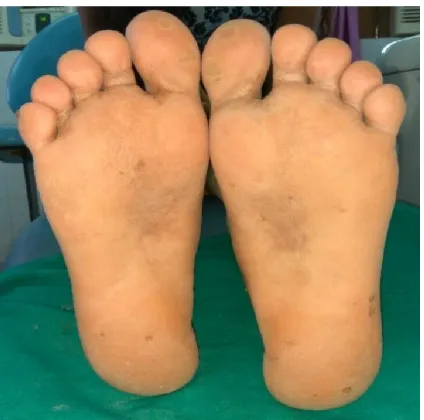
protuberant. The skin was dry, scaly and wrinkled. The palms and soles were hyperkeratotic. The nails were either poorly developed or absent in some of the toes. There was extensive pigmentation of skin around mouth and eyes. She had a concave profile with a pointed chin. The neck of the patient showed numerous tubercular scars indicative of previous tuberculosis infection suggestive of poor immune system.
[image:2.595.57.274.142.520.2]Fig.: Frontal view of the patient
Fig. Profile view of the patient
[image:2.595.61.268.145.315.2]Fig. Poorly Developed Nails
Fig. Palmar Keratosis
[image:2.595.326.545.279.489.2]Fig. Toes Without Nails
[image:2.595.329.541.523.733.2] [image:2.595.55.271.548.738.2]Intra-oral examination
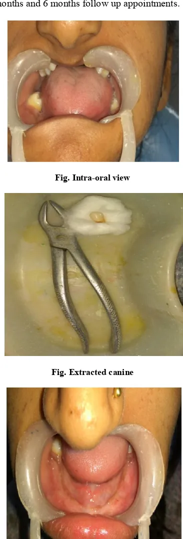
revealed dry mucosa and large tongue. Saliva was thick and ropy. Maxillary arch has three teeth-2 molars and a canine while mandibular arch had two standing molars-one on either side of the arch. Maxillary canine was mobile. The alveolar processes were poorly developed. The deficient growth of alveolar process and mandibular over closure resulted in a distinctly aged, prognathic appearance similar to an edentulous person. Orthopantamogram x-ray revaled the presence of a
permanent tooth bud of maxillary right second molar.
Prosthodontic management
In this case, the mobile right maxillary canine was first of all extracted. The root was almost resorbed. In order to preserve whatever alveolar bone was present teeth supported maxillary overdenture and a removal partial denture for mandibular arch were considered.
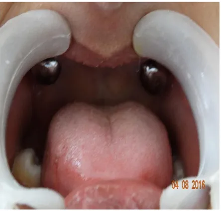
Both the maxillary molars were reduced to serve as
overdenture abutments without performing endodontic treatment as the roots were not properly developed (with an immature root apex). Alginate impression of prepared teeth was made to fabricate metal copings.
Metal copings were cemented on to the reduced teeth in
the next appointment to protect them from caries.
Preliminary impressions of both the arches were made.
Custom trays were prepared using auto polymerizing
acrylic resin after giving spacer to the relief areas like incisive papilla, mid-palatine raphe and crest of mandibular ridge.
Border moulding was done using green stick
compound.
Secondary impression were made using silicone
impression material and master casts were obtained.
Record bases were made on those master casts to
establish maxilla-mandibular relation. Fullness of face, appearance of lips, height of lower face compared to upper, freeway space and phonetics test were taken into
consideration. The cast were articulated once
satisfactory maxilla-mandibular relation was made.
While arranging teeth, basic denture guidelines for teeth
placement were followed. Due to reduced arch length and width, teeth of small size and reduced occlusal table were used. While arranging teeth neutral zone concept was followed.
Trial dentures were checked for retention, occlusion,
phonetics and esthetics.
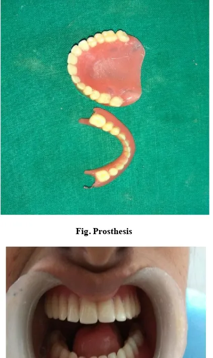
Trial dentures after approval by the patient and her
father, were processed using heat cure acrylic resin. After polishing, the dentures were ready for insertion.
The prosthesis were inserted. Fit was verified. The girl
was taught about insertion and removal of prosthesis and post-operative instructions on hygiene and maintenance were given.
After 24 hours, patient was recalled for the first
post-insertion follow-up. Problem areas were corrected.
Afterwards she was scheduled for 1 week, 1 month, 3
[image:3.595.51.277.192.340.2]months and 6 months follow up appointments.
[image:3.595.340.523.198.737.2]Fig. Intra-oral view
Fig. Extracted canine
Fig. Cemented metal copings
[image:4.595.55.272.289.508.2]Fig. Primary impression
[image:4.595.333.538.320.584.2]Fig. Primary cast
Fig. Wax spacer with relief
Fig. Custom trays
[image:4.595.62.268.537.737.2]Fig. Prosthesis
Fig. Dentures in mouth
Fig. Confident and smiling
DISCUSSION
This clinical report aims to describe the prosthodontic management of a 13-year old girl affected by ectodermal dysplasia. The mobile tooth with poor prognosis was extracted. Maxillary molars were modified to serve as over denture abutments while in lower jaw a RPD was given as lower molar
teeth were not fully erupted. They may serve as (remained) as potential overdenture abutment. Underdevelopment of alveolar ridges and xerostomia in ED patients make denture retention and stability difficult. Due to presence of erupting teeth and continued growth and development of jaws, periodic recall of patient is important for modification and/or replacement of prosthesis. Endosseous implants can be considered once the full growth is achieved. Early dental intervention can improve appearance and build confidence of the patient. Psychological scarring can thus be minimized.
REFERENCES
Buyse M. 1990. Birth deects encyclopedia. St.Louis, Mosby year book, p 597-8
Buyse ML. 1990. Birth defects encyclopedia. Cambridge: Blackwell Scientific Publications, 596-605
Champlin TL, Mallory SB. 1989. Hypohidrotic Ectodermal Dysplasia: a review. J Ark Med Soc., 86:115-7
Clarke a, Phillips DI, Brown R, Harper PS. 1987. Clinical aspects of X-linked Hypohidrotic Ectodermal Dysplasia. Arch Dis Child., 62:989-96
Clarke A. 1987. Hypohidrotic Ectodermal Dysplasia. J Med Genet, 24:659-63
Clarke A. 1987. Hypohidrotic Ectodermal Dysplasias. J Med Genet, 24:659-63
Cronin RJ Jr, Oesterlo LJ, Ranly DM. 1994. Mandibular implants and the gowing patients. Int J Oral Maxillofacial Implants, 9:55-62
Crwford PJ, Aldred ML, Clarke A. 1991. Clinical and radiographic dental findings in X-linked hypohidrotic Ectodermal Dysplasia. J Med Genet, 28:181-5
Freire-Maia. 1994. ECtodermal Dysplasias- a clinical and genetic study. NewYork: Alan R.Liss Inc.
Gorlin RJ, Cohen MM, Hennekam RC. 2001. editors.
Syndromes of head and neck 4th edition. NewYork :Oxford
university press, p 540-5
Levin LS. 1988. Dental and oral abnormalities in selected ectodermal dysplasia syndromes. Birth defects, 24:205-27 Nowak AJ. 1988. Dental treatment for patients with
ectodermal dysplasias. Birth defects, 24:243-52
Peterson-Falzone SJ, Caldarelli DD, Landahl KL. 1981. Abnormal laryngeal voice quality in Ectodermal Dysplasia, Arch Otolaryngol., 107:300-4
Pinheiro M, Freire-Maria N. 1994. Ectodermal Dysplasia: A clinical classification and a casual review. Am J Med Genet, 53:153-62
Rimoin DL, Connor JM, Pyeritz RE, Emery and Rimoin’s
1996. Principles and practice of medical genetics.3rd
edition.London. Churchill livingstone, p.1279-83
Sanjay Prasad, Tapan K Giri. Oral rehabilitation of a hypohidrotic ectodermal dysplasia patient. The Journal of Indian Prosthodontic Society/july 2009/vol9/issue
Sarnat BG, Brodie AG, Kuback WH. 1953. Fourteen year report of facial growth in case of complete anodontia with ectodermal dysplasia. AMA Am J Dis Child, 86:162-9 Soderholm AL, Kaitila J. 1985. Expansion of X-linked
hypohidrotic Ectodermal Dysplasia in six males and in their mothers. Clin Genet, 28:136-44
[image:5.595.59.270.59.413.2]Tape MW, 1995. Type E. Ectodermal Dysplasia:literature review and a case report. Compend Contin Educ Dent, 16:524-8.
Wynbrandt J, Ludman MD. 1990. The encyclopedia of genetic disorders and birth defects. NewYork:Facts on File, 110-1