COMPARISON OF RETROPERITONEAL & TRANSPERITONEAL LAPAROSCOPIC NEPHRECTOMY
IN MANAGEMENT OF PYONEPHROSIS IN TERMS OF CONVERSION TO OPEN
*Milind Patil
Department of Surgery, Government Medical College Baroda, Vadodara, India
ARTICLE INFO ABSTRACT
Background: open approaches to Aim and Objectives:
surgery in pyonephrosis using retroperitonel and trans peritoneal laproscopic nephrectomy. Methods:
College & SSG Hospital, vadodara.We performed a retrospective review of a maintained database of 219 consecutive laparoscopic simple
February 2015. Results:
access using three was used in 54(24.6%) patients. In our study total 163 (74.4%
(percutaneous nephrostomy) in situ, 79.3% in lap transperitoneal group and 59.2% in lap retro peritoneal group. 27(12.3%) patients required conversion to open surgery. Adhesion 13(5.9%) and bleeding 9(4.1%) were the main factors for conversion, w
due to bowel injury and limited space in 3(1.3%) patients. Conversion rate was 12.1 % (20/165) for transperitoneal procedures while 12.9 % (7/54) for retroperitoneal approach. Laparoscopic approach requires proper
help in identifying renal hilar anatomy as well as the relationship with the surrounding structures. Conclusion:
transperitoneal laproscopic nephrectomy in terms of conversion to open surgery.
Copyright © 2016, Milind Patil and Ashvin Kankotiya.
unrestricted use, distribution, and reproduction in any medium, provided the original work is properly cited.
INTRODUCTION
Laparoscopic simple nephrectomy is often far from “simple” even for the most experienced laparoscopic surgeon because the conditions for which it is performed often result in significant perinephric scarring. Since the mid
been an evolution in surgical practice from tra
approaches toward minimally invasive means of treating operative lesions. Although these changes have been made possible through advances in video technology and instrumentation design, the primary driver has been an increasingly educated patient population seeking less painful means of treatment. Over a century ago, gynecologic colleagues introduced laparoscopic surgery primarily as a diagnostic tool. Only recently has it become a practical and acceptable alternative to treat complex surgical
development of then laparoscopic pelvic lymphadenectomy for patients with prostate cancer inaugurated the role of
*Corresponding author: Milind Patil,
Department of Surgery, Government Medical College Baroda, Vadodara, India.
ISSN: 0975-833X
Article History: Received 10th July, 2016
Received in revised form 15th August, 2016
Accepted 28th September, 2016
Published online 30th October,2016
Key words:
Laproscopy, Nephrectomy, Pyonephrosis.
Citation: Milind Patil and Ashvin Kankotiya.2016.
specific learning disorder”, International Journal of Current Research
CASE STUDY
COMPARISON OF RETROPERITONEAL & TRANSPERITONEAL LAPAROSCOPIC NEPHRECTOMY
IN MANAGEMENT OF PYONEPHROSIS IN TERMS OF CONVERSION TO OPEN
Milind Patil and Ashvin Kankotiya
Department of Surgery, Government Medical College Baroda, Vadodara, India
ABSTRACT
Background: Since the mid-1990s, there has been an evolution in surgical practice from traditional open approaches to minimally invasive means of treating operative lesions
Aim and Objectives: This study is carried out to study rate & indication of conversion to open surgery in pyonephrosis using retroperitonel and trans peritoneal laproscopic nephrectomy.
Methods: This study was retrospective study done in Department of Urology Baroda Medical College & SSG Hospital, vadodara.We performed a retrospective review of a maintained database of 219 consecutive laparoscopic simple nephrectomies done for pyonephrosis between July 2001 to February 2015.
Results: In study transperitoneal route using four ports was used in 165 (75.3%) while retroperitoneal access using three was used in 54(24.6%) patients. In our study total 163 (74.4%
(percutaneous nephrostomy) in situ, 79.3% in lap transperitoneal group and 59.2% in lap retro peritoneal group. 27(12.3%) patients required conversion to open surgery. Adhesion 13(5.9%) and bleeding 9(4.1%) were the main factors for conversion, while 2 (0.9%) patients required conversion due to bowel injury and limited space in 3(1.3%) patients. Conversion rate was 12.1 % (20/165) for transperitoneal procedures while 12.9 % (7/54) for retroperitoneal approach. Laparoscopic approach requires proper placement of ports for meticulous surgical dissection. Preoperative plain and CECT help in identifying renal hilar anatomy as well as the relationship with the surrounding structures. Conclusion: In our study, retroperitoneal laparoscopic nephrectomy has
transperitoneal laproscopic nephrectomy in terms of conversion to open surgery.
.This is an open access article distributed under the Creative Commons Att
use, distribution, and reproduction in any medium, provided the original work is properly cited.
is often far from “simple” even for the most experienced laparoscopic surgeon because the conditions for which it is performed often result in significant perinephric scarring. Since the mid-1990s, there has been an evolution in surgical practice from traditional open approaches toward minimally invasive means of treating operative lesions. Although these changes have been made possible through advances in video technology and instrumentation design, the primary driver has been an ient population seeking less painful Over a century ago, gynecologic colleagues introduced laparoscopic surgery primarily as a diagnostic tool. Only recently has it become a practical and acceptable alternative to treat complex surgical diseases. The development of then laparoscopic pelvic lymphadenectomy for patients with prostate cancer inaugurated the role of
Department of Surgery, Government Medical College Baroda,
laparoscopy in treating urologic lesions (Griffith In June 1990, Clayman and coworkers at
overcame the barriers to laparoscopic solid organ removal by performing the first laparoscopic nephrectomy
1991). In less than 7 hours, an elderly patient with a 3
renal mass underwent laparoscopic radical nephrectomy through five trocar sites. This accomplishment represents one of the milestones in minimally invasive surgery because it provided the solution for removing a large solid organ without the need for an incision (Clayman
report, many institutions have verified the utility of laparoscopic approach to address the diseases of the kidney. Retroperitoneal laparoscopic nephrecto
Gauer et al in 1993. Laparoscopic nephrectomy has proven to be beneficial as compared to open surgery in terms of lesser postoperative pain, a shorter hospital stay and a more rapid return of full activity (Gaur
1999).
MATERIALS AND METHODS
This study was done in department of urology, College & SSG Hospital, v
International Journal of Current Research
Vol. 8, Issue, 10, pp.40584-40588, October, 2016
2016. “Therapeutic play techniques in the management of anger and
International Journal of Current Research, 8, (10), 40584-40588.
COMPARISON OF RETROPERITONEAL & TRANSPERITONEAL LAPAROSCOPIC NEPHRECTOMY
IN MANAGEMENT OF PYONEPHROSIS IN TERMS OF CONVERSION TO OPEN SURGERY
Department of Surgery, Government Medical College Baroda, Vadodara, India
1990s, there has been an evolution in surgical practice from traditional means of treating operative lesions.
This study is carried out to study rate & indication of conversion to open surgery in pyonephrosis using retroperitonel and trans peritoneal laproscopic nephrectomy.
This study was retrospective study done in Department of Urology Baroda Medical College & SSG Hospital, vadodara.We performed a retrospective review of a maintained database of nephrectomies done for pyonephrosis between July 2001 to
In study transperitoneal route using four ports was used in 165 (75.3%) while retroperitoneal access using three was used in 54(24.6%) patients. In our study total 163 (74.4%) had PCN (percutaneous nephrostomy) in situ, 79.3% in lap transperitoneal group and 59.2% in lap retro peritoneal group. 27(12.3%) patients required conversion to open surgery. Adhesion 13(5.9%) and hile 2 (0.9%) patients required conversion due to bowel injury and limited space in 3(1.3%) patients. Conversion rate was 12.1 % (20/165) for transperitoneal procedures while 12.9 % (7/54) for retroperitoneal approach. Laparoscopic approach placement of ports for meticulous surgical dissection. Preoperative plain and CECT help in identifying renal hilar anatomy as well as the relationship with the surrounding structures.
In our study, retroperitoneal laparoscopic nephrectomy has to be considered equal to transperitoneal laproscopic nephrectomy in terms of conversion to open surgery.
is an open access article distributed under the Creative Commons Attribution License, which permits
laparoscopy in treating urologic lesions (Griffith et al, 1990). 1990, Clayman and coworkers at washington university overcame the barriers to laparoscopic solid organ removal by performing the first laparoscopic nephrectomy (Clayman et al., In less than 7 hours, an elderly patient with a 3-cm solid renal mass underwent laparoscopic radical nephrectomy through five trocar sites. This accomplishment represents one of the milestones in minimally invasive surgery because it for removing a large solid organ without Clayman et al., 1991). Since this report, many institutions have verified the utility of laparoscopic approach to address the diseases of the kidney. Retroperitoneal laparoscopic nephrectomy was introduced by in 1993. Laparoscopic nephrectomy has proven to be beneficial as compared to open surgery in terms of lesser postoperative pain, a shorter hospital stay and a more rapid et al., 1993; Rozenberg et al.,
MATERIALS AND METHODS
This study was done in department of urology, Baroda Medical College & SSG Hospital, vadodara. We performed a
OF CURRENT RESEARCH
retrospective review of a maintained database of 219 consecutive laparoscopic simple nephrectomies done for pyonephrosis between July 2001 to February 2015.
Inclusion Criteria
All patients who went for laparoscopic simple
nephrectomy for pyoneprosis
Exclusion Criteria
Laparoscopic simple nephrectomy performed for other than pyoneprosis that include
Obstructive or reflux nephropathy Renal tuberculosis
Multicystic dysplastic kidney Reno vascular
hypertension
Acquired renal cystic disease Nephrosclerosis
Symptomatic patients with autosomal dominant
polycystic kidney disease.
Patient Evaluation and Preparation
All the patients were given light diet in the previous evening and polyethylene glycol preparation for bowel wash out.informed consent obtained with discussion of possible complications.
Operative procedure
For laparoscopic nephrectomy, the patient is initially positioned supine for induction of general anesthesia. A bladder catheter and nasogastric tube is placed for decompression of the bladder and stomach prior to insufflation. The subsequent steps and positioning of the patient depends on the approach for the procedure.
Retroperitoneoscopic Nephrectomy
Patient is placed in the lateral flank position with elevation of the kidney bridge. Further, the table may be tilted anteriorly to allow the peritoneum and bowel to fall away from the proposed port site. The primary port is placed using a 1.5-cm incision, 2 cm below and posterior to the tip of the 12th rib in the posterior axillary line, deepened down to the thoracolumbar fascia A balloon dilator was constructed as described by Gaur (1993). The balloon dilator was used to displace the adjacent fat and peritoneum. A 10mm port was then placed in this opening and used as the camera port. The 2nd and 3rd ports were inserted under direct vision. An
automatic insufflator was used to maintain the CO2 pressure at
14mm Hg. The hilar vessels are dissected first and divided. The ureter is dissected and divided. The kidney is mobilized all round and delivered intact by extending a port or by joining two ports. A 18-F ryles tube drain is left behind in the retroperitoneal space through the 5-mm port site at the discretion of the surgeon. The patient positioned in a modified lateral decubitus position and the umbilicus is placed over the break in the operating table. An axillary roll is placed and padding used to support the buttocks and flank. The table is rolled toward the surgeon to assist with retraction of the bowel. The abdomen is insufflated using a Veress needle. Trocars are usually inserted near the umbilicus, midway between the iliac crest and umbilicus, just below the costal
margin in the midclavicular line, and 4th port at the anterior
axillary line midway between the twelfth rib and the iliac crest.
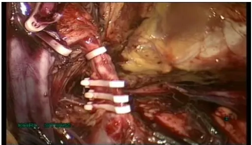
[image:2.595.306.562.97.245.2]In general, 10/12 mm ports are used at the umbilicus and lower quadrant, whereas 5 mm ports are used at the costal and lateral margins.
Fig. 1. Intra-Operate Photo Hilar Clipping
Fig. 2. Port Site Laparoscopic Right Simple Nephrectomy
For a left nephrectomy, the white line of told is incised from the level of the iliac vessels to above the spleen including the lienocolic ligament. During a right-sided nephrectomy, the peritoneal incision is carried cephalad, above the hepatic flexure including the right triangular and right anterior coronary ligaments. Medial traction on the colon reveals colorenal attachments that must be divided to complete the colon dissection. Adequate mobilization of the colon reveals the psoas muscle followed by the gonadal vessels and the ureter. The ureter is elevated and followed proximally to the lower pole and hilum of the kidney. The ureter is not divided at this time because it can be used to help elevate the kidney and identify the hilar vessels which are clipped and divided individually after a meticulous hilar dissection. Once the hilar vessels have been divided, the dissection continues posteriorly and superiorly to the upper pole and the adrenal gland is preserved. The ureter is divided and the kidney is removed intact by extending a 10 mm port. The muscle layer of the 10 mm trocar sites is closed with 2-0 vicryl sutures.
Post Operative Care
The nasogastric tube is removed at the completion of the procedure. The patient can begin oral diet as tolerated after the bowel sounds return or next day morning. The foley catheter is removed within 24 hour the patient is ambulating and a drain be removed within 24 hour or when the output is less than 50 ml in 24 hrs. The patient is discharged when tolerating a diet.
[image:2.595.309.559.266.425.2]RESULTS
Number of Patients
In our study there were 219 simple nephrectomies of which transperitoneal route was used in 165 (75.3%) while retroperitoneal access was used in 54(24.6%) patients.
Demographics
In our study there was male predominance. Nephrectomy was perfored in 120 male patients and 99 female patients, out of 120 male 85 underwent lap transperitoneal nephrectomy and 35 underwent lap retro peritoneal nephrectomy and in female 89, and 19 respectively. In present study right side simple nephrectomy was done in 128 and left side in 91 cases. The mean age at surgery was 55 years (rang 3-77 years).
Etiology
Etiology was Renal stone/Pelviuretric junction stone in 98 (44.75%), Uretric stone 87 (39.72%), Pelviureteric junction obstruction in 23(10.5%), Uretric stricture in 9 (4.1%), vesicoureteric reflux in 2 (0.9%).
DISCUSSION
The rate of conversion to open surgery in laparoscopic
tranceperitonealsimple nephrectomy is range from (5-11.1 %)
and (6-16 %) in laparoscopic retroperitoneal simple
nephrectomy in most of the published series. In some series reports higher conversion rate in pyonephrosis up to 80%.
No differences were observed regarding age, body mass index (BMI) or gender distribution between the conversion and no conversion groups and right-sided nephrectomy were associated with higher chances of conversion into an open procedure 8.6% (19/219) as compare to left-sided
nephrectomy 3.6%(8/219). High conversion rate in rt nephrectomy are generally associated with difficulty in progressing due to severe perirenal adhesions and fibrosis around short renal vein and inferior vena cava. As, many simple nephrectomies are far from simple owing to the scarring associated with the pathologic process. Indeed the underlying renal pathology has been shown to have a direct
correlation to the incidence of conversion with renal tuberculosis, post-traumatic renal atrophy, infarcted kidneys, and xanthogranulomatous pyelonephritis having an open conversion rate of 89% in one large multi-institutional german
study(McDougall et al., 1994).
Side Distribution of Conversion to Open
Side distribution of coversion RT Side LT Side
Lap transperitoneal nephrectomy 12 7
Lap retroperitoneal nephrectomy 7 3
Total 27 patients required conversion to open surgery (12.3%) with 18 % (18) of these conversions occurring during the first 100 cases only 9 patient required conversion to open in next 129 patients (6.9%). Our rate of conversion is comparable to other series despite that we are performing in difficult pyonephrotic patients.
Etiology
Renal/ puj stone Uretric stone Puj obstruction Uretric stricture Vesicoureteric reflux
98(44.75%) 87(39.72%) 23 (10.5%), 9 (4.1%) 2 (0.9%)
Conversions to open
Lap transperitoneal group Lap retroperitoneal group
S no
1 Eraky et al 9 (8%) Hemal et al 30 (16.2%)
2 Keeley et al 4 (5%) Gaur 6(16%)
3 Ono et al 3 (11.1%) Doublet et al 0
4 Kerbl et al 1 (5%) Ono et al 0
5 Rassweiler et al 2 (11.1%) Rassweiler et al –1 (5.9%)
6 Parra et al 1 (8.0%) Mcdougall et al 0
Indication for Conversion to open
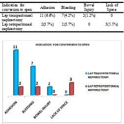
In present study 27(12.3%) patients requiring conversion were having adhesion 13(5.9%) and bleeding 9(4.1%) were the main factors for conversion, while 2 (0.9%) patients required conversion due to bowel injury and limited space in 3(1.3%)
patients. Conversion rate was 12.1% (20/165) for
transperitoneal procedures while 12.9 %(7/54) for
[image:4.595.37.290.188.440.2]retroperitoneal approach.
Table. Indication for conversion to open
Indication for
conversion to open Adhesion Bleeding
Bowel Injury
Lack of Space Lap transperitoneal
nephrectomy
11 (6.6%) 7(4.2%) 2(1.2%) 0
Lap retroperitoneal nephrectomy
2(3.7%) 2(3.7%) 0 3(5.5%)
The role of laparoscopic surgery in patients with pyonephrosis has been controversial in earlier laparoscopic series, with higher open conversion and complication rates (Keeley et al., 1991; Rassweiler et al., 1998 and Bercowsky et al., 1999). Reported open conversion rate is 16-33% and complication rate is 20-50% in a contemporary series (Kapoor et al., 2006; Vanderbrink et al., 2007; Rosoff et al., 2006; Manohar et al., 2007 and Khaira et al., 2005). With increase in advanced laparoscopic experience and skills, LN can be offered in selected patients with acceptable morbidity, decreased blood
loss and shorter convalescence (Guzzo et al., 2009).Katz r. e et
al reported an overall conversion rate of 5% in a series of 185
laparoscopic nephrectomies with 7% and 15% major and minor Complications rate (Katz et al., 2004). Ricardo j et et al reported an overall conversion rate of 28% of 50 laparoscopic nephrectomies cases with 9.0% and 12% major and minor Complications rate (Ricardo et al., 2008). Lee et al reported an overall conversion rate of 17% in a series of 31 laparoscopic nephrectomies with 8% and 16% major and minor Complications rate. Zaidi z et al reported an overall conversion rate of 11.6% in a series of 60 laparoscopic nephrectomies with 3% and 16% major and minor Complications rate (Zaidi
et al., 2007). Wayland hsiao et al reported an overall
conversion rate of 6.6% in a series of 100 laparoscopic nephrectomies with 20% and 11% major and minor Complications (Wayland Hsiao and John, 2008). Hemal ak
et al reported an overall conversion rate of 9.8% in a series of
185 laparoscopic nephrectomies with 9.8% and 3.8% major and minor Complication. M. tobias-machado et al reported an overall conversion rate of 11.7% in a series of 17 laparoscopic
nephrectomies with 6% and 13% major and minor Complications (Tobias-Machado et al., 2005). Present study- Our data suggest that overall conversion rate of 12.1% in a series of 219 laparoscopic nephrectomies with 6.8 % and 12.7 % major and minor Complications. Our overall rate of conversion and complications was comparable with other series performing lap nephrectomies for nonfunctioning pyonephrotic kidney.
Conclusion
In our study, retroperitoneal laparoscopic nephrectomy has to
be considered equal to transperitoneal laproscopic
nephrectomy in terms of conversion to open surgery. In retroperitonel group lack of space is major coencern while in transperitoneal group poor control of bleeding and injury to bowel is main problem. Adhensions are handled better by retroperitoneal laproscpoic surgery.
REFERENCES
Bercowsky, E., Shalhav, A.L., Portis, A., Elbahnasy, A.M., McDougall, E.M., Clayman, R.V. et al. 1999. Is the
laparoscopic approach justified in patients with
Xanthogranulomatous Pyelonephritis? Urology. 54:437– 42. (PubMed)
Clayman, R.V., Kavoussi, L.R., Soper, N.J., Dierks, SM, meretyk s, darcy md, et al. Laparoscopic nephrectomy: initial case report. J urol., 146:278-82.
Gaur, D. D., Agrawal, D. K., Purohit, K. C. 1993. Retroperitoneal laparoscopic nephrectomy: initial case report. J. Urol., 149:103-5.
Guzzo, T.J., Bivalacqua, T.J., Pierorazio, P.M., Varkarakis, J., Schaeffer, E.M., Allaf, M.E. 2009. Xanthogranulomatous Pyelonephritis: Presentation and management in the era of laparoscopy. BJU international. 104:1265–8. (PubMed) Kapoor, R., Vijjan, V., Singh, K., Goyal, R, Mandhani, A,
Dubey, D, et al. 2006. Is laparoscopic nephrectomy the
preferred approach in Xanthogranulomatous
Pyelonephritis? Urology, 68:952–5. (PubMed)
Katz, R., Pode, D., Golijanin, D., Gofrit, O.N., Shenfeld O.Z., Shapiro, A., Reissman, P. 2004. Departments of Urology and General Surgery, Hadassah and Hebrew University Medical Center, Jerusalem, Israel. Dec
Keeley, F.X., Tolley, D.A. 1998. A review of our first 100 cases of laparoscopic nephrectomy defining risk factors for complications. Br J Urol., 82:615–8. (PubMed)
Khaira, H.S., Shah, R.B., Wolf, J.S. 2005. Laparoscopic and open surgical nephrectomy for Xanthogranulomatous Pyelonephritis. J Endourol., 19:813–7. (PubMed)
Manohar, T., Desai, M., Desai, M. 2007. Laparoscopic nephrectomy for benign and inflammatory condition. J
Endourol. 21:1323–8.(PubMed)
McDougall, E.M., Clayman, R.V., Fadden, P.T. 1994. Retroperitoneoscopy the Washington University Medical School experience. Urology, 43:446–52.
Rassweiler, J., Fornara, P., Weber, M., Janetschek, G., Fahlenkamp, D., Henkel, T, et al. 1998. Laparoscopic nephrectomy the experience of the laparoscopy working group of the German Urologic Association. J Urol. 160:18–21. (PubMed)
Ricardo, J. 2008. Duarte, Anuar I. Mitre, José L. Chambô, Marco A. Arap, and Miguel Srougi. Journal of
Endourology. April 2008, 22(4): 681-686. doi:10.1089/
Rosoff, J., Raman, J.D., Del Pizzo, J.J. 2006. Feasibility of
laparoscopic approach in management of
Xanthogranulomatous Pyelonephritis. Urology. 68:711– 4. (PubMed)
Rozenberg, H, Bruyere, F., abdelkader, T, Husset, A., Hamoura, H. 1999. Transperitoneal laparoscopic nephrectomy. Prog Urol, 9:1034-8.
Tobias-Machado; M., Marco, T. Lasmar; Lucas T. Batista; Pedro H. Forseto Jr; Roberto V. Juliano; Eric R. 2005. Wroclawski Division of Urology, ABC Medical School, Santo André, Sao Paulo, Brazil Int. braz j urol., vol.31 no.1 Rio de Janeiro Jan./Feb.
Vanderbrink, B.A., Ost, M.C., Rastinehad, A., Anderson, A., Badlani, G., Smith, A., et al. 2007. Laparoscopic versus open radical nephrectomy for Xanthogranulomatous
Pyelonephritis: Contemporary outcomes analysis. J
Endourol, 21:65–70. (PubMed)
Wayland Hsiao and John G. 2008. Pattaras. Journal of Endourology. October, 22(10): 2285-2290. doi:10.1089/ end.2008.9718.Volume: 22 Issue 10: October 20, 2008 Zaidi, Z., Samad, L., Aquil, S. 2007. The Indus Hospital,
C-76, Sector 31/5, Korangi Crossing, Korangi, Karachi. J Pak
Med Assoc. Jul;57(7):355-9.