Pyridine-2,5-diamine
Sergiu Draguta,a* Victor N. Khrustalev,bMarina S. Fonari,cMikhail Yu. Antipindand Tatiana V. Timofeevad
a
D. Ghitu Institute of Electronic Engineering and Nanotechnologies, 3/3 Academy str., MD-2028 Chisinau, Moldova,bX-Ray Structural Centre, A.N. Nesmeyanov Institute of Organoelement Compounds, Russian Academy of Sciences, 28 Vavilov St, B-334, Moscow 119991, Russian Federation,cInstitute of Applied Physics
Academy of Sciences of Moldova, 5 Academy str., MD-2028 Chisinau, Moldova, anddDepartment of Chemistry & Biology, New Mexico Highlands University, 803
University Avenue, Las Vegas, NM 87701, USA Correspondence e-mail: [email protected]
Received 6 November 2012; accepted 8 November 2012
Key indicators: single-crystal X-ray study;T= 296 K; mean(C–C) = 0.002 A˚; Rfactor = 0.042;wRfactor = 0.109; data-to-parameter ratio = 18.8.
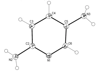
In the title molecule, C5H7N3, intracyclic angles cover the
range 117.15 (10)–124.03 (11). The N atoms of the amino groups have trigonal–pyramidal configurations deviating slightly from the pyridine plane by 0.106 (2) and
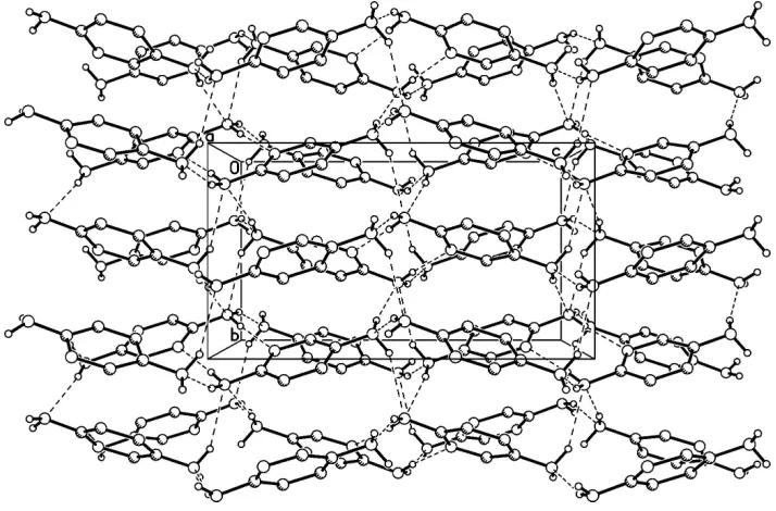
0.042 (2) A˚ . In the crystal, the pyridine N atom serves as an acceptor of an N—H N hydrogen bond which links two molecules into a centrosymmetric dimer. Intermolecular N— H N hydrogen bonds between the amino groups further consolidate the crystal packing, forming a three-dimensional network.
Related literature
For general background, see: Domenicano et al. (1975); Domenicano & Vaciago (1979); Mootz & Wussow (1981); Crawford et al.(2009). For the crystal structures of isomeric diaminopyridines, see: Schwalbe et al. (1987); Rubin-Preminger & Englert (2007); Al-Dajaniet al.(2009); Betzet al. (2011).
Experimental
Crystal data
C5H7N3 Mr= 109.14
a= 11.4447 (11) A˚
b= 7.1447 (7) A˚
c= 12.8030 (12) A˚
V= 1046.89 (17) A˚3
MoKradiation
= 0.09 mm1
T= 296 K
0.300.250.20 mm
Data collection
Bruker APEXII CCD diffractometer
Absorption correction: multi-scan (SADABS; Sheldrick, 2003)
Tmin= 0.973,Tmax= 0.982
13022 measured reflections 1595 independent reflections 1240 reflections withI> 2(I)
Rint= 0.049
Refinement
R[F2> 2(F2)] = 0.042 wR(F2) = 0.109 S= 1.01 1595 reflections 85 parameters
H atoms treated by a mixture of independent and constrained refinement
max= 0.36 e A˚ 3 min=0.24 e A˚ 3
Table 1
Hydrogen-bond geometry (A˚ ,).
D—H A D—H H A D A D—H A
N2—H2A N1i 0.874 (17) 2.183 (17) 3.0541 (15) 175.1 (10) N2—H2B N3ii
0.879 (17) 2.309 (17) 3.1457 (16) 159.3 (10) N3—H3A N2iii
0.894 (16) 2.397 (17) 3.2150 (16) 152.2 (10) N3—H3B N2iv
0.898 (17) 2.593 (17) 3.4803 (16) 170.0 (10)
Symmetry codes: (i) xþ1;yþ1;zþ1; (ii) xþ1
2;yþ1;zþ 1 2; (iii)
x;yþ3 2;z
1
2; (iv)xþ1;y 1 2;zþ
1 2.
Data collection:APEX2(Bruker, 2005); cell refinement:SAINT (Bruker, 2001); data reduction:SAINT; program(s) used to solve structure: SHELXTL (Sheldrick, 2008); program(s) used to refine structure:SHELXTL; molecular graphics:SHELXTL; software used to prepare material for publication:SHELXTL.
The authors are grateful for NSF supportvia DMR grant 0934212 (PREM) and CHE 0832622.
Supplementary data and figures for this paper are available from the IUCr electronic archives (Reference: CV5359).
References
Al-Dajani, M. T. M., Salhin, A., Mohamed, N., Loh, W.-S. & Fun, H.-K. (2009).
Acta Cryst.E65, o2931–o2932.
Betz, R., Gerber, T., Hosten, E. & Schalekamp, H. (2011).Acta Cryst.E67, o2154.
Bruker (2001).SAINT. Bruker AXS Inc., Madison, Wisconsin, USA. Bruker (2005).APEX2. Bruker AXS Inc., Madison, Wisconsin, USA. Crawford, S., Kirchner, M. T., Blaser, D., Boese, R., David, W. I. F., Dawson,
A., Gehrke, A., Ibberson, R. M., Marshall, W. G., Parsons, S. & Yamamuro, O. (2009).Angew. Chem. Int. Ed.48, 755–757.
Domenicano, A. & Vaciago, A. (1979).Acta Cryst.B35, 1382–1388. Domenicano, A., Vaciago, A. & Coulson, C. A. (1975).Acta Cryst.B31, 221–
234.
Mootz, D. & Wussow, H.-G. (1981).J. Chem. Phys.75, 1517–1522. Rubin-Preminger, J. M. & Englert, U. (2007).Acta Cryst.E63, o757–o758. Schwalbe, C. H., Williams, G. J. B. & Koetzle, T. F. (1987).Acta Cryst.C43,
2191–2195.
Sheldrick, G. M. (2003).SADABS. Bruker AXS Inc., Madison, Wisconsin, USA.
Sheldrick, G. M. (2008).Acta Cryst.A64, 112–122.
Structure Reports Online
supporting information
Acta Cryst. (2012). E68, o3353 [doi:10.1107/S1600536812046260]
Pyridine-2,5-diamine
Sergiu Draguta, Victor N. Khrustalev, Marina S. Fonari, Mikhail Yu. Antipin and Tatiana V.
Timofeeva
S1. Comment
Polydentate ligands have found widespread use in coordination chemistry due to the increased thermodynamic stability
of resultant coordination compounds as compared to that of such compounds with monodentate ligands. In this aspect,
the title compound can be considered as a versatile polydentate ligand in metal-organic synthesis. Furthermore, owing to
the presence of three donor atoms, the title compound might play a role of the building block in the formation of
metal-organic frameworks as well as for cocrystals.
In this work, we determined the crystal structure of the title compound (I), C5H7N3 (Figure 1), to enable comparative
studies of its geometrical parameters in metal-organic complexes. Yet, the crystal structure of I can be helpful in the
future investigations.
The geometry of aromatic molecules is known to be sensitive to the electronic effects of substituents. Based on the
crystallographic analysis of monosubstituted arenes, it was concluded (Domenicano et al., 1975; Domenicano & Vaciago,
1979) that the endocyclic angle at the ipso-C atom is > 120° for a electron-withdrawing substituent and < 120° for a
σ-electron-donating substituent. Moreover, in the nitrogen-containing heterocyclic aromatic molecules, the endocyclic
angle at the nitrogen atom is < 120°, and those at the carbon atoms in ortho-positions to the heteroatom are > 120°
(Mootz & Wussow, 1981; Crawford et al., 2009). Similarly to the related diaminopyridines (Schwalbe et al., 1987;
Rubin-Preminger & Englert, 2007; Al-Dajani et al., 2009; Betz et al., 2011), these effects are also manifested in the
investigated compound. So, the smallest endocyclic angle in I (117.13 (10)°) is observed at the C5 carbon atom bearing
the σ-electron-donating amino group, whereas the largest endocyclic angle in I (124.03 (11)°) is observed at the C6
carbon atom in ortho-position to the N1 heteroatom. The value of the endocyclic angle at the second (C2) carbon atom in
ortho-position to the N1 heteroatom (121.83 (11)°) is significantly smaller than that at the C6 carbon atom because the
C2 carbon atom bears the σ-electron-donating amino group, i.e., the C2 carbon atom is subjected to the influence of the
two opposed electronic effects. The value of the endocyclic angle at the N1 heteroatom (118.15 (10)°) is also in good
agreement with the above mentioned electronic effects.
The N2 and N3 nitrogen atoms of the amino groups have the trigonal-pyramidal configurations. It is worthy to note that
these atoms are slightly out of the plane defined the aromatic system (r.m.s. deviation is 0.003 Å) by 0.106 (2) and
-0.042 (2) Å, respectively. Apparently, this fact is explained by the developed hydrogen bonding system with the
participation of the both amino groups.
In the crystal of I, the amino groups act both as proton donors and proton acceptors upon formation of the
intermolecular N—H···N hydrogen bonds (Table 1). The N1 heteroatom serves as the acceptor for a hydrogen atom of
one of the two amino groups (Table 1). In total, the molecules of I are linked by the intermolecular N—H···N hydrogen
The compound I was obtained commercially (Aldrich) as a fine-crystalline powder and purified additionally by filtration.
Crystals suitable for the X-ray diffraction study were grown by slow evaporation from chloroform solution.
S3. Refinement
The hydrogen atoms of the amino groups were localized in the difference-Fourier map and refined isotropically with
fixed isotropic displacement parameters [Uiso(H) = 1.2Ueq(N)]. C-bound H atoms were placed in calculated positions [C—
[image:3.610.112.493.189.470.2]H = 0.93 Å], and refined as riding, with Uiso(H) = 1.2Ueq(C).
Figure 1
Molecular structure of I. Displacement ellipsoids are shown at the 50% probability level. H atoms are presented as small
Figure 2
A portion of the crystal packing showing intermolecular N—H···N hydrogen bonds as dashed lines.
Pyridine-2,5-diamine
Crystal data
C5H7N3 Mr = 109.14
Orthorhombic, Pbca Hall symbol: -P 2ac 2ab a = 11.4447 (11) Å b = 7.1447 (7) Å c = 12.8030 (12) Å V = 1046.89 (17) Å3 Z = 8
F(000) = 464 Dx = 1.385 Mg m−3
Mo Kα radiation, λ = 0.71073 Å Cell parameters from 2334 reflections θ = 3.2–28.8°
µ = 0.09 mm−1 T = 296 K Prism, red
0.30 × 0.25 × 0.20 mm
Data collection
Bruker APEXII CCD diffractometer
Radiation source: fine-focus sealed tube Graphite monochromator
φ and ω scans
Absorption correction: multi-scan (SADABS; Sheldrick, 2003) Tmin = 0.973, Tmax = 0.982
13022 measured reflections 1595 independent reflections 1240 reflections with I > 2σ(I) Rint = 0.049
θmax = 30.5°, θmin = 3.2° h = −16→16
k = −10→10 l = −18→18
Refinement
Refinement on F2 Least-squares matrix: full R[F2 > 2σ(F2)] = 0.042 wR(F2) = 0.109 S = 1.01 1595 reflections 85 parameters
0 restraints
Primary atom site location: structure-invariant direct methods
Secondary atom site location: difference Fourier map
and constrained refinement w = 1/[σ2(F
o2) + (0.0475P)2 + 0.63P] where P = (Fo2 + 2Fc2)/3
Δρmax = 0.36 e Å−3 Δρmin = −0.24 e Å−3
Special details
Geometry. All e.s.d.'s (except the e.s.d. in the dihedral angle between two l.s. planes) are estimated using the full covariance matrix. The cell e.s.d.'s are taken into account individually in the estimation of e.s.d.'s in distances, angles and torsion angles; correlations between e.s.d.'s in cell parameters are only used when they are defined by crystal symmetry. An approximate (isotropic) treatment of cell e.s.d.'s is used for estimating e.s.d.'s involving l.s. planes.
Refinement. Refinement of F2 against ALL reflections. The weighted R-factor wR and goodness of fit S are based on F2, conventional R-factors R are based on F, with F set to zero for negative F2. The threshold expression of F2 > σ(F2) is used only for calculating R-factors(gt) etc. and is not relevant to the choice of reflections for refinement. R-factors based on F2 are statistically about twice as large as those based on F, and R- factors based on ALL data will be even larger.
Fractional atomic coordinates and isotropic or equivalent isotropic displacement parameters (Å2)
x y z Uiso*/Ueq
N1 0.46050 (8) 0.50612 (15) 0.35845 (8) 0.0150 (2)
N2 0.36236 (10) 0.66538 (15) 0.49098 (8) 0.0167 (2)
H2A 0.4111 (14) 0.610 (2) 0.5333 (13) 0.020*
H2B 0.2925 (15) 0.674 (2) 0.5190 (12) 0.020*
N3 0.38898 (10) 0.41571 (15) 0.07954 (8) 0.0164 (2)
H3A 0.3841 (13) 0.510 (2) 0.0337 (12) 0.020*
H3B 0.4559 (15) 0.352 (2) 0.0701 (12) 0.020*
C2 0.36402 (10) 0.59703 (16) 0.38919 (9) 0.0139 (2)
C3 0.27102 (10) 0.63298 (17) 0.32018 (9) 0.0148 (2)
H3 0.2045 0.6952 0.3433 0.018*
C4 0.28001 (10) 0.57463 (16) 0.21780 (9) 0.0150 (2)
H4 0.2192 0.5971 0.1712 0.018*
C5 0.38092 (10) 0.48140 (16) 0.18414 (9) 0.0139 (2)
C6 0.46769 (10) 0.45101 (17) 0.25771 (9) 0.0148 (2)
H6 0.5350 0.3889 0.2364 0.018*
Atomic displacement parameters (Å2)
U11 U22 U33 U12 U13 U23
N1 0.0139 (5) 0.0159 (5) 0.0154 (5) −0.0001 (4) −0.0006 (4) 0.0002 (4)
N2 0.0162 (5) 0.0189 (5) 0.0150 (5) 0.0022 (4) −0.0009 (4) −0.0008 (4)
N3 0.0163 (5) 0.0190 (5) 0.0137 (5) 0.0027 (4) 0.0005 (4) 0.0003 (4)
C2 0.0148 (5) 0.0120 (5) 0.0148 (5) −0.0018 (4) 0.0007 (4) 0.0006 (4)
C3 0.0130 (5) 0.0138 (5) 0.0176 (5) 0.0010 (4) 0.0003 (4) 0.0012 (4)
C4 0.0134 (5) 0.0138 (5) 0.0177 (5) 0.0001 (4) −0.0024 (4) 0.0018 (4)
C5 0.0148 (5) 0.0124 (5) 0.0143 (5) −0.0013 (4) 0.0008 (4) 0.0008 (4)
C6 0.0122 (5) 0.0154 (5) 0.0169 (5) 0.0007 (4) 0.0009 (4) 0.0003 (4)
Geometric parameters (Å, º)
N1—C2 1.3401 (15) C2—C3 1.4069 (16)
N2—C2 1.3919 (15) C3—H3 0.9300
N2—H2A 0.874 (17) C4—C5 1.4011 (16)
N2—H2B 0.879 (17) C4—H4 0.9300
N3—C5 1.4221 (15) C5—C6 1.3859 (16)
N3—H3A 0.894 (16) C6—H6 0.9300
N3—H3B 0.898 (17)
C2—N1—C6 118.15 (10) C4—C3—H3 120.5
C2—N2—H2A 114.3 (11) C2—C3—H3 120.5
C2—N2—H2B 114.8 (10) C3—C4—C5 119.83 (11)
H2A—N2—H2B 111.2 (14) C3—C4—H4 120.1
C5—N3—H3A 111.4 (10) C5—C4—H4 120.1
C5—N3—H3B 110.4 (10) C6—C5—C4 117.13 (10)
H3A—N3—H3B 110.2 (14) C6—C5—N3 122.81 (11)
N1—C2—N2 117.15 (10) C4—C5—N3 120.01 (10)
N1—C2—C3 121.83 (11) N1—C6—C5 124.03 (11)
N2—C2—C3 120.90 (11) N1—C6—H6 118.0
C4—C3—C2 119.02 (11) C5—C6—H6 118.0
C6—N1—C2—N2 −175.08 (11) C3—C4—C5—C6 0.54 (16)
C6—N1—C2—C3 0.96 (17) C3—C4—C5—N3 177.98 (11)
N1—C2—C3—C4 −0.66 (17) C2—N1—C6—C5 −0.51 (17)
N2—C2—C3—C4 175.23 (11) C4—C5—C6—N1 −0.23 (17)
C2—C3—C4—C5 −0.12 (17) N3—C5—C6—N1 −177.60 (11)
Hydrogen-bond geometry (Å, º)
D—H···A D—H H···A D···A D—H···A
N2—H2A···N1i 0.874 (17) 2.183 (17) 3.0541 (15) 175.1 (10)
N2—H2B···N3ii 0.879 (17) 2.309 (17) 3.1457 (16) 159.3 (10)
N3—H3A···N2iii 0.894 (16) 2.397 (17) 3.2150 (16) 152.2 (10)
N3—H3B···N2iv 0.898 (17) 2.593 (17) 3.4803 (16) 170.0 (10)