research communications
Acta Cryst.(2019). E75, 1399–1402 https://doi.org/10.1107/S2056989019011964
1399
Received 24 July 2019 Accepted 29 August 2019
Edited by K. Fejfarova, Institute of Biotechnology CAS, Czech Republic
Keywords:crystal structure; chiral-amino alcohol; tetrahydroisoquinoline; hydrogen bond; Hirshfeld surface analysis.
CCDC reference:1950166
Supporting information:this article has supporting information at journals.iucr.org/e
Crystal structure of
(1S,2R)-2-[(3R,4S)-3-methyl-4-
phenyl-1,2,3,4-tetrahydroisoquinolin-2-yl]-1,2-di-phenylethanol
Karim Ben Aliaand Pascal Retailleaub*
a
Laboratoire de Recherche en Energie et Matie`re pour le De´veloppement des Sciences Nucle´aires, Centre National des Sciences et Technologies Nucle´aires, Poˆle Technologique, 2020 Sidi-Thabet, Tunisia, andbInstitut de Chimie des Substances Naturelles, CNRS UPR 2301, Universite´ Paris-Sud, Paris-Saclay University, 1, av. de la Terrasse, 91198 Gif-sur-Yvette, France. *Correspondence e-mail: pascal.retailleau@cnrs.fr
The synthesis and crystal structure of the title compound, C30H29NO, are
described. This compound is a member of the chiral dihydroisoquinoline-derived family, used as building blocks for functional materials and as source of chirality in asymmetric synthesis, and was isolated as one of two diastereomeric
-amino alcohols, the title molecule being found to be the (S,R) diastereoi-somer. In the crystal, molecules are packed in a herringbone manner parallel to (103) and (103)viaweak C—H O and C—H (ring) interactions. Hirshfeld surface analysis showed that the surface contacts are predominantly H H interactions (ca 75%). The crystal studied was refined as a two-component inversion twin.
1. Chemical context
-amino alcohols exhibit a broad spectrum of biological activities and are used as antibacterial and tuberculostatic agents (Yendapally & Lee, 2008). In particular, chiral-amino alcohols are very important chiral molecules that are used as building blocks and structural motifs in pharmaceutically active molecules and natural products and which serve as the main sources of chirality in asymmetric synthesis (Leeet al., 2003; Malkovet al., 2007; Guoet al., 2017).
Among this family of chiral amino-alcohols is the title compound, (I), which we prepared through the alkylation of tetrahydroisoquinoline by the opening racemictrans-stilbene oxide reaction. Two diastereoisomers were obtained in a 1:1 ratio as determined by1H NMR analysis on the crude mixture. These diastereoisomers were separated by column chroma-tography. The title molecule was found to be the (S,R) diastereoisomer.
2. Structural commentary
The structure of (I) was confirmed using single crystal X-ray diffraction. The asymmetric unit of the orthorhombic unit cell comprises a single molecule, shown in Fig. 1. The tetra-hydroisoquinoline unit is substituted by a methyl group in position 3, a phenyl substituent in position 4 and a-alcohol substituent at the N atom. The heterocyclic ring exhibits a half-chair conformation, with atom C3 deviating by 0.706 (3) A˚ from the plane formed by atoms C1/N2/C4/C9/ C10. The substituents in positions 3 and 4 of the heterocyclic ring are in axial positions. The molecular structure of (I) is stabilized by an intramolecular hydrogen bond between the hydroxy O19—H19 group and atom N2, and to a lesser extent, between the aromatic C21—H21 and the phenyl group in position 4 (Table 1). By reference to two unchanging chiral C18 and C19 atoms, the molecule was found to be the (18R,19S) diastereoisomer resulting from the reaction of tetrahydroisoquinoline and the (S,S) trans-stilbene oxide enantiomer.
This structure was confirmed through the means of usual 1D and 2D NMR experiments. NMR data show that the trans
diequatorial arrangement of H3 and H4 is suggested by the coupling constant between H3 and H4 in 1H NMR (J3,4
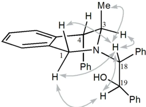
0 Hz), so the substituents C3-methyl and C4-phenyl are in an axial disposition. The absolute configurations of carbon atoms C18 and C19 were deduced from the NOESY maps to beR
andS, respectively (Fig. 2).
3. Supramolecular features
In the crystal, molecules of (I) pack with no classical hydrogen bonds: the potential donor hydroxyl group is involved in an intramolecular interaction with the N atom. However, the oxygen atom acts as an acceptor in the short contact C6— H6 O19 (x, 1
2 + y, 1
2 z) with an O19 H distance of
2.57 A˚ , which is of the same order of magnitude of the H O van der Waals distance (2.60 A˚ ), whereas C—H O contacts are frequently reported with H O separations shorter than 2.4 A˚ (Taylor & Kennard, 1982). The N atom does not play a role in the packing as it is buried inside the structure. Nevertheless, these directed C—H O interactions make an
1400
Ben Ali and Retailleau C30H29NO Acta Cryst.(2019). E75, 1399–1402
[image:2.610.313.565.69.253.2]research communications
Table 1
Hydrogen-bond geometry (A˚ ,).
Cg2,Cg3,Cg4 andCg5 are the centroids of the C5–C10, C12-C17, C20–C25, and C26–C31 rings, respectively.
D—H A D—H H A D A D—H A
O19—HOH N2 0.86 (3) 2.18 (3) 2.737 (2) 123 (2)
C27—H27 O19 0.93 2.48 2.798 (3) 100
C21—H21 Cg3 0.93 3.14 3.930 (4) 144
C6—H6 O19i 0.93 2.57 3.492 (3) 170
C14—H14 Cg5ii 0.93 2.95 3.770 (4) 147
C16—H16 Cg4iii 0.93 2.92 3.743 (3) 148
C31—H31 Cg2iv 0.93 2.96 3.803 (3) 152
Symmetry codes: (i) x;yþ1
2;zþ12; (ii) x;yþ1;z; (iii) x1;y;z; (iv)
xþ1;y1 2;zþ
[image:2.610.44.295.119.203.2]1 2.
Figure 2
[image:2.610.316.564.535.709.2]Selected NOESY correlations observed for compound (I).
Figure 3
The ribbon structure of (I) formed along theb-axis direction viaC— H O interactions (cyan dashed lines) and C—H interactions (blue dashed lines). The red spheres indicate the centroids of the phenyl rings. Figure 1
[image:2.610.46.297.554.695.2]important contribution to the packing: zigzagging along the [010] direction, they pair molecules in ribbons, placing the isoquinoline moieties parallel to the (103) plane on both sides but without overlapping. The ribbon cohesion is reinforced by C—H interactions involving the phenyl group in position 4 and those attached to the-alcohol part and which flank the ribbon, as shown in Fig. 3. They stack in the [100] direction as columns arranged in a herringbone manner but avoiding- -stacking (Fig. 4).
4. Database survey
A search of the Cambridge Structural Database, CSD (Version 5.40; ConQuest1.21; Groomet al., 2016) found 495 structures of tetrahydroisoquinoline derivatives. Limiting the search to compounds with tri-substitutions on positions C3, C4 and the secondary amine N reduces the number of structures to seven: ADAGOC (Gzellaet al., 2006), JIPKEZ (Whiteet al., 2007), TIBPIE (Ben Aliet al., 2007), VAHJOG (Davieset al., 2016), XOSDUE (Gzellaet al., 2002), YEKKIK (Shiet al., 2012) and ZIFSUE (Guoet al., 2013). Except for the racemic VAHJOG, they all crystallize in the sameP212121space group.
The structures of ZIFSUE, TIBPIE, VAHJOG, JIPKEZ and (I) superimpose well over the heterobicycle with the same conformation, unlike ADAGOC and XOSDUE which have a different half-chair configuration. The amino alcohol TIBPIE is obviously the closest related structure, differing in the N substitution of a cyclohexane carrying the hydroxyl group which is involved in the intramolecular hydrogen bond.
5. Hirshfeld surface analysis
The intermolecular interactions were quantified using Hirsh-feld surface analysis and the associated two-dimensional fingerprint plots using CrystalExplorer17.5 (Turner et al., 2017). The electrostatic potentials were calculated using
TONTO, integrated within CrystalExplorer. The analysis of intermolecular interactions through the mapping of dnorm
presented in Fig. 5 compares the contact distancesdiandde
from the Hirshfeld surface to the nearest atom inside and outside, respectively, with their respective van der Waals radii. The blue, white and red colour conventions recognize the interatomic contacts as longer, at van der Waals separations and short interatomic contacts. The C—H O contacts are
identified in the dnorm-mapped surface as two red spots showing the interaction between the neighbouring molecules (Fig. 5a). The overall two-dimensional fingerprint plot derived form the Hirshfeld surface is a useful method to summarize the frequency of each combination of de and di across the surface of the studied molecule, encompassing all inter-molecular contacts (Fig. 5b). The delineated fingerprint plots (Fig. 5b and 6a,c) focus on specific interactions, providing information about the major and minor percentage contribu-tion of interatomic contacts in the compound. The H H interactions account for the three quarters of the total (73.7%) with an evident sting at aboutdi= de= 1.1 A˚ (Fig. 5b). The C H/H C plot, which refers to the C—H interactions previously described (22.7%,) shows two broad symmetrical wings at aboutdi+de= 2.8 A˚ (Fig. 6a). These interactions are
research communications
Acta Cryst.(2019). E75, 1399–1402 Ben Ali and Retailleau C
[image:3.610.42.313.72.156.2]30H29NO
1401
Figure 5(a) View of the three-dimensional Hirshfeld surface mapped overdnorm,
[image:3.610.317.565.72.249.2]over the range0.1345 and +1.8231 arbitrary units, (b) the full two-dimensional fingerprint plot for (I) and the two-two-dimensional fingerprint plots for the O H/H O interactions and the H H interactions
Figure 6
(a) The Hirshfeld surface mapped over the shape-index property, (b) the two-dimensional fingerprint plot for the H C/C H interactions, (c) the Hirshfeld surface mapped over curvedness and (d) the two-dimensional fingerprint plot for the C C interactions in the title compound. Figure 4
[image:3.610.313.564.514.694.2]observed as red regions on the shape-index surface (Fig. 6b). The absence of C C contacts, highlighted by the Hirshfeld surface with high curvedness delineated by dark-blue edges, confirms that no– stacking interactions take place in the crystal packing (Fig. 6c,d). The third marginal contribution is O H/H O (3.6%) with a pair of sharp spikes at about
di + de = 2.4 A˚ , symmetrically disposed with respect to the diagonal, indicating the presence of intermolecular C—H O interactions, which play a role in ordering the molecules inside the crystal.
6. Synthesis and crystallization
The title -amino alcohol was obtained by mixing racemic
trans-stilbene oxide (5.1g, 26mmol) with (3R,4S )-3-methyl-4-phenyl-1,2,3,4-tetrahydroisoquinoleine (3g, 13mmol), which was prepared according to the method of Bohe´et al.(1999).
The mixture was heated at 353.15 K for 48 h in CF3CH2OH
(65 ml), the reaction being monitored by TLC. Two diaster-eoisomers were obtained in a 1:1 ratio. These diasterdiaster-eoisomers were separated by column chromatography. Only the title compound (white solid) was successfully recrystallized. Crys-tals were grown by placing this dastereoisomer in a minimum
amount of hot heptane. [] D 25
= 23.6 (c 1, CHCl3), m.p.
425 K.
7. Refinement
Crystal data, data collection and structure refinement details are summarized in Table 2. H atoms were placed in calculated positions (C—H = 0.93–0.98 A˚ ) and refined as riding with
Uiso(H) = 1.2Ueq(C). The crystal studied was refined as a two-component inversion twin.
Acknowledgements
The authors are indebted to Dr Mathias Meyer (Rigaku) for his invaluable help in converting ancient KappaCCD images into a format readable byCrysAlis PROsoftware.
References
Ben Ali, K., Chiaroni, A. & Bohe´, L. (2007).Acta Cryst.E63, o1719– o1720.
Bohe´, L., Lusinchi, M. & Lusinchi, X. (1999).Tetrahedron,55, 141– 154.
Davies, S. G., Fletcher, A. M., Frost, A. B., Kennedy, M. S., Roberts, P. M. & Thomson, J. E. (2016).Tetrahedron,72, 2139–2163. Groom, C. R., Bruno, I. J., Lightfoot, M. P. & Ward, S. C. (2016).Acta
Cryst.B72, 171–179.
Guo, J., Zhu, M., Wu, T., Hao, C., Wang, K., Yan, Z., Huang, W., Wang, J., Zhao, D. & Cheng, M. (2017).Bioorg. Med. Chem. 25, 3500–3511.
Guo, R.-N., Cai, X.-F., Shi, L., Ye, Z.-S., Chen, M.-W. & Zhou, Y.-G. (2013).Chem. Commun.49, 8537–8539.
Gzella, A., Bro´zda, D., Koroniak, L. & Rozwadowska, M. D. (2002). Acta Cryst.C58, o503–o506.
Gzella, A., Chrzanowska, M., Dreas, A., Kaczmarek, M. S. & Wozniak, Z. (2006).Acta Cryst.E62, o1774–o1776.
Hooft, R. W. W. (1998). COLLECT. Nonius BV, Delft, The Netherlands.
Lee, R. E., Protopopova, M., Crooks, E., Slayden, R. A., Terrot, M. & Barry, C. E. (2003).J. Comb. Chem.5, 172–187.
Malkov, A. V., Kabeshov, M. A., Bella, M., Kysilka, O., Malyshev, D. A., Pluha´cˇkova´, K. & Kocˇovsky´, P. (2007).Org. Lett.9, 5473– 5476.
Otwinowski, Z. & Minor, W. (1997).Methods in Enzymology, Vol. 276, Macromolecular Crystallography, Part A, edited by C. W. Carter Jr & R. M. Sweet, pp. 307–326. New York: Academic Press. Rigaku OD (2019). CrysAlis PRO. Rigaku Oxford Diffraction,
Yarnton, England.
Sheldrick, G. M. (2015a).Acta Cryst.A71, 3–8. Sheldrick, G. M. (2015b).Acta Cryst.C71, 3–8.
Shi, L., Ye, Z.-S., Cao, L.-L., Guo, R.-N., Hu, Y. & Zhou, Y.-G. (2012). Angew. Chem. Int. Ed.51, 8286–8289.
Taylor, R. & Kennard, O. (1982).J. Am. Chem. Soc.104, 5063–5070. Turner, M. J., McKinnon, J. J., Wolff, S. K., Grimwood, D. J., Spackman, P. R., Jayatilaka, D. & Spackman, M. A. (2017). CrystalExplorer17. University of Western Australia.
White, J., Hulme, A. & Parsons, S. (2007). Private communication (refcode JIPKEZ). CCDC, Cambridge, England.
Yendapally, R. & Lee, R. E. (2008).Bioorg. Med. Chem. Lett. 18, 1607–1611.
1402
Ben Ali and Retailleau C30H29NO Acta Cryst.(2019). E75, 1399–1402
[image:4.610.43.295.90.376.2]research communications
Table 2
Experimental details.
Crystal data
Chemical formula C30H29NO
Mr 419.54
Crystal system, space group Orthorhombic,P212121
Temperature (K) 293
a,b,c(A˚ ) 7.3009 (8), 11.0552 (11), 30.006 (3)
V(A˚3) 2421.8 (4)
Z 4
Radiation type MoK
(mm1) 0.07
Crystal size (mm) 0.590.450.35
Data collection
Diffractometer Nonius KappaCCD area detector
Absorption correction Multi-scan (CrysAlis PRO; Rigaku
OD, 2019)
Tmin,Tmax 0.844, 1.000
No. of measured, independent and observed [I> 2(I)] reflections
21751, 4427, 3948
Rint 0.027
(sin/)max(A˚
1
) 0.602
Refinement
R[F2> 2(F2)],wR(F2),S 0.038, 0.083, 1.07
No. of reflections 4425
No. of parameters 295
H-atom treatment H atoms treated by a mixture of
independent and constrained refinement
max, min(e A˚
3
) 0.11,0.11
Absolute structure Refined as an inversion twin.
Computer programs:DENZO(Otwinowski & Minor, 1997);COLLECT(Hooft, 1998),
CrysAlis PRO(Rigaku OD, 2019),SHELXT2014/5(Sheldrick, 2015a),SHELXL2018/3
supporting information
sup-1
Acta Cryst. (2019). E75, 1399-1402
supporting information
Acta Cryst. (2019). E75, 1399-1402 [https://doi.org/10.1107/S2056989019011964]
Crystal structure of (1
S
,2
R
)-2-[(3
R
,4
S
)-3-methyl-4-phenyl-1,2,3,4-tetrahydro-isoquinolin-2-yl]-1,2-diphenylethanol
Karim Ben Ali and Pascal Retailleau
Computing details
Data collection: DENZO (Otwinowski & Minor, 1997); COLLECT (Hooft, 1998); cell refinement: CrysAlis PRO (Rigaku
OD, 2019); data reduction: CrysAlis PRO (Rigaku OD, 2019); program(s) used to solve structure: SHELXT2014/5
(Sheldrick, 2015a); program(s) used to refine structure: SHELXL2018/3 (Sheldrick, 2015b); software used to prepare
material for publication: SHELXL2018/3 (Sheldrick, 2015b).
(1S,2R)-2-[(3R,4S)-3-Methyl-4-phenyl-1,2,3,4-tetrahydroisoquinolin-2-yl]-1,2-diphenylethanol
Crystal data
C30H29NO
Mr = 419.54
Orthorhombic, P212121
a = 7.3009 (8) Å b = 11.0552 (11) Å c = 30.006 (3) Å V = 2421.8 (4) Å3
Z = 4 F(000) = 896
Dx = 1.151 Mg m−3
Mo Kα radiation, λ = 0.71073 Å Cell parameters from 6302 reflections θ = 2.0–24.2°
µ = 0.07 mm−1
T = 293 K Prism, colorless 0.59 × 0.45 × 0.35 mm
Data collection
Nonius KappaCCD area detector diffractometer
Radiation source: 1.5kW sealed tube Graphite monochromator
ω and φ scans
Absorption correction: multi-scan (CrysAlisPro; Rigaku OD, 2019) Tmin = 0.844, Tmax = 1.000
21751 measured reflections 4427 independent reflections 3948 reflections with I > 2σ(I) Rint = 0.027
θmax = 25.4°, θmin = 2.7°
h = −8→8 k = −13→13 l = −36→36
Refinement
Refinement on F2 Least-squares matrix: full R[F2 > 2σ(F2)] = 0.038
wR(F2) = 0.083
S = 1.07 4425 reflections 295 parameters 0 restraints
Primary atom site location: dual
Secondary atom site location: difference Fourier map
Hydrogen site location: mixed
H atoms treated by a mixture of independent and constrained refinement
w = 1/[σ2(F
o2) + (0.0304P)2 + 0.3321P] where P = (Fo2 + 2Fc2)/3
(Δ/σ)max < 0.001 Δρmax = 0.11 e Å−3 Δρmin = −0.11 e Å−3
Extinction correction: SHELXL-2018/3 (Sheldrick 2015b),
supporting information
sup-2
Acta Cryst. (2019). E75, 1399-1402
Extinction coefficient: 0.0139 (11) Absolute structure: Refined as an inversion twin.
Special details
Geometry. All esds (except the esd in the dihedral angle between two l.s. planes) are estimated using the full covariance matrix. The cell esds are taken into account individually in the estimation of esds in distances, angles and torsion angles; correlations between esds in cell parameters are only used when they are defined by crystal symmetry. An approximate (isotropic) treatment of cell esds is used for estimating esds involving l.s. planes.
Refinement. Refined as a 2-component inversion twin.
Fractional atomic coordinates and isotropic or equivalent isotropic displacement parameters (Å2)
x y z Uiso*/Ueq
supporting information
sup-3
Acta Cryst. (2019). E75, 1399-1402
H19 0.338016 −0.182861 0.163143 0.048* O19 0.2046 (2) −0.08765 (16) 0.11933 (6) 0.0560 (5) HOH 0.198 (4) −0.016 (3) 0.1307 (9) 0.067* C20 0.5896 (3) 0.02111 (19) 0.10855 (7) 0.0394 (5) C21 0.4942 (4) 0.0997 (2) 0.08094 (8) 0.0553 (7) H21 0.377223 0.125320 0.088719 0.064* C22 0.5742 (5) 0.1398 (3) 0.04165 (9) 0.0730 (8) H22 0.510686 0.192712 0.023154 0.084* C23 0.7467 (5) 0.1018 (3) 0.02999 (10) 0.0778 (10) H23 0.798359 0.127746 0.003312 0.089* C24 0.8428 (4) 0.0263 (3) 0.05729 (10) 0.0685 (8) H24 0.960600 0.001927 0.049589 0.079* C25 0.7644 (3) −0.0137 (2) 0.09627 (8) 0.0502 (6) H25 0.830335 −0.065258 0.114775 0.058* C26 0.4523 (3) −0.22470 (19) 0.10333 (7) 0.0434 (5) C27 0.4150 (4) −0.2197 (2) 0.05843 (9) 0.0648 (8) H27 0.330932 −0.163529 0.047712 0.075* C28 0.5013 (5) −0.2974 (3) 0.02915 (9) 0.0816 (10) H28 0.474735 −0.293390 −0.001130 0.094* C29 0.6252 (5) −0.3798 (3) 0.04431 (11) 0.0786 (9) H29 0.684240 −0.431168 0.024431 0.090* C30 0.6622 (4) −0.3865 (2) 0.08887 (11) 0.0693 (8) H30 0.746120 −0.442975 0.099374 0.080* C31 0.5752 (4) −0.3097 (2) 0.11835 (9) 0.0533 (6) H31 0.599963 −0.315488 0.148673 0.061*
Atomic displacement parameters (Å2)
U11 U22 U33 U12 U13 U23
supporting information
sup-4
Acta Cryst. (2019). E75, 1399-1402
O19 0.0414 (9) 0.0551 (10) 0.0715 (12) 0.0037 (8) −0.0092 (9) −0.0069 (9) C20 0.0415 (13) 0.0403 (11) 0.0364 (12) −0.0010 (10) 0.0015 (10) −0.0042 (9) C21 0.0617 (17) 0.0595 (14) 0.0448 (14) 0.0047 (14) 0.0015 (12) 0.0049 (12) C22 0.097 (2) 0.0752 (19) 0.0467 (15) −0.0039 (19) −0.0026 (17) 0.0129 (14) C23 0.095 (3) 0.091 (2) 0.0469 (16) −0.024 (2) 0.0244 (17) −0.0042 (16) C24 0.0622 (18) 0.082 (2) 0.0616 (18) −0.0138 (17) 0.0209 (15) −0.0176 (16) C25 0.0452 (14) 0.0532 (14) 0.0522 (15) −0.0035 (12) 0.0073 (11) −0.0090 (11) C26 0.0456 (14) 0.0384 (11) 0.0461 (13) −0.0055 (11) −0.0012 (11) −0.0046 (10) C27 0.086 (2) 0.0577 (15) 0.0507 (16) 0.0103 (16) −0.0131 (15) −0.0060 (12) C28 0.122 (3) 0.076 (2) 0.0477 (16) 0.007 (2) −0.0009 (18) −0.0158 (15) C29 0.095 (2) 0.0622 (18) 0.078 (2) 0.0071 (19) 0.0216 (19) −0.0239 (16) C30 0.069 (2) 0.0568 (16) 0.082 (2) 0.0137 (15) 0.0011 (17) −0.0151 (15) C31 0.0581 (16) 0.0453 (12) 0.0563 (15) 0.0058 (12) −0.0049 (13) −0.0066 (11)
Geometric parameters (Å, º)
supporting information
sup-5
Acta Cryst. (2019). E75, 1399-1402
C15—H15 0.9300
supporting information
sup-6
Acta Cryst. (2019). E75, 1399-1402
C13—C12—C4 119.3 (2) C27—C28—H28 119.8 C17—C12—C4 122.7 (2) C28—C29—C30 119.7 (3) C12—C13—C14 121.1 (3) C28—C29—H29 120.2 C12—C13—H13 119.5 C30—C29—H29 120.2 C14—C13—H13 119.5 C29—C30—C31 120.2 (3) C15—C14—C13 120.2 (3) C29—C30—H30 119.9 C15—C14—H14 119.9 C31—C30—H30 119.9 C13—C14—H14 119.9 C26—C31—C30 120.7 (3) C14—C15—C16 119.7 (3) C26—C31—H31 119.7 C14—C15—H15 120.2 C30—C31—H31 119.7
supporting information
sup-7
Acta Cryst. (2019). E75, 1399-1402 Hydrogen-bond geometry (Å, º)
Cg2, Cg3, Cg4 and Cg5 are the centroids of the C5–C10, C12-C17, C20–C25, and C26–C31 rings, respectively.
D—H···A D—H H···A D···A D—H···A
O19—HOH···N2 0.86 (3) 2.18 (3) 2.737 (2) 123 (2) C27—H27···O19 0.93 2.48 2.798 (3) 100 C21—H21···Cg3 0.93 3.14 3.930 (4) 144 C6—H6···O19i 0.93 2.57 3.492 (3) 170 C14—H14···Cg5ii 0.93 2.95 3.770 (4) 147 C16—H16···Cg4iii 0.93 2.92 3.743 (3) 148 C31—H31···Cg2iv 0.93 2.96 3.803 (3) 152