Received 17 July 2019 Accepted 18 July 2019
Edited by W. T. A. Harrison, University of Aberdeen, Scotland
‡ Additional correspondence author, e-mail: julio@power.ufscar.br.
Keywords:crystal structure; acetylene; hydrogen bonding; Hirshfeld surface analysis; NCI plots; computational chemistry.
CCDC reference:1941466
Supporting information:this article has supporting information at journals.iucr.org/e
2-Methyl-4-(4-nitrophenyl)but-3-yn-2-ol: crystal
structure, Hirshfeld surface analysis and
computational chemistry study
Ignez Caracelli,aJulio Zukerman-Schpector,b‡ Ricardo S. Schwab,bEverton M. da Silva,bMukesh M. Jotanicand Edward R. T. Tiekinkd*
aDepartamento de Fı´sica, Universidade Federal de Sa˜o Carlos, 13565-905 Sa˜o Carlos, SP, Brazil,bDepartamento de
Quı´mica, Universidade Federal de Sa˜o Carlos, 13565-905 Sa˜o Carlos, SP, Brazil,cDepartment of Physics, Bhavan’s Sheth
R. A. College of Science, Ahmedabad, Gujarat 380001, India, anddResearch Centre for Crystalline Materials, School of
Science and Technology, Sunway University, 47500 Bandar Sunway, Selangor Darul Ehsan, Malaysia. *Correspondence e-mail: edwardt@sunway.edu.my
The di-substituted acetylene residue in the title compound, C11H11NO3, is capped at either end by di-methylhydroxy and 4-nitrobenzene groups; the nitro substituent is close to co-planar with the ring to which it is attached [dihedral angle = 9.4 (3)]. The most prominent feature of the molecular packing is the
formation, via hydroxy-O—H O(hydroxy) hydrogen bonds, of hexameric clusters about a site of symmetry 3. The aggregates are sustained by 12-membered { OH}6 synthons and have the shape of a flattened chair. The clusters are connected into a three-dimensional architecture by benzene-C— H O(nitro) interactions, involving both nitro-O atoms. The aforementioned interactions are readily identified in the calculated Hirshfeld surface. Computational chemistry indicates there is a significant energy, primarily electrostatic in nature, associated with the hydroxy-O—H O(hydroxy) hydrogen bonds. Dispersion forces are more important in the other identified but, weaker intermolecular contacts.
1. Chemical context
compound, (I), previously reported (Bleicheret al., 1998), was isolated and crystallized. Herein, the crystal and molecular structures of (I) are described along with a detailed analysis of the molecular packing by Hirshfeld surface analysis, non-covalent interaction plots and computational chemistry.
2. Structural commentary
The molecular structure of (I), Fig. 1, features a di-substituted acetylene residue. At one end, the acetylene terminates with a di-methylhydroxy substituent and at the other end, with a 4-nitrobenzene group. The nitro group is slightly inclined out of the plane of the benzene ring to which it is connected, with the dihedral angle between the planes being 9.4 (3).
3. Supramolecular features
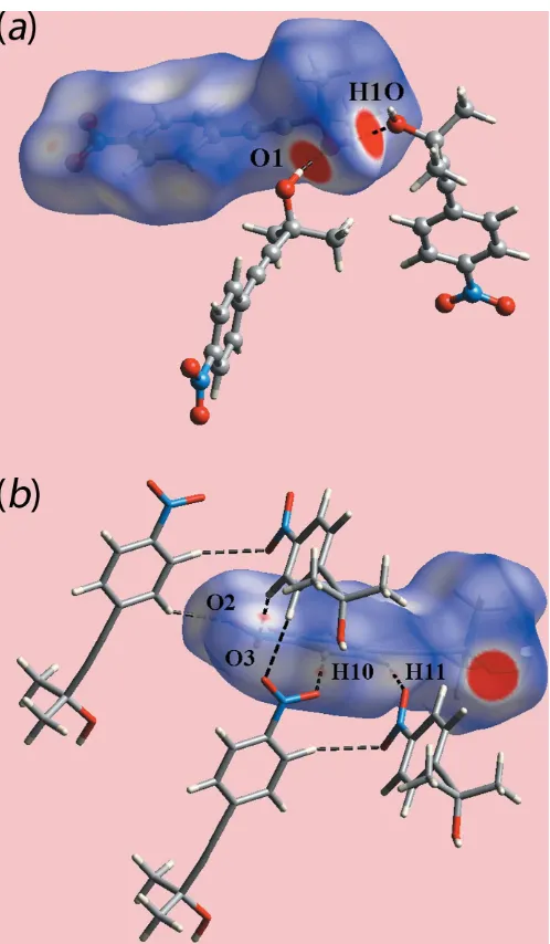
The spectacular feature of the molecular packing of (I) is the presence of hexameric clusters connected by hydroxy-O— H O(hydroxy) hydrogen bonds, Table 1. As seen from Fig. 2(a), the six-molecule aggregates are sustained by 12-membered { OH}6 synthons. The aggregates are disposed about a site of symmetry 3 so the rings have the shape of a flattened chair, Fig. 2(b). The crystal also features weak benzene-C—H O(nitro) interactions, involving both nitro-O atoms. In essence, one nitro group of one molecule forms two such interactions with two symmetry-related molecules to form a supramolecular chain along the c-axis direction with helical symmetry (31screw axis), Fig. 3(a). An end-on view of the chain is shown in Fig. 3(b). These weak benzene-C— H O(nitro) interactions serve to link the six-molecule aggregates into a three-dimensional architecture, Fig. 4.
4. Hirshfeld surface analysis
The Hirshfeld surface calculations for (I) were performed in accord with protocols described in a recently published paper (Tanet al., 2019) employingCrystal Explorer 17(Turneret al., 2017). On the Hirshfeld surfaces mapped over dnorm in
Fig. 5(a), the donors and acceptors of O—H O hydrogen bond involving the atoms of the hydroxyl group are char-acterized as bright-red spots. The faint-red spots near the phenyl-H10, H11 and nitro-O2, O3 atoms on the dnorm -mapped Hirshfeld surface in Fig. 5(b) represent the effect of Table 1
Hydrogen-bond geometry (A˚ ,).
D—H A D—H H A D A D—H A
O1—H1O O1i 0.82 1.87 2.682 (2) 173
C10—H10 O3ii 0.93 2.67 3.548 (3) 157
C11—H11 O2iii 0.93 2.68 3.467 (3) 143
Symmetry codes: (i) xyþ1 3;x
1 3;zþ
5
3; (ii) xþyþ 2 3;xþ
4 3;zþ
1 3; (iii) yþ4
3;xyþ 2 3;zþ
2 3.
Figure 1
[image:2.610.82.258.136.187.2] [image:2.610.316.560.256.692.2]The molecular structure of (I), showing the atom-labelling scheme and displacement ellipsoids at the 35% probability level.
Figure 2
[image:2.610.47.296.621.716.2]weak C—H O interactions as listed in Table 1. The Hirsh-feld surface mapped over electrostatic potential in Fig. 6 also illustrates the donors and acceptors of the indicated inter-actions through blue and red regions corresponding to positive and negative electrostatic potentials, respectively. In the view
Figure 4
[image:3.610.42.293.66.396.2] [image:3.610.316.565.67.494.2]A view of the unit-cell contents of (I) shown in projection down thecaxis. The hydroxy-O—H O(hydroxy) hydrogen bonding and benzene-C— H O(nitro) interactions are shown as orange and blue dashed lines, respectively.
Figure 3
Details of benzene-C—H O(nitro) interactions (shown as blue dashed lines) in the crystal of (I): (a) a view of the supramolecular chain along the
[image:3.610.47.297.523.697.2] [image:3.610.315.566.571.689.2]c-axis direction and (b) an end-on view of the chain.
Figure 5
Two views of the Hirshfeld surface for (I) mapped overdnorm: (a) in the
range0.202 to +1.400 arbitrary units and (b) in the range0.102 to +1.400 arbitrary units, highlighting, respectively, intermolecular O— H O and C—H O interactions through black dashed lines.
Figure 6
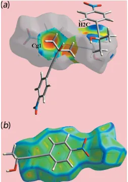
of a surface mapped with the shape-index property, Fig. 7(a), the C—H / H—C contacts listed in Table 2 are evident as the blue bump and a bright-orange region about the participating atoms. The overlap between benzene (C6–C11) ring of a reference molecule within the Hirshfeld surface mapped over curvedness and the symmetry related ring,
Fig. 7(b) is an indication of the – stacking interaction between them [centroid–centroid distance = 3.7873 (14) A˚ ; symmetry operation: 1x, 1y, 1z].
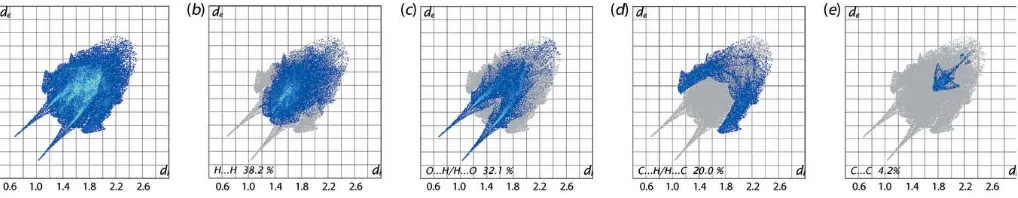
The overall two-dimensional fingerprint plot for (I), Fig. 8(a), and those delineated into H H, O H/H O, C H/H C and C C contacts (McKinnonet al., 2007) are illustrated in Fig. 8(b)–(e), respectively, and provide more information on the influence of short interatomic contacts upon the molecular packing. The percentage contributions from the different interatomic contacts to the Hirshfeld surface are summarized in Table 3. The greatest contribution to the Hirshfeld surface of 38.2% are derived from H H contacts but these exert a negligible influence on the packing, at least in terms of directional interactions, as the interatomic distances are greater than sum of their van der Waals radii. The pair of long spikes with their tips atde+di1.8 A˚ in the fingerprint plot delineated into O H/H O contacts, Fig. 8(c), are due to the presence of the O—H O hydrogen bond, whereas the points corresponding to comparatively weak intermolecular C—H O interactions, Table 1, and the short interatomic O H/H O contacts are merged within the plot, Table 2. The presence of the C—H contact, formed by the methyl-H2Catom and the benzene (C6–C11) ring, results in short interatomic C H/H C contacts, Table 2 and Fig. 7(a), and by the pair of forceps-like tips at de+di2.8 A˚ in Fig. 8(d). The points corresponding to other such short interatomic contacts involving the acetylene-C5 and methyl-C3—H3c atoms at longer separations are merged within the plot. The arrow-shaped distribution of points aroundde+di3.6 A˚ in the fingerprint plot delineated into C C contacts, Fig. 8(e), indicate – overlap between symmetry-related benzene (C6–C11) rings, as illustrated in Fig. 7(b). The small percentage contributions from the other interatomic contacts listed in Table 3 have negligible influence upon the molecular packing as their separations are greater than the sum of the respective van der Waals radii.
5. Interaction energies
[image:4.610.313.567.102.205.2]The pairwise interaction energies between the molecules within the crystal were calculated by summing up four energy components comprising electrostatic (Eele), polarization (Epol), dispersion (Edis) and exchange-repulsion (Erep) terms
Table 3
Percentage contributions of interatomic contacts to the Hirshfeld surface for (I).
Contact Percentage contribution
H H 38.2
O H/H O 32.1
C H/H C 20.0
C C 4.2
N O/O N 1.7
O O 1.6
C N/N C 1.0
N H/H N 0.8
[image:4.610.43.296.118.202.2]C O/O C 0.4
Table 2
Summary of short interatomic contacts (A˚ ) in (I).
The interatomic distances are calculated inCrystal Explorer 17(Turneret al., 2017) whereby theX—H bond lengths are adjusted to their neutron values.
Contact Distance Symmetry operation
O1 H3A 2.71 1
3+y, 2 3x+y,
5 3z
O2 H2B 2.69 2
3y, 1
3+xy, 2 3+z
O3 H2A 2.69 1x, 1y, 1z
C1 H1O 2.85 1
3+y, 2 3x+y,
5 3z
C5 H3C 2.79 1
3+y, 2 3x+y,
2 3z
C7 H2C 2.85 1
3+y, 2 3x+y,
2 3z
C8 H2C 2.80 1
3+y, 2 3x+y,
2 3z
Figure 7
(a) A view of the Hirshfeld surface for (I) mapped with the shape-index property, highlighting intermolecular C—H / H—C contacts by blue bumps and bright-orange concave regions, respectively, and (b) a view of the Hirshfeld surface mapped over curvedness, highlighting—
[image:4.610.44.297.313.673.2]after applying relevant scale factors (Turneret al., 2017). These energies were obtained by using the wave function calculated at the B3LYP/6-31G(d,p) level. The strength and the nature of intermolecular interactions in terms of their energies are qu-antitatively summarized in Table 4. The energies calculated for the different intermolecular interactions indicate that the electrostatic contribution is dominant in the O—H O hydrogen bond whereas the dispersive component has a significant influence due to the presence of short interatomic C H/H C and O H/H O contacts occurring between the same pair of molecules. The C—H O2(nitro) interaction has almost the same contributions from the electrostatic and dispersive components. This is in contrast to a major contri-bution only from the dispersive component for the analogous
contact involving the nitro-O3 atom. The dispersion energy component makes the major contribution to the relevant pairs of molecules involved in other short interatomic contacts, Table 4, as well as in C—H and–stacking interactions. It is also evident from a comparison of the total energies of intermolecular interactions, Table 4, that the O—H O hydrogen bond and–stacking interaction are stronger than the other interactions, and, of these, the intermolecular C— H O contacts are weaker than the C—H interactions.
The magnitudes of intermolecular energies are represented graphically by energy frameworks to view the supramolecular architecture of the crystal through the cylinders joining centroids of molecular pairs by using red, green and blue colour codes for the componentsEele, EdispandEtot, respec-tively, Fig. 9. The radius of the cylinder is proportional to the magnitude of interaction energy, which are adjusted to the same scale factor of 30 with a cut-off value of 3 kJ mol1 within 222 unit cells.
6. Non-covalent interaction plots
[image:5.610.44.296.92.211.2]Non-covalent interaction plot (NCIplot) analyses provide a visual representation of the nature of the contact between specified species in crystals (Johnsonet al., 2010; Contreras-Garca´ et al., 2011). This method is based on the electron density (and derivatives) and was employed in the present study to confirm the nature of some of the specified inter-molecular contacts. The colour-based isosurfaces generated
Figure 9
A comparison of the energy frameworks calculated for (I) and viewed down thecaxis showing (a) electrostatic potential force, (b) dispersion force and (c) total energy. The energy frameworks were adjusted to the same scale factor of 30 with a cut-off value of 3 kJ mol1within 222 unit cells.
Table 4
Summary of interaction energies (kJ mol1) calculated for (I).
Contact R(A˚ ) Eele Epol Edis Erep Etot
O1—H1O O1i
H3A O1i 8.80 52.3 12.0 18.8 72.7 35.7 H1O C1i
C10—H10 O3ii 8.28 3.7 1.4 9.2 4.9 9.8 C11—H11 O2iii 9.51 5.8 1.7 5.7 5.0 9.6
O3 H2Aiv
(C6–C11) (C6–C11)iv 4.25 9.4 1.8 47.1 28.9 34.4
H3C C5v
H2C C7v
H2C C8v 5.78 2.1 0.7 28.6 18.2 16.4
[image:5.610.57.566.420.519.2]C2—H2C (C6–C11)v
Figure 8
[image:5.610.43.568.567.721.2]correspond to the values of sign(2
)(r), whereis the elec-tron density and 2
is the second eigenvalue of the Hessian matrix of . Crucially, through a three-colour scheme, a specific interaction can be identified as being attractive or otherwise. Thus, a green isosurface indicates a weakly attrac-tive interaction whereas a blue isosurface indicates an attractive interaction; a repulsive interaction appears red. The isosurfaces for three identified intermolecular interactions are given in the upper view of Fig. 10. Thus, in Fig. 10(a), a green isosurface is apparent for the conventional hydroxy-O— H O(hydroxy) hydrogen bond. Similarly, green isosurfaces are seen between the interacting atoms involved in the phenyl-C—H O(nitro), Fig. 10(b), and the methyl-C—H (C11– C16), Fig. 10(c), interactions.
The lower views of Fig. 10, show the plots of the RDG versus sign(2
)(r). The non-covalent interaction peaks appear at density values less than 0.0 atomic units, consistent with their being weakly attractive interactions.
7. Database survey
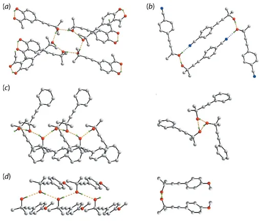
There are four literature precedents for (I) with varying substitution patterns in the appended benzene ring. These are the unsubstituted ‘parent’ compound [(II); FESMEV; Singe-lenberg & van Eijck, 1987], and the 4-cyano [(III}; HEFDAA; Clegg, 2017], 4-methoxy [(IV); YUQPEG; Eissmann et al., 2010] and 3-acetyl-4-hydroxy [(V); UVETAS; Hu¨bscheret al., 2016] derivatives. Selected geometric parameters for (I)–(IV)
[image:6.610.45.554.92.205.2]are collated in Table 5. Of particular interest in the mode of supramolecular association in their crystals. As seen from Fig. 11, four distinct patterns appear. In (V), three indepen-dent molecules comprise the asymmetric unit and these associate about a centre of inversion in space groupP21/cto form a hexameric clusters via hydroxy-O—H O(hydroxy) hydrogen bonds as seen in (I), Fig. 11(a); intramolecular hy-droxy-O—H O(carbonyl) hydrogen bonds are also apparent. In (III), the two independent molecules comprising the asymmetric unit associate about a centre of inversion in space group P21/n into a supramolecular dimer viapairs of hydroxy-O—H O(hydroxy) and hydroxy-O—H N(cyano) hydrogen bonds as shown in Fig. 11(b). In this case, one independent hydroxy-oxygen atom and one cyano-nitrogen atom do not accept a hydrogen-bonding interaction. Three crystallographically independent molecules are also found in (II) (space group Pca21) and these self-associate to form a
Table 5
Geometric data (A˚ ,) for related 2-methyl-4-(aryl)but-3-yn-2-ol molecules.
Compound Z0
Cring—Cacetylene Cacetylene—Cacetylene Cacetylene—Cquaternary Supramolecular motif Reference
(I) 1 1.438 (3) 1.189 (3) 1.471 (3) hexamer This work
(II) 3 1.443 (5) 1.211 (5) 1.454 (5) chain Singelenberg & van Eijck (1987) 1.437 (6) 1.192 (6) 1.479 (6)
1.437 (5) 1.189 (5) 1.479 (5)
(III) 2 1.441 (2) 1.193 (2) 1.490 (2) dimer Clegg (2017)
1.435 (2) 1.1895 (2) 1.480 (2)
(IV) 1 1.4377 (16) 1.2000 (16) 1.4791 (16) chain Eissmannet al.(2010) (V) 3 1.4418 (18) 1.1951 (19) 1.4764 (19) hexamer Hu¨bscheret al.(2016)
[image:6.610.312.565.452.670.2]1.444 (2) 1.194 (2) 1.4859 (19) 1.4402 (19) 1.1904 (19) 1.4723 (18)
Figure 10
Non-covalent interaction plots for (a) hydroxy-O—H O(hydroxy) hydrogen bonding, (b) the phenyl-C—H O(nitro) interactions and (c) the methyl-C—H (C11–C16) interactions.
Figure 11
[image:6.610.45.297.579.696.2]supramolecular chain via hydroxy-O—H O(hydroxy) hydrogen bonds with non-crystallographic threefold symmetry, Fig. 11(c). Finally, zigzag supramolecular chains sustained by hydroxy-O—H O(hydroxy) hydrogen bonds are found in the crystal of (IV), Fig. 11(d) in space groupPbca.
8. Synthesis and crystallization
The title compound was prepared as per the literature procedure (Bleicher et al., 1998). Yield: 87%. Yellow solid, m.p. 377–379 K.1H NMR (400 MHz, CDCl3):= 8.16 (dt,J= 8.9, 2.2 Hz, 2H), 7.54 (dt,J= 8.9, 2.2 Hz, 2H), 2.24 (s, 1H) and 1.63 (s, 6H) ppm. 13C NMR (101 MHz, CDCl3): = 147.2, 132.5, 129.8, 123.6, 99.2, 80.5, 66.7 and 31.3 ppm. Irregular colourless crystals of (I) for the X-ray study were grown by slow evaporation of its ethyl acetate solution.
9. Refinement details
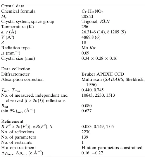
Crystal data, data collection and structure refinement details are summarized in Table 6. The carbon-bound H atoms were placed in calculated positions (C—H = 0.93–0.96 A˚ ) and were included in the refinement in the riding-model approximation, withUiso(H) set to 1.2–1.5Ueq(C). The O-bound H atom was refined with a distance restraint of 0.820.01 A˚ , and with Uiso(H) = 1.5Ueq(O).
Acknowledgements
We thank Professor Regina H. A. Santos from IQSC-USP for the X-ray data collection.
Funding information
Funding for this research was provided by GlaxoSmithKline (GSK) and the Brazilian agencies: The National Council for Scientific and Technological Development are thanked for fellowships (CNPq: 308480/2016-3 to IC; 303207/2017-5 to JZS), Sa˜o Paulo Research Foundation (FAPESP, grants 2013/ 06558-3 and 2014/50249-8) and Coordenac¸a˜o de Aperfeic¸oa-mento de Pessoal de Nı´vel Superior – Brasil (CAPES) – Finance Code 001. Sunway University Sdn Bhd is also thanked for funding (grant No. STR-RCTR-RCCM-001-2019).
References
Bleicher, L. S., Cosford, N. D. P., Herbaut, A., McCallum, J. S. & McDonald, I. A. (1998).J. Org. Chem.63, 1109–1118.
Brandenburg, K. (2006).DIAMOND. Crystal Impact GbR, Bonn, Germany.
Bruker (2009). APEX2 and SAINT. Bruker AXS Inc., Madison, Wisconsin, USA.
Burla, M. C., Caliandro, R., Carrozzini, B., Cascarano, G. L., Cuocci, C., Giacovazzo, C., Mallamo, M., Mazzone, A. & Polidori, G. (2015).J. Appl. Cryst.48, 306–309.
ChemAxon (2010).Marvinsketch. http://www.chemaxon.com. Clegg, W. (2017). Private communication (refcode: HEFDAA).
CCDC, Cambridge, England.
Contreras-Garcı´a, J., Johnson, E. R., Keinan, S., Chaudret, R., Piquemal, J.-P., Beratan, D. N. & Yang, W. (2011).J. Chem. Theory Comput.7, 625–632.
Eissmann, F., Kafurke, U. & Weber, E. (2010).Acta Cryst.E66, o1866. Erde´lyi, M. & Gogoll, A. (2001).J. Org. Chem.66, 4165–4169. Farrugia, L. J. (2012).J. Appl. Cryst.45, 849–854.
Hu¨bscher, J., Rosin, R., Seichter, W. & Weber, E. (2016).Acta Cryst. E72, 1370–1373.
Hundertmark, T., Littke, A. F., Buchwald, S. L. & Fu, G. C. (2000). Org. Lett.2, 1729–1731.
Johnson, E. R., Keinan, S., Mori-Sa´nchez, P., Contreras-Garcı´a, J., Cohen, A. J. & Yang, W. (2010).J. Am. Chem. Soc. 132, 6498– 6506.
Li, X., Sun, S., Yang, F., Kang, J., Wu, Y. & Wu, Y. (2015). Org. Biomol. Chem.13, 2432–2436.
McKinnon, J. J., Jayatilaka, D. & Spackman, M. A. (2007).Chem. Commun. pp. 3814–3816.
Sheldrick, G. M. (1996). SADABS. University of Go¨ttingen, Germany.
Sheldrick, G. M. (2015).Acta Cryst.C71, 3–8.
Singelenberg, F. A. J. & van Eijck, B. P. (1987).Acta Cryst.C43, 693– 695.
Tan, S. L., Jotani, M. M. & Tiekink, E. R. T. (2019).Acta Cryst.E75, 308–318.
Tan, X., Kong, L., Dai, H., Cheng, X., Liu, F. & Tschierske, C. (2013). Chem. Eur. J.19, 16303–16313.
Turner, M. J., Mckinnon, J. J., Wolff, S. K., Grimwood, D. J., Spackman, P. R., Jayatilaka, D. & Spackman, M. A. (2017).Crystal Explorer 17. The University of Western Australia.
[image:7.610.312.560.90.358.2]Westrip, S. P. (2010).J. Appl. Cryst.43, 920–925. Table 6
Experimental details.
Crystal data
Chemical formula C11H11NO3
Mr 205.21
Crystal system, space group Trigonal,R3:H
Temperature (K) 296
a,c(A˚ ) 26.3146 (14), 8.1205 (5)
V(A˚3) 4869.8 (6)
Z 18
Radiation type MoK
(mm1) 0.09
Crystal size (mm) 0.340.280.16
Data collection
Diffractometer Bruker APEXII CCD
Absorption correction Multi-scan (SADABS; Sheldrick, 1996)
Tmin,Tmax 0.440, 0.745
No. of measured, independent and observed [I> 2(I)] reflections
10643, 2230, 1513
Rint 0.080
(sin /)max(A˚
1
) 0.627
Refinement
R[F2> 2(F2)],wR(F2),S 0.053, 0.149, 1.05
No. of reflections 2230 No. of parameters 139
No. of restraints 1
H-atom treatment H-atom parameters constrained
max,min(e A˚
3) 0.16,0.27
sup-1 Acta Cryst. (2019). E75, 1232-1238
supporting information
Acta Cryst. (2019). E75, 1232-1238 [https://doi.org/10.1107/S2056989019010284]
2-Methyl-4-(4-nitrophenyl)but-3-yn-2-ol: crystal structure, Hirshfeld surface
analysis and computational chemistry study
Ignez Caracelli, Julio Zukerman-Schpector, Ricardo S. Schwab, Everton M. da Silva, Mukesh M.
Jotani and Edward R. T. Tiekink
Computing details
Data collection: APEX2 (Bruker, 2009); cell refinement: SAINT (Bruker, 2009); data reduction: SAINT (Bruker, 2009);
program(s) used to solve structure: SIR2014 (Burla et al., 2015); program(s) used to refine structure: SHELXL2018/3
(Sheldrick, 2015); molecular graphics: ORTEP-3 for Windows (Farrugia, 2012), DIAMOND (Brandenburg, 2006);
software used to prepare material for publication: MarvinSketch (ChemAxon, 2010) and publCIF (Westrip, 2010).
2-Methyl-4-(4-nitrophenyl)but-3-yn-2-ol
Crystal data
C11H11NO3 Mr = 205.21
Trigonal, R3:H a = 26.3146 (14) Å c = 8.1205 (5) Å V = 4869.8 (6) Å3 Z = 18
F(000) = 1944
Dx = 1.260 Mg m−3
Mo Kα radiation, λ = 0.71073 Å Cell parameters from 2006 reflections θ = 2.7–23.9°
µ = 0.09 mm−1 T = 296 K
Irregular, colourles 0.34 × 0.28 × 0.16 mm
Data collection
Bruker APEXII CCD diffractometer φ and ω scans
Absorption correction: multi-scan (SADABS; Sheldrick, 1996) Tmin = 0.440, Tmax = 0.745 10643 measured reflections
2230 independent reflections 1513 reflections with I > 2σ(I) Rint = 0.080
θmax = 26.4°, θmin = 1.6° h = −32→32
k = −32→32 l = −9→10
Refinement
Refinement on F2 Least-squares matrix: full R[F2 > 2σ(F2)] = 0.053 wR(F2) = 0.149 S = 1.05 2230 reflections 139 parameters 1 restraint
Primary atom site location: structure-invariant direct methods
Secondary atom site location: difference Fourier map
Hydrogen site location: inferred from neighbouring sites
H-atom parameters constrained w = 1/[σ2(F
o2) + (0.0511P)2 + 3.9317P] where P = (Fo2 + 2Fc2)/3
sup-2 Acta Cryst. (2019). E75, 1232-1238
Special details
Geometry. All esds (except the esd in the dihedral angle between two l.s. planes) are estimated using the full covariance matrix. The cell esds are taken into account individually in the estimation of esds in distances, angles and torsion angles; correlations between esds in cell parameters are only used when they are defined by crystal symmetry. An approximate (isotropic) treatment of cell esds is used for estimating esds involving l.s. planes.
Fractional atomic coordinates and isotropic or equivalent isotropic displacement parameters (Å2)
x y z Uiso*/Ueq
O1 0.56993 (7) 0.33529 (6) 0.78650 (17) 0.0513 (4) H1O 0.569241 0.305973 0.823659 0.077* O2 0.54778 (9) 0.61319 (8) 0.0578 (2) 0.0811 (6) O3 0.62154 (8) 0.66736 (8) 0.2126 (3) 0.0785 (6) N1 0.58062 (9) 0.61978 (9) 0.1723 (3) 0.0565 (5) C1 0.53437 (9) 0.31996 (8) 0.6425 (2) 0.0388 (5) C2 0.47123 (10) 0.27726 (10) 0.6889 (3) 0.0612 (7) H2A 0.459150 0.293892 0.775102 0.092* H2B 0.468058 0.241200 0.726620 0.092* H2C 0.446510 0.269637 0.594458 0.092* C3 0.55665 (12) 0.29361 (11) 0.5140 (3) 0.0634 (7) H3A 0.551638 0.257044 0.553954 0.095* H3B 0.597504 0.320235 0.493279 0.095* H3C 0.534860 0.286951 0.413769 0.095* C4 0.54001 (9) 0.37468 (9) 0.5772 (2) 0.0439 (5) C5 0.54464 (10) 0.41762 (9) 0.5145 (2) 0.0458 (5) C6 0.55281 (9) 0.46950 (9) 0.4317 (2) 0.0407 (5) C7 0.51138 (9) 0.46599 (9) 0.3192 (2) 0.0424 (5) H7 0.477559 0.430184 0.300298 0.051* C8 0.52018 (9) 0.51543 (9) 0.2351 (2) 0.0441 (5) H8 0.492523 0.513284 0.159933 0.053* C9 0.57045 (9) 0.56768 (9) 0.2648 (2) 0.0416 (5) C10 0.61185 (10) 0.57274 (9) 0.3773 (3) 0.0515 (6) H10 0.645262 0.608821 0.396842 0.062* C11 0.60276 (10) 0.52332 (10) 0.4602 (3) 0.0509 (6) H11 0.630422 0.526000 0.536242 0.061*
Atomic displacement parameters (Å2)
U11 U22 U33 U12 U13 U23
sup-3 Acta Cryst. (2019). E75, 1232-1238
C6 0.0535 (12) 0.0444 (11) 0.0311 (9) 0.0297 (10) 0.0052 (8) 0.0026 (8) C7 0.0452 (11) 0.0423 (11) 0.0400 (10) 0.0223 (10) 0.0024 (9) 0.0009 (8) C8 0.0480 (12) 0.0543 (13) 0.0379 (10) 0.0317 (11) 0.0011 (9) 0.0045 (9) C9 0.0486 (12) 0.0431 (11) 0.0413 (10) 0.0291 (10) 0.0088 (9) 0.0080 (8) C10 0.0491 (13) 0.0422 (12) 0.0598 (13) 0.0203 (10) −0.0055 (10) −0.0006 (10) C11 0.0566 (14) 0.0538 (13) 0.0468 (11) 0.0310 (11) −0.0109 (10) −0.0005 (10)
Geometric parameters (Å, º)
O1—C1 1.424 (2) C3—H3C 0.9600 O1—H1O 0.8200 C4—C5 1.189 (3) O2—N1 1.221 (3) C5—C6 1.438 (3) O3—N1 1.219 (2) C6—C11 1.387 (3) N1—C9 1.466 (3) C6—C7 1.390 (3) C1—C4 1.471 (3) C7—C8 1.382 (3) C1—C2 1.516 (3) C7—H7 0.9300 C1—C3 1.523 (3) C8—C9 1.371 (3) C2—H2A 0.9600 C8—H8 0.9300 C2—H2B 0.9600 C9—C10 1.376 (3) C2—H2C 0.9600 C10—C11 1.375 (3) C3—H3A 0.9600 C10—H10 0.9300 C3—H3B 0.9600 C11—H11 0.9300
C1—O1—H1O 109.5 H3B—C3—H3C 109.5 O3—N1—O2 123.3 (2) C5—C4—C1 175.7 (2) O3—N1—C9 118.5 (2) C4—C5—C6 176.5 (2) O2—N1—C9 118.2 (2) C11—C6—C7 119.25 (18) O1—C1—C4 106.76 (15) C11—C6—C5 120.46 (18) O1—C1—C2 109.11 (16) C7—C6—C5 120.27 (19) C4—C1—C2 110.61 (18) C8—C7—C6 120.36 (19) O1—C1—C3 110.10 (17) C8—C7—H7 119.8 C4—C1—C3 108.98 (16) C6—C7—H7 119.8 C2—C1—C3 111.20 (18) C9—C8—C7 118.75 (18) C1—C2—H2A 109.5 C9—C8—H8 120.6 C1—C2—H2B 109.5 C7—C8—H8 120.6 H2A—C2—H2B 109.5 C8—C9—C10 122.25 (18) C1—C2—H2C 109.5 C8—C9—N1 118.80 (18) H2A—C2—H2C 109.5 C10—C9—N1 118.95 (19) H2B—C2—H2C 109.5 C11—C10—C9 118.6 (2) C1—C3—H3A 109.5 C11—C10—H10 120.7 C1—C3—H3B 109.5 C9—C10—H10 120.7 H3A—C3—H3B 109.5 C10—C11—C6 120.79 (19) C1—C3—H3C 109.5 C10—C11—H11 119.6 H3A—C3—H3C 109.5 C6—C11—H11 119.6
sup-4 Acta Cryst. (2019). E75, 1232-1238
C7—C8—C9—C10 −1.2 (3) N1—C9—C10—C11 −178.21 (19) C7—C8—C9—N1 178.28 (17) C9—C10—C11—C6 −0.3 (3) O3—N1—C9—C8 171.3 (2) C7—C6—C11—C10 −0.7 (3) O2—N1—C9—C8 −9.2 (3) C5—C6—C11—C10 177.72 (19)
Hydrogen-bond geometry (Å, º)
D—H···A D—H H···A D···A D—H···A
O1—H1O···O1i 0.82 1.87 2.682 (2) 173 C10—H10···O3ii 0.93 2.67 3.548 (3) 157 C11—H11···O2iii 0.93 2.68 3.467 (3) 143