858
https://doi.org/10.1107/S2056989019007072 Acta Cryst.(2019). E75, 858–862research communications
Received 15 April 2019 Accepted 15 May 2019
Edited by W. T. A. Harrison, University of Aberdeen, Scotland
Keywords:crystal structure; hydrogen bonding; low temperature; zymonic; pyruvic.
CCDC references:1916323; 1916322
Supporting information:this article has supporting information at journals.iucr.org/e
Crystal structure of zymonic acid and a
redetermination of its precursor, pyruvic acid
Dominik Heger,a,b* Alexis J. Eugene,aSean R. Parkina* and Marcelo I. Guzmana*
aDepartment of Chemistry, University of Kentucky, Lexington, Kentucky 40506, USA, andbDepartment of Chemistry, Faculty of Science, Masaryk University, Kamenice 5, 625 00 Brno, Czech Republic. *Correspondence e-mail: [email protected], [email protected], [email protected]
The structure of zymonic acid (systematic name: 4-hydroxy-2-methyl-5-oxo-2,5-dihydrofuran-2-carboxylic acid), C6H6O5, which had previously eluded
crystal-lographic determination, is presented here for the first time. It forms by intramolecular condensation of parapyruvic acid, which is the product of aldol condensation of pyruvic acid. A redetermination of the crystal structure of pyruvic acid (systematic name: 2-oxopropanoic acid), C3H4O3, at low
temperature (90 K) and with increased precision, is also presented [for the previous structure, see: Harata et al. (1977). Acta Cryst. B33, 210–212]. In zymonic acid, the hydroxylactone ring is close to planar (r.m.s. deviation = 0.0108 A˚ ) and the dihedral angle between the ring and the plane formed by the bonds of the methyl and carboxylic acid carbon atoms to the ring is 88.68 (7). The torsion angle of the carboxylic acid group relative to the ring is 12.04 (16). The pyruvic acid molecule is almost planar, having a dihedral angle between the carboxylic acid and methyl-ketone groups of 3.95 (6). Intermolecular inter-actions in both crystal structures are dominated by hydrogen bonding. The commonR2
2
(8) hydrogen-bonding motif links carboxylic acid groups on adjacent molecules in both structures. In zymonic acid, this results in dimers about a crystallographic twofold of space groupC2/c, which forces the carboxylic acid group to be disordered exactly 50:50, which scrambles the carbonyl and hydroxyl groups and gives an apparent equalization of the C—O bond lengths [1.2568 (16) and 1.2602 (16) A˚ ]. The other hydrogen bonds in zymonic acid (O—H O and weak C—H O), link molecules across a 21-screw axis, and
generate an R22(9) motif. These hydrogen-bonding interactions propagate to
form extended pleated sheets in the abplane. Stacking of these zigzag sheets alongcinvolves only van der Waals contacts. In pyruvic acid, inversion-related molecules are linked intoR2
2
(8) dimers, with van der Waals interactions between dimers as the only other intermolecular contacts.
1. Chemical context
The Human Metabolome Database (Wishartet al., 2007, 2009, 2013, 2018) lists the compound 4-hydroxy-2-methyl-5-oxo-furan-2-carboxylic acid (C6H6O5), commonly named zymonic
acid, with the metabocard HMDB0031210. Zymonic acid is used as a flavor constituent for confectionery and tobacco products (Yannai, 2004). The generation of zymonic acid can proceed by condensation of parapyruvic acid, which itself forms by aldol condensation of pyruvic acid (IUPAC name 2-oxopropanoic acid, C3H4O3; Bloomeret al., 1970). Therefore,
zymonic acid is directly derived from pyruvic acid, and is thus related to the compounds present in the tricarboxylic acid (Krebs) cycle (Nelson & Cox, 2004) and its reductive version (Guzman, 2011; Guzman & Martin, 2008; Zhou & Guzman, 2016). As an intermediate in central metabolism, zymonic acid
is produced in the cytoplasm at very low concentration, from where it can be excreted to the extracellular region.
The electron-impact mass spectrum (MS) and electrospray ionization fragmentation of zymonic acid following gas and liquid chromatography, respectively, have been reported (Allen et al., 2015, 2016). The use of 13C-zymonic acid has enabled mapping of pH changes, independently of concen-tration, in mammalian organs and tumorsviahyperpolarized magnetic resonance (Du¨welet al., 2017). Thus, zymonic acid is a non-invasive extracellular imaging sensor to localize and quantify pHin vivo(Du¨welet al., 2017; Hundshammeret al., 2017), with many possible applications in medical diagnosis (Schillinget al., 2016). As part of the process resulting in the aforementioned invention, the detailed 1H and 13C NMR spectra of pure zymonic acid have been reported (Hund-shammeret al., 2017). Herein, we contribute new information to characterize zymonic acid by reporting for the first time its crystal structure, along with a low-temperature redetermina-tion of pyruvic acid.
2. Structural commentary
Aside from the effects on the geometry of the carboxylic acid group in zymonic acid that stem from disorder about the twofold axis (see below), there are no unusual bond lengths or angles in either compound.
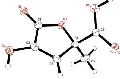
In zymonic acid (Fig. 1), the hydroxylactone ring is essen-tially planar (r.m.s. deviation = 0.0108 A˚ ), with the largest deviation from planarity [0.0171 (8) A˚ ] for the ring oxygen atom, O3. The plane defined by the ring carbon atom C4, the methyl carbon atom C6, and the carboxylic acid carbon atom
C5, is almost perpendicular to the mean plane of the ring atoms [dihedral angle = 88.68 (7)]. Lastly, the orientation of the carboxylic acid group relative to the ring, as defined by the torsion angle O4—C5—C4—O3, is 12.04 (16). For the carb-oxylic acid group, disorder about the crystallographic twofold axis effectively averages the C O double and C—O single bonds, rendering them equivalent [the C5—O4 and C5—O5 distances are 1.2568 (16) and 1.2602 (16) A˚ , respectively], and requires modeling of half-occupancy hydrogens (H4O and H5O) on each.
In spite of increased precision resulting from much lower temperature (90 K versus 266 K) and data collection on modern equipment, the redetermined structure of pyruvic acid (Fig. 2) is largely unchanged from that reported by Harataet al.(1977). For example, the dihedral angle between the planes defined by atoms C1/C2/C3/O3 and C1/C2/O1/O2 is 3.95 (6) at 90.00 (2) Kversus3.5at 266 (1) K.
3. Supramolecular features
The main intermolecular interactions in the crystals of both zymonic and pyruvic acids are hydrogen bonds. In zymonic acid, the carboxylic acid groups of adjacent molecules are related by a crystallographic twofold axis to form hydrogen bonds [O4—H4O O4ii and O5—H5O O5ii; symmetry code: (ii) 1x,y,3
2z] givingR 2
2(8) dimer motifs (Table 1).
This common supramolecular construct in carboxylic acids usually occurs between inversion-related or symmetry-inde-pendent molecules. Here, the orientation of the dimer relative to the crystallographic twofold axis forces the average
struc-research communications
Acta Cryst.(2019). E75, 858–862 Hegeret al. C
[image:2.610.313.567.93.152.2] [image:2.610.63.278.111.195.2]6H6O5and C3H4O3
859
Figure 2The molecular structure of pyruvic acid, with displacement ellipsoids drawn at the 50% probability level.
Table 1
Hydrogen-bond geometry (A˚ ,) for zymonic acid.
D—H A D—H H A D A D—H A
O2—H2 O1i 0.84 1.96 2.7103 (14) 148 C3—H3 O2i 0.95 2.48 3.0720 (16) 120 O4—H4O O4ii 1.09 1.52 2.607 (2) 176 O5—H5O O5ii 0.99 1.63 2.624 (2) 179
Symmetry codes: (i)xþ3 2;yþ
1 2;zþ
3
[image:2.610.343.533.583.716.2]2; (ii)xþ1;y;zþ 3 2.
Figure 1
[image:2.610.70.269.588.719.2]ture to be statistically disordered (Fig. 3). Another pair of
hydrogen bonds [O2—H2 O1i and C3—H3 O2i;
symmetry code: (i)3 2x,
1 2+y,
3
2z], link molecules related by
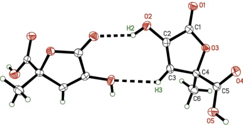
a 21-screw axis, into R22(9) motifs (Fig. 4). These
hydrogen-bonding interactions combine to form extended pleated sheets that propagate in the abplane (Fig. 5), which in turn, stack along the c-axis direction. In pyruvic acid, inversion-related molecules form the common R2
2(8) dimer motif (Fig. 6,
Table 2). In accordance with the work of Harataet al.(1977), there are no other noteworthy intermolecular interactions.
4. Database survey
A search of the Cambridge Crystal Structure Database (Version 5.40, Nov. 2018; Groomet al., 2016) for zymonic acid gave no hits for searches on either ‘zymonic’ or on the structural formula. A search on the structural formula of pyruvic acid gave two hits. CSD entry PRUVAC (Harataet al., 1977) describes the pure compound at 266 K, and is similar to the present pyruvic acid structure (after transformation to a common cell setting). CSD entry FAFGUR (Prohens et al., 2016) describes a co-crystal of pyruvic acid with the drug agomelatine. The CSD does contain structures for derivatives of both zymonic and pyruvic acids, but none of these have features that are especially relevant to the current work.
5. Synthesis and crystallization
Vacuum distillation of pyruvic acid (Sigma–Aldrich, 98.5%) was used for purification (Eugene & Guzman, 2017a,b). Freshly distilled pyruvic acid was crystallized in a closed vial in a freezer at 253 K. The tail of this distillation, a viscous yellowish residue enriched in parapyruvic and zymonic acids, was isolated in a vial, and the headspace filled with N2(g)
before sealing it with a cap. Crystals of zymonic acid were produced slowly from this isolated residue kept at 275 K inside a refrigerator. The easily identifiable transparent crystals of zymonic acid appear above the level of the viscous solution within two weeks. Pyruvic acid crystals are deliquescent in air, even at 263 K (Harataet al., 1977), so they had to be kept cold, with minimal exposure to ambient air. Thus, throughout all experimental stages from initial inspection through data collection, special techniques for crystal handling at low temperature (Parkin & Hope, 1998) were employed.
860
Hegeret al. C6H6O5and C3H4O3 Acta Cryst.(2019). E75, 858–862
[image:3.610.49.298.72.188.2]research communications
Figure 4 TheR2
2(9) dimer of zymonic acid. Unlabeled atoms are related to their
labeled counterparts by a crystallographic 21-screw axis (32x, 1 2+y,
3 2z).
[image:3.610.46.295.394.522.2]Disorder of the carboxylic acid H atoms is omitted to enhance clarity.
Figure 5
[image:3.610.47.293.590.706.2]A packing plot of zymonic acid viewed down the baxis, showing the stacking alongcof zigzag pleated assemblies of molecules. Disorder of the carboxylic acid hydrogen atoms is omitted to enhance clarity.
Figure 6 TheR2
2(8) dimer of pyruvic acid. Unlabeled atoms are related to their
labeled counterparts by crystallographic inversion symmetry (1x, 1y, 1z).
Table 2
Hydrogen-bond geometry (A˚ ,) for pyruvic acid.
D—H A D—H H A D A D—H A
O1—H1 O2i 0.913 (14) 1.742 (14) 2.6536 (8) 175.5 (12)
Symmetry code: (i)xþ1;yþ1;zþ1.
Figure 3 TheR2
2(8) dimer of zymonic acid. Unlabeled atoms are related to their
labeled counterparts by a crystallographic twofold axis (1x,y,3 2z).
This uncommon symmetry [for anR2
2(8) dimer] forces the O—H O
[image:3.610.315.564.607.707.2]6. Refinement
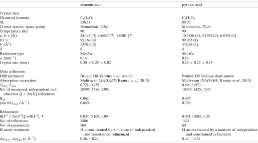
Crystal data, data collection, and structure refinement details are summarized in Table 3. Non-disordered hydrogen atoms were found in difference Fourier maps. For pyruvic acid, the hydroxyl hydrogen-atom coordinates were refined freely, while methyl hydrogen C—H distances used a riding model that allowed the C—H distance to refine. For zymonic acid, riding models were used for all hydrogen atoms apart from those disordered about the twofold axis, which were modeled in accordance with the recommendations of Fa´bry (2018).
Uiso(H) parameters of non-disordered hydrogens were set to
either 1.2Ueqor 1.5Ueq(for the methyl and hydroxyl groups,
respectively) of the attached atom. To ensure stable refine-ment of disordered groups in the zymonic acid structure,
constraints (SHELXL command EADP) were used to
equalize displacement parameters of superimposed atoms.
Funding information
We are thankful for research funding from the National Science Foundation under NSF CAREER award CHE-1255290 to MIG, and the MRI program, grants CHE-0319176 and CHE-1625732. An International Visiting Scholar grant from the College of Arts and Sciences at UK is gratefully acknowledged.
References
Allen, F., Greiner, R. & Wishart, D. (2015).Metabolomics,11, 98–110. Allen, F., Pon, A., Greiner, R. & Wishart, D. (2016).Anal. Chem.88,
7689–7697.
Bloomer, J. L., Gross, M. A., Kappler, F. E. & Pandey, G. N. (1970).J. Chem. Soc. D, p. 1030a.
Bruker (2016).APEX3.Bruker AXS Inc., Madison, Wisconsin, USA. Du¨wel, S., Hundshammer, C., Gersch, M., Feuerecker, B., Steiger, K., Buck, A., Walch, A., Haase, A., Glaser, S. J., Schwaiger, M. & Schilling, F. (2017).Nat. Commun.8, 15126.
Eugene, A. J. & Guzman, M. I. (2017a).J. Phys. Chem. A,121, 2924– 2935.
Eugene, A. J. & Guzman, M. I. (2017b).J. Phys. Chem. A,121, 8741– 8744.
Fa´bry, J. (2018).Acta Cryst.E74, 1344–1357.
Groom, C. R., Bruno, I. J., Lightfoot, M. P. & Ward, S. C. (2016).Acta Cryst.B72, 171–179.
Guzman, M. I. (2011).Origins of Life: The Primal Self-Organization, edited by R. Egel, D.-H. Lankenau & A. Y. Mulkidjanian, pp. 85– 105: Springer Berlin Heidelberg.
Guzman, M. I. & Martin, S. T. (2008).Int. J. Astrobiology,7, 271–278. Harata, K., Sakabe, N. & Tanaka, J. (1977).Acta Cryst.B33, 210–212. Hundshammer, C., Du¨wel, S., Ko¨cher, S. S., Gersch, M., Feuerecker, B., Scheurer, C., Haase, A., Glaser, S. J., Schwaiger, M. & Schilling, F. (2017).ChemPhysChem,18, 2422–2425.
Krause, L., Herbst-Irmer, R., Sheldrick, G. M. & Stalke, D. (2015).J. Appl. Cryst.48, 3–10.
Nelson, D. L. & Cox, M. (2004).Lehninger Principles of Biochemistry, 4th ed. New York: W. H. Freeman.
Parkin, S. (2013). CIFFIX, https://xray.uky.edu/Resources/scripts/ ciffix
research communications
Acta Cryst.(2019). E75, 858–862 Hegeret al. C
[image:4.610.46.561.91.377.2]6H6O5and C3H4O3
861
Table 3Experimental details.
zymonic acid pyruvic acid
Crystal data
Chemical formula C6H6O5 C3H4O3
Mr 158.11 88.06
Crystal system, space group Monoclinic,C2/c Monoclinic,P21/c
Temperature (K) 90 90
a,b,c(A˚ ) 24.145 (3), 6.6523 (7), 8.6201 (7) 10.7486 (3), 5.1925 (2), 6.8302 (2)
(
) 95.169 (4) 99.063 (1)
V(A˚3) 1378.9 (3) 376.45 (2)
Z 8 4
Radiation type MoK MoK
(mm1) 0.14 0.14
Crystal size (mm) 0.300.250.02 0.260.220.18
Data collection
Diffractometer Bruker D8 Venture dual source Bruker D8 Venture dual source
Absorption correction Multi-scan (SADABS; Krauseet al., 2015) Multi-scan (SADABS; Krauseet al., 2015)
Tmin,Tmax 0.721, 0.959 0.890, 0.971
No. of measured, independent and observed [I> 2(I)] reflections
18595, 1586, 1392 10479, 1425, 1242
Rint 0.062 0.025
(sin/)max(A˚
1) 0.650 0.796
Refinement
R[F2> 2(F2)],wR(F2),S 0.037, 0.100, 1.09 0.031, 0.082, 1.08
No. of reflections 1586 1425
No. of parameters 104 60
H-atom treatment H atoms treated by a mixture of independent and constrained refinement
H atoms treated by a mixture of independent and constrained refinement
max,min(e A˚
3) 0.36,0.24 0.40,0.21
Parkin, S. & Hope, H. (1998).J. Appl. Cryst.31, 945–953.
Prohens, R., Barbas, R., Portell, A., Font-Bardia, M., Alcobe´, X. & Puigjaner, C. (2016).Cryst. Growth Des.16, 1063–1070.
Schilling, F., Glaser, S., Du¨wel, S. & Gersch, M. (2016).pH-Biosensors Based on Compounds Produced From Pyruvic Acid For Magnetic Resonance Imaging and Spectroscopy and Their Uses. Google Patents.
Sheldrick, G. M. (2008).Acta Cryst.A64, 112–122. Sheldrick, G. M. (2015a).Acta Cryst.A71, 3–8. Sheldrick, G. M. (2015b).Acta Cryst.C71, 3–8.
Wishart, D. S., Feunang, Y. D., Marcu, A., Guo, A. C., Liang, K., Va´zquez-Fresno, R., Sajed, T., Johnson, D., Li, C., Karu, N., Sayeeda, Z., Lo, E., Assempour, N., Berjanskii, M., Singhal, S., Arndt, D., Liang, Y., Badran, H., Grant, J., Serra-Cayuela, A., Liu, Y., Mandal, R., Neveu, V., Pon, A., Knox, C., Wilson, M., Manach, C. & Scalbert, A. (2018).Nucleic Acids Res.46, D608–D617. Wishart, D. S., Jewison, T., Guo, A. C., Wilson, M., Knox, C., Liu, Y.,
Djoumbou, Y., Mandal, R., Aziat, F., Dong, E., Bouatra, S., Sinelnikov, I., Arndt, D., Xia, J., Liu, P., Yallou, F., Bjorndahl, T., Perez-Pineiro, R., Eisner, R., Allen, F., Neveu, V., Greiner, R. & Scalbert, A. (2013).Nucleic Acids Res.41, D801–D807.
Wishart, D. S., Knox, C., Guo, A. C., Eisner, R., Young, N., Gautam, B., Hau, D. D., Psychogios, N., Dong, E., Bouatra, S., Mandal, R., Sinelnikov, I., Xia, J., Jia, L., Cruz, J. A., Lim, E., Sobsey, C. A., Shrivastava, S., Huang, P., Liu, P., Fang, L., Peng, J., Fradette, R., Cheng, D., Tzur, D., Clements, M., Lewis, A., De Souza, A., Zuniga, A., Dawe, M., Xiong, Y., Clive, D., Greiner, R., Nazyrova, A., Shaykhutdinov, R., Li, L., Vogel, H. J. & Forsythe, I. (2009).Nucleic Acids Res.37, D603–D610.
Wishart, D. S., Tzur, D., Knox, C., Eisner, R., Guo, A. C., Young, N., Cheng, D., Jewell, K., Arndt, D., Sawhney, S., Fung, C., Nikolai, L., Lewis, M., Coutouly, M.-A., Forsythe, I., Tang, P., Shrivastava, S., Jeroncic, K., Stothard, P., Amegbey, G., Block, D., Hau, D. D., Wagner, J., Miniaci, J., Clements, M., Gebremedhin, M., Guo, N., Zhang, Y., Duggan, G. E., Macinnis, G. D., Weljie, A. M., Dowlatabadi, R., Bamforth, F., Clive, D., Greiner, R., Li, L., Marrie, T., Sykes, B. D., Vogel, H. J. & Querengesser, L. (2007). Nucleic Acids Res.35, D521–D526.
Yannai, S. (2004). Dictionary of food compounds with CD-ROM: Additives, flavors, and ingredients. Boca Raton: Chapman and Hall/ CRC.
Zhou, R. & Guzman, M. I. (2016).J. Phys. Chem. C,120, 7349–7357.
862
Hegeret al. C6H6O5and C3H4O3 Acta Cryst.(2019). E75, 858–862
supporting information
sup-1 Acta Cryst. (2019). E75, 858-862
supporting information
Acta Cryst. (2019). E75, 858-862 [https://doi.org/10.1107/S2056989019007072]
Crystal structure of zymonic acid and a redetermination of its precursor,
pyruvic acid
Dominik Heger, Alexis J. Eugene, Sean R. Parkin and Marcelo I. Guzman
Computing details
For both structures, data collection: APEX3 (Bruker, 2016); cell refinement: APEX3 (Bruker, 2016); data reduction:
APEX3 (Bruker, 2016); program(s) used to solve structure: SHELXT (Sheldrick, 2015a). Program(s) used to refine
structure: SHELXL2018/3 (Sheldrick, 2015b) for zymonic; SHELXL2018 (Sheldrick, 2015b) for pyruvic. For both
structures, molecular graphics: XP in SHELXTL (Sheldrick, 2008); software used to prepare material for publication:
SHELXL (Sheldrick, 2008) and CIFFIX (Parkin, 2013).
4-Hydroxy-2-methyl-5-oxo-2,5-dihydrofuran-2-carboxylic acid (zymonic)
Crystal data
C6H6O5
Mr = 158.11
Monoclinic, C2/c
a = 24.145 (3) Å
b = 6.6523 (7) Å
c = 8.6201 (7) Å
β = 95.169 (4)°
V = 1378.9 (3) Å3
Z = 8
F(000) = 656
Dx = 1.523 Mg m−3
Mo Kα radiation, λ = 0.71073 Å
Cell parameters from 9925 reflections
θ = 3.2–27.5°
µ = 0.14 mm−1
T = 90 K
Thin plate, colourless 0.30 × 0.25 × 0.02 mm
Data collection
Bruker D8 Venture dual source diffractometer
Radiation source: microsource
Detector resolution: 5.6 pixels mm-1
φ and ω scans
Absorption correction: multi-scan (SADABS; Krause et al., 2015)
Tmin = 0.721, Tmax = 0.959
18595 measured reflections 1586 independent reflections 1392 reflections with I > 2σ(I)
Rint = 0.062
θmax = 27.5°, θmin = 3.2°
h = −31→31
k = −8→8
l = −11→10
Refinement
Refinement on F2
Least-squares matrix: full
R[F2 > 2σ(F2)] = 0.037
wR(F2) = 0.100
S = 1.09
1586 reflections 104 parameters 0 restraints
Primary atom site location: structure-invariant direct methods
Secondary atom site location: difference Fourier map
Hydrogen site location: mixed
H atoms treated by a mixture of independent and constrained refinement
w = 1/[σ2(F
o2) + (0.0428P)2 + 1.4377P]
supporting information
sup-2 Acta Cryst. (2019). E75, 858-862
(Δ/σ)max = 0.001
Δρmax = 0.36 e Å−3
Δρmin = −0.24 e Å−3
Extinction correction: SHELXL2018 (Sheldrick, 2015a),
Fc*=kFc[1+0.001xFc2λ3/sin(2θ)]-1/4
Extinction coefficient: 0.0057 (13)
Special details
Experimental. The crystal was mounted using polyisobutene oil on the tip of a fine glass fibre, which was fastened in a copper mounting pin with electrical solder. It was placed directly into the cold gas stream of a liquid-nitrogen based cryostat (Hope, 1994; Parkin & Hope, 1998).
Diffraction data were collected with the crystal at 90K, which is standard practice in this laboratory for the majority of flash-cooled crystals.
Geometry. All esds (except the esd in the dihedral angle between two l.s. planes) are estimated using the full covariance matrix. The cell esds are taken into account individually in the estimation of esds in distances, angles and torsion angles; correlations between esds in cell parameters are only used when they are defined by crystal symmetry. An approximate (isotropic) treatment of cell esds is used for estimating esds involving l.s. planes.
Refinement. Refinement progress was checked using Platon (Spek, 2009) and by an R-tensor (Parkin, 2000). The final
model was further checked with the IUCr utility checkCIF.
Fractional atomic coordinates and isotropic or equivalent isotropic displacement parameters (Å2)
x y z Uiso*/Ueq Occ. (<1)
C1 0.67036 (5) 0.40914 (19) 0.56905 (14) 0.0170 (3)
O1 0.68738 (4) 0.24154 (14) 0.54864 (11) 0.0209 (2)
C2 0.69378 (5) 0.5659 (2) 0.67706 (14) 0.0178 (3)
O2 0.74026 (4) 0.51656 (15) 0.76588 (12) 0.0240 (3)
H2 0.752557 0.618162 0.815574 0.036*
C3 0.66149 (5) 0.7275 (2) 0.66335 (15) 0.0182 (3)
H3 0.667502 0.849554 0.719574 0.022*
O3 0.62412 (4) 0.48283 (13) 0.48901 (11) 0.0188 (2)
C4 0.61371 (5) 0.68442 (19) 0.54353 (15) 0.0186 (3)
O4 0.54061 (4) 0.51162 (15) 0.66013 (12) 0.0249 (3)
H4O 0.507244 0.517998 0.736346 0.109 (14)* 0.5
O5 0.54177 (4) 0.84824 (16) 0.66262 (14) 0.0303 (3)
H5O 0.509866 0.849228 0.727610 0.109 (14)* 0.5
C5 0.56056 (5) 0.67919 (19) 0.62725 (15) 0.0188 (3)
C6 0.60945 (7) 0.8285 (2) 0.40645 (17) 0.0257 (3)
H6A 0.644218 0.825746 0.355945 0.039*
H6B 0.602833 0.964985 0.443442 0.039*
H6C 0.578557 0.787894 0.331471 0.039*
Atomic displacement parameters (Å2)
U11 U22 U33 U12 U13 U23
C1 0.0184 (6) 0.0180 (6) 0.0151 (6) −0.0007 (5) 0.0038 (5) 0.0010 (5)
O1 0.0238 (5) 0.0173 (5) 0.0213 (5) 0.0015 (4) 0.0010 (4) −0.0013 (4)
C2 0.0165 (6) 0.0204 (6) 0.0165 (6) −0.0023 (5) 0.0016 (5) −0.0020 (5)
O2 0.0186 (5) 0.0249 (5) 0.0274 (5) 0.0035 (4) −0.0050 (4) −0.0073 (4)
C3 0.0168 (6) 0.0190 (6) 0.0190 (6) −0.0032 (5) 0.0020 (5) −0.0036 (5)
O3 0.0213 (5) 0.0155 (5) 0.0192 (5) 0.0003 (3) −0.0013 (4) −0.0027 (3)
supporting information
sup-3 Acta Cryst. (2019). E75, 858-862
O4 0.0225 (5) 0.0207 (5) 0.0314 (6) −0.0031 (4) 0.0027 (4) 0.0013 (4)
O5 0.0252 (5) 0.0209 (5) 0.0457 (7) 0.0027 (4) 0.0074 (5) −0.0040 (4)
C5 0.0165 (6) 0.0171 (6) 0.0217 (6) 0.0004 (5) −0.0042 (5) −0.0001 (5)
C6 0.0351 (8) 0.0192 (7) 0.0223 (7) −0.0013 (6) −0.0008 (6) 0.0026 (5)
Geometric parameters (Å, º)
C1—O1 1.2067 (16) C4—C6 1.5179 (19)
C1—O3 1.3505 (15) C4—C5 1.5282 (19)
C1—C2 1.4763 (18) O4—C5 1.2568 (16)
C2—C3 1.3268 (18) O4—H4O 1.0854
C2—O2 1.3411 (16) O5—C5 1.2602 (16)
O2—H2 0.8400 O5—H5O 0.9926
C3—C4 1.5051 (17) C6—H6A 0.9800
C3—H3 0.9500 C6—H6B 0.9800
O3—C4 1.4505 (15) C6—H6C 0.9800
O1—C1—O3 122.52 (12) O3—C4—C5 108.03 (10)
O1—C1—C2 128.95 (12) C3—C4—C5 107.73 (11)
O3—C1—C2 108.53 (11) C6—C4—C5 112.41 (11)
C3—C2—O2 134.75 (12) C5—O4—H4O 115.0
C3—C2—C1 109.12 (11) C5—O5—H5O 117.2
O2—C2—C1 116.12 (11) O4—C5—O5 125.69 (13)
C2—O2—H2 109.5 O4—C5—C4 118.80 (11)
C2—C3—C4 108.35 (11) O5—C5—C4 115.45 (11)
C2—C3—H3 125.8 C4—C6—H6A 109.5
C4—C3—H3 125.8 C4—C6—H6B 109.5
C1—O3—C4 109.24 (10) H6A—C6—H6B 109.5
O3—C4—C3 104.7 (1) C4—C6—H6C 109.5
O3—C4—C6 109.47 (11) H6A—C6—H6C 109.5
C3—C4—C6 114.07 (11) H6B—C6—H6C 109.5
O1—C1—C2—C3 −179.25 (13) C1—O3—C4—C5 −112.19 (11)
O3—C1—C2—C3 1.17 (15) C2—C3—C4—O3 −1.69 (14)
O1—C1—C2—O2 −0.3 (2) C2—C3—C4—C6 −121.33 (13)
O3—C1—C2—O2 −179.92 (10) C2—C3—C4—C5 113.14 (12)
O2—C2—C3—C4 −178.23 (14) O3—C4—C5—O4 12.04 (16)
C1—C2—C3—C4 0.39 (15) C3—C4—C5—O4 −100.56 (13)
O1—C1—O3—C4 178.13 (12) C6—C4—C5—O4 132.92 (13)
C2—C1—O3—C4 −2.26 (13) O3—C4—C5—O5 −170.61 (11)
C1—O3—C4—C3 2.43 (13) C3—C4—C5—O5 76.79 (14)
C1—O3—C4—C6 125.11 (12) C6—C4—C5—O5 −49.72 (16)
Hydrogen-bond geometry (Å, º)
D—H···A D—H H···A D···A D—H···A
O2—H2···O1i 0.84 1.96 2.7103 (14) 148
supporting information
sup-4 Acta Cryst. (2019). E75, 858-862
O4—H4O···O4ii 1.09 1.52 2.607 (2) 176
O5—H5O···O5ii 0.99 1.63 2.624 (2) 179
Symmetry codes: (i) −x+3/2, y+1/2, −z+3/2; (ii) −x+1, y, −z+3/2.
2-Oxopropanoic acid (pyruvic)
Crystal data
C3H4O3
Mr = 88.06
Monoclinic, P21/c
a = 10.7486 (3) Å
b = 5.1925 (2) Å
c = 6.8302 (2) Å
β = 99.063 (1)°
V = 376.45 (2) Å3
Z = 4
F(000) = 184
Dx = 1.554 Mg m−3
Mo Kα radiation, λ = 0.71073 Å
Cell parameters from 6955 reflections
θ = 3.8–34.3°
µ = 0.14 mm−1
T = 90 K
Well-facetted block, colourless 0.26 × 0.22 × 0.18 mm
Data collection
Bruker D8 Venture dual source diffractometer
Radiation source: microsource
Detector resolution: 5.6 pixels mm-1
φ and ω scans
Absorption correction: multi-scan (SADABS; Krause et al., 2015)
Tmin = 0.890, Tmax = 0.971
10479 measured reflections 1425 independent reflections 1242 reflections with I > 2σ(I)
Rint = 0.025
θmax = 34.5°, θmin = 3.8°
h = −16→16
k = −7→8
l = −10→10
Refinement
Refinement on F2
Least-squares matrix: full
R[F2 > 2σ(F2)] = 0.031
wR(F2) = 0.082
S = 1.08
1425 reflections 60 parameters 0 restraints
Primary atom site location: structure-invariant direct methods
Secondary atom site location: difference Fourier map
Hydrogen site location: mixed
H atoms treated by a mixture of independent and constrained refinement
w = 1/[σ2(F
o2) + (0.0299P)2 + 0.1264P]
where P = (Fo2 + 2Fc2)/3 (Δ/σ)max < 0.001
Δρmax = 0.40 e Å−3
Δρmin = −0.21 e Å−3
Special details
Experimental. The crystal was mounted using polyisobutene oil on the tip of a fine glass fibre, which was fastened in a copper mounting pin with electrical solder. It was placed directly into the cold gas stream of a liquid-nitrogen based cryostat (Parkin & Hope, 1998).
Diffraction data were collected with the crystal at 90K, which is standard practice in this laboratory for the majority of flash-cooled crystals.
Geometry. All esds (except the esd in the dihedral angle between two l.s. planes) are estimated using the full covariance matrix. The cell esds are taken into account individually in the estimation of esds in distances, angles and torsion angles; correlations between esds in cell parameters are only used when they are defined by crystal symmetry. An approximate (isotropic) treatment of cell esds is used for estimating esds involving l.s. planes.
Refinement. Refinement progress was checked using Platon (Spek, 2009) and by an R-tensor (Parkin, 2000). The final
supporting information
sup-5 Acta Cryst. (2019). E75, 858-862
Fractional atomic coordinates and isotropic or equivalent isotropic displacement parameters (Å2)
x y z Uiso*/Ueq
O1 0.41673 (5) 0.30370 (12) 0.30191 (9) 0.01968 (14)
H1 0.4903 (13) 0.320 (3) 0.3883 (19) 0.030*
C1 0.33963 (6) 0.48448 (14) 0.34005 (11) 0.01316 (14)
O2 0.36317 (5) 0.65673 (11) 0.46175 (8) 0.01589 (13)
C2 0.20853 (6) 0.46732 (14) 0.21028 (10) 0.01271 (14)
C3 0.11464 (7) 0.65949 (15) 0.25725 (11) 0.01520 (15)
H3A 0.1499 (3) 0.8314 (12) 0.255 (1) 0.023*
H3B 0.0940 (5) 0.6245 (9) 0.388 (1) 0.023*
H3C 0.0389 (6) 0.6478 (9) 0.1594 (9) 0.023*
O3 0.18824 (5) 0.30508 (11) 0.08250 (9) 0.01774 (14)
Atomic displacement parameters (Å2)
U11 U22 U33 U12 U13 U23
O1 0.0132 (2) 0.0218 (3) 0.0222 (3) 0.0041 (2) −0.0027 (2) −0.0084 (2)
C1 0.0121 (3) 0.0138 (3) 0.0136 (3) −0.0002 (2) 0.0018 (2) 0.0003 (2)
O2 0.0135 (2) 0.0155 (3) 0.0176 (3) −0.00038 (19) −0.00071 (19) −0.0034 (2)
C2 0.0119 (3) 0.0139 (3) 0.0121 (3) −0.0011 (2) 0.0010 (2) 0.0009 (2)
C3 0.0140 (3) 0.0163 (3) 0.0148 (3) 0.0023 (2) 0.0008 (2) −0.0013 (3)
O3 0.0167 (3) 0.0181 (3) 0.0173 (3) −0.0002 (2) −0.0008 (2) −0.0049 (2)
Geometric parameters (Å, º)
O1—C1 1.3053 (9) C2—C3 1.4896 (10)
O1—H1 0.913 (14) C3—H3A 0.971 (6)
C1—O2 1.2201 (9) C3—H3B 0.971 (6)
C1—C2 1.5446 (10) C3—H3C 0.971 (6)
C2—O3 1.2079 (9)
C1—O1—H1 108.4 (8) C2—C3—H3A 109.5
O2—C1—O1 126.37 (7) C2—C3—H3B 109.5
O2—C1—C2 120.38 (6) H3A—C3—H3B 109.5
O1—C1—C2 113.24 (6) C2—C3—H3C 109.5
O3—C2—C3 124.85 (7) H3A—C3—H3C 109.5
O3—C2—C1 119.96 (7) H3B—C3—H3C 109.5
C3—C2—C1 115.19 (6)
O2—C1—C2—O3 175.81 (7) O2—C1—C2—C3 −4.59 (10)
O1—C1—C2—O3 −3.34 (10) O1—C1—C2—C3 176.26 (6)
Hydrogen-bond geometry (Å, º)
D—H···A D—H H···A D···A D—H···A
supporting information
sup-6 Acta Cryst. (2019). E75, 858-862