4-Nitrophenol–piperazine (2/1)
Perumal Nagapandiselvi,aSrinivasan Muralidharan,a Thothadri Srinivasan,bRengaswamy Goplalakrishnanaand Devadasan Velmuruganb*
aDepartment of Physics, Anna University, Chennai 600 025, India, andbCentre of
Advanced Study in Crystallography and Biophysics, University of Madras, Guindy Campus, Chennai 600 025, India
Correspondence e-mail: [email protected]
Received 20 May 2013; accepted 3 June 2013
Key indicators: single-crystal X-ray study;T= 293 K; mean(C–C) = 0.002 A˚; Rfactor = 0.037;wRfactor = 0.111; data-to-parameter ratio = 14.0.
In the title adduct, C6H5NO30.5C4H10N2, the piperazine ring possesses inversion symmetry and has a chair conformation. Its mean plane makes a dihedral angle of 65.45 (7)with the
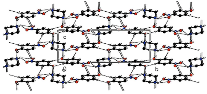
4-nitrophenol ring. In the crystal, the piperazine ring is linked to two 4-nitrophenol moleculesviaO—H N hydrogen bonds. The molecules are also linked via bifurcated N—H (O,O) hydrogen bonds involving the NO2O atoms, forming a two-dimensional network lying parallel to (102). The networks are linked via C—H O hydrogen bonds, forming a three-dimensional structure.
Related literature
For the biological properties of piperazine compounds, see: Foroumadiet al.(2007); Upadhayayaet al.(2004); Chenet al.
(2006); Cunicoet al. (2009); Smitset al.(2008); Beckeret al.
(2006).
Experimental
Crystal data
C6H5NO30.5C4H10N2 Mr= 182.18
Monoclinic,P21=c a= 6.1879 (2) A˚ b= 19.9274 (7) A˚ c= 6.9846 (2) A˚
= 91.199 (1)
V= 861.07 (5) A˚3 Z= 4
MoKradiation
= 0.11 mm1 T= 293 K
0.300.250.20 mm
Data collection
Bruker SMART APEXII area-detector diffractometer Absorption correction: multi-scan
(SADABS; Bruker, 2008) Tmin= 0.968,Tmax= 0.979
12570 measured reflections 1763 independent reflections 1437 reflections withI> 2(I) Rint= 0.024
Refinement
R[F2> 2(F2)] = 0.037 wR(F2) = 0.111 S= 1.04 1763 reflections 126 parameters
H atoms treated by a mixture of independent and constrained refinement
max= 0.19 e A˚
3
min=0.17 e A˚
3
Table 1
Hydrogen-bond geometry (A˚ ,).
D—H A D—H H A D A D—H A
O3—H3A N2i
0.82 1.82 2.6210 (16) 167
N2—H2A O1 0.796 (19) 2.58 (2) 3.2437 (17) 141.4 (17) N2—H2A O2 0.796 (19) 2.557 (19) 3.2273 (17) 142.8 (19) C2—H2 O1i
0.93 2.51 3.3428 (17) 149
C6—H6 O3ii
0.93 2.57 3.5035 (17) 179
Symmetry codes: (i)x1;yþ1 2;zþ
1
2; (ii)xþ1;yþ1;zþ1.
Data collection:APEX2(Bruker, 2008); cell refinement:SAINT (Bruker, 2008); data reduction:SAINT; program(s) used to solve structure:SHELXS97(Sheldrick, 2008); program(s) used to refine structure: SHELXL97 (Sheldrick, 2008); molecular graphics: ORTEP-3 for Windows(Farrugia, 2012) andPLATON(Spek, 2009); software used to prepare material for publication:SHELXL97and PLATON(Spek, 2009).
The authors thank the TBI X-ray facility, CAS in Crystal-lography and Biophysics, University of Madras, India, for the data collection. TS and DV thank the UGC (SAP–CAS) for the departmental facilities. TS also thanks DST Inspire for a fellowship.
Supplementary data and figures for this paper are available from the IUCr electronic archives (Reference: SU2605).
References
Becker, O. M., Dhanoa, D. S., Marantz, Y., Chen, D., Shacham, S., Cheruku, S., Heifetz, A., Mohanty, P., Fichman, M., Sharadendu, A., Nudelman, R., Kauffman, M. & Noiman, S. (2006).J. Med. Chem.49, 3116–3135. Bruker (2008).APEX2, SAINTandSADABS. Bruker AXS Inc., Madison,
Wisconsin, U. S. A.
Chen, J. J., Lu, M., Jing, Y. K. & Dong, J. H. (2006).Bioorg. Med. Chem.14, 6539–6547.
Cunico, W., Gomes, C. R. B., Moreth, M., Manhanini, D. P., Figueiredo, I. H., Penido, C., Henriques, M. G. M. O., Varotti, F. P. & Krettli, A. U. (2009). Eur. J. Med. Chem.44, 1363–1368.
Farrugia, L. J. (2012).J. Appl. Cryst.45, 849–854.
Foroumadi, A., Emami, S., Mansouri, S., Javidnia, A., Saeid-Adeli, N., Shirazi, F. H. & Shafiee, A. (2007).Eur. J. Med. Chem.42, 985–992.
Sheldrick, G. M. (2008).Acta Cryst.A64, 112–122.
Smits, R. A., Lim, H. D., Hanzer, A., Zuiderveld, O. P., Guaita, E., Adami, M., Coruzzi, G., Leurs, R. & Esch, I. J. P. (2008).J. Med. Chem.51, 2457–2467. Spek, A. L. (2009).Acta Cryst.D65, 148–155.
Upadhayaya, R. S., Sinha, N., Jain, S., Kishore, N., Chandra, R. & Arora, S. K. (2004).Bioorg. Med. Chem.12, 2225–2238.
Acta Crystallographica Section E
Structure Reports
Online
supporting information
Acta Cryst. (2013). E69, o1044 [https://doi.org/10.1107/S1600536813015328]
4-Nitrophenol
–
piperazine (2/1)
Perumal Nagapandiselvi, Srinivasan Muralidharan, Thothadri Srinivasan, Rengaswamy
Goplalakrishnan and Devadasan Velmurugan
S1. Comment
Piperazine-based research has attracted considerable attention in recent years. A broad range of compounds displaying
antibacterial (Foroumadi et al., 2007), antifungal (Upadhayaya et al., 2004), anticancer (Chen et al., 2006), antiparasitic
(Cunico et al., 2009), antihistamin (Smits et al., 2008), and antidepressive activities (Becker et al., 2006) have been found
to contain this versatile core. In view of these important properties, we have undertaken the X-ray diffraction study of the
title compound.
In the title adduct, C6H5N1O3, 0.5(C4H10N2), the piperazine ring (N2/C7/C8/N2a/C7a/C8a) possesses inversion
symmetry. It adopts a chair conformation and its mean plane makes a dihedral angle of 65.45 (7)° with the 4-nitrophenol
ring (C1-C6).
In the crystal, the piperazine ring is linked to two 4-nitrophenol molecules via O-H···N hydrogen bonds (Table 1 and Fig
2). The molecules are also linked via bifurcated N-H···O/O hydrogen bonds, involving the NO2 O atoms, forming a
two-dimensional network lying parallel to (102). These networks are linked via C-H···O hydrogen bonds forming a
three-dimensional structure (Table 1).
S2. Experimental
Piperazine 4-nitrophenol was synthesized by mixing an equimolar mixture (1:1) of anhydrous piperazine and
4-nitro-phenol in methanol. The resultant solution was stirred magnetically at room temperature and filtered into a clean beaker.
The filtrate was kept in a constant temperature bath at 308 K. Yellow block-like crystals suitable for x-ray diffraction
were harvested from the solution within a day.
S3. Refinement
The OH and C-bound H atoms were positioned geometrically and refined using a riding model: O—H = 0.82 Å, C—H =
Figure 1
The molecular structure of the title compound, with atom labelling. Displacement ellipsoids are drawn at the 30%
probability level (symmetry code: (a) = -x+2, -y, -z+1).
Figure 2
The crystal packing of the title compound viewed along the a axis. The N-H···O, O-H···N and C-H···O hydrogen bonds
are shown as dashed lines; see Table 1 for details. The H atoms not involved in hydrogen bonding have been omitted for
clarity.
4-Nitrophenol–piperazine (2/1)
Crystal data
C6H5NO3·0.5C4H10N2 Mr = 182.18
Monoclinic, P21/c Hall symbol: -P 2ybc a = 6.1879 (2) Å b = 19.9274 (7) Å c = 6.9846 (2) Å β = 91.199 (1)° V = 861.07 (5) Å3 Z = 4
F(000) = 384 Dx = 1.405 Mg m−3
Mo Kα radiation, λ = 0.71073 Å Cell parameters from 1763 reflections θ = 2.0–26.4°
µ = 0.11 mm−1 T = 293 K Block, yellow
0.30 × 0.25 × 0.20 mm
Data collection
Bruker SMART APEXII area-detector diffractometer
Radiation source: fine-focus sealed tube Graphite monochromator
ω and φ scans
[image:3.610.131.477.256.413.2]12570 measured reflections 1763 independent reflections 1437 reflections with I > 2σ(I) Rint = 0.024
θmax = 26.4°, θmin = 2.0° h = −7→7
k = −24→24 l = −8→8
Refinement
Refinement on F2 Least-squares matrix: full R[F2 > 2σ(F2)] = 0.037 wR(F2) = 0.111 S = 1.04 1763 reflections 126 parameters 0 restraints
Primary atom site location: structure-invariant direct methods
Secondary atom site location: difference Fourier map
Hydrogen site location: inferred from neighbouring sites
H atoms treated by a mixture of independent and constrained refinement
w = 1/[σ2(F
o2) + (0.0534P)2 + 0.1751P] where P = (Fo2 + 2Fc2)/3
(Δ/σ)max < 0.001 Δρmax = 0.19 e Å−3 Δρmin = −0.17 e Å−3
Extinction correction: SHELXL, Fc*=kFc[1+0.001xFc2λ3/sin(2θ)]-1/4 Extinction coefficient: 0.022 (4)
Special details
Geometry. All esds (except the esd in the dihedral angle between two l.s. planes) are estimated using the full covariance matrix. The cell esds are taken into account individually in the estimation of esds in distances, angles and torsion angles; correlations between esds in cell parameters are only used when they are defined by crystal symmetry. An approximate (isotropic) treatment of cell esds is used for estimating esds involving l.s. planes.
Refinement. Refinement of F2 against ALL reflections. The weighted R-factor wR and goodness of fit S are based on F2, conventional R-factors R are based on F, with F set to zero for negative F2. The threshold expression of F2 > 2sigma(F2) is used only for calculating R-factors(gt) etc. and is not relevant to the choice of reflections for refinement. R-factors based on F2 are statistically about twice as large as those based on F, and R- factors based on ALL data will be even larger.
Fractional atomic coordinates and isotropic or equivalent isotropic displacement parameters (Å2)
x y z Uiso*/Ueq
C1 0.3284 (2) 0.39314 (6) 0.57841 (18) 0.0431 (3)
C2 0.2308 (2) 0.33121 (6) 0.61501 (19) 0.0446 (3)
H2 0.0929 0.3300 0.6654 0.053*
C3 0.3365 (2) 0.27233 (6) 0.57715 (19) 0.0436 (3)
H3 0.2711 0.2313 0.6019 0.052*
C4 0.5409 (2) 0.27456 (6) 0.50202 (18) 0.0412 (3)
C5 0.6389 (2) 0.33516 (7) 0.45997 (18) 0.0460 (3)
H5 0.7756 0.3359 0.4072 0.055*
C6 0.5330 (2) 0.39403 (7) 0.4966 (2) 0.0474 (3)
H6 0.5974 0.4348 0.4670 0.057*
C7 0.8041 (2) −0.00321 (8) 0.3919 (2) 0.0604 (4)
H7A 0.7274 −0.0085 0.2703 0.073*
H7B 0.6982 0.0024 0.4911 0.073*
C8 1.0620 (3) 0.06407 (7) 0.5674 (2) 0.0615 (4)
H8A 0.9605 0.0707 0.6697 0.074*
H8B 1.1545 0.1033 0.5618 0.074*
N1 0.65536 (19) 0.21262 (6) 0.46944 (16) 0.0506 (3)
N2 0.9439 (2) 0.05619 (6) 0.38520 (18) 0.0515 (3)
O2 0.56486 (19) 0.15891 (5) 0.50020 (17) 0.0672 (3)
O3 0.23383 (18) 0.45125 (5) 0.61778 (16) 0.0628 (3)
H3A 0.1330 0.4447 0.6898 0.094*
H2A 0.867 (3) 0.0873 (10) 0.362 (3) 0.071 (5)*
Atomic displacement parameters (Å2)
U11 U22 U33 U12 U13 U23
C1 0.0508 (7) 0.0362 (6) 0.0425 (7) 0.0028 (5) 0.0080 (5) −0.0010 (5) C2 0.0394 (6) 0.0439 (7) 0.0507 (7) 0.0000 (5) 0.0092 (5) −0.0019 (5) C3 0.0464 (7) 0.0370 (6) 0.0473 (7) −0.0033 (5) 0.0015 (6) −0.0022 (5) C4 0.0439 (7) 0.0420 (7) 0.0376 (6) 0.0076 (5) −0.0014 (5) −0.0052 (5) C5 0.0397 (7) 0.0547 (8) 0.0438 (7) 0.0013 (5) 0.0079 (5) −0.0017 (6) C6 0.0521 (8) 0.0420 (7) 0.0486 (7) −0.0058 (6) 0.0106 (6) 0.0011 (5) C7 0.0445 (7) 0.0688 (10) 0.0680 (9) −0.0099 (7) 0.0014 (6) 0.0101 (8) C8 0.0785 (10) 0.0362 (7) 0.0703 (10) −0.0139 (7) 0.0146 (8) −0.0050 (6) N1 0.0530 (7) 0.0529 (7) 0.0458 (6) 0.0152 (5) −0.0031 (5) −0.0079 (5)
N2 0.0537 (7) 0.0356 (6) 0.0658 (8) 0.0096 (5) 0.0141 (6) 0.0094 (5)
O1 0.0564 (7) 0.0762 (8) 0.0972 (10) 0.0210 (6) 0.0138 (6) −0.0163 (6) O2 0.0812 (8) 0.0428 (6) 0.0776 (8) 0.0125 (5) 0.0034 (6) −0.0018 (5)
O3 0.0740 (7) 0.0385 (5) 0.0772 (8) 0.0080 (5) 0.0329 (6) 0.0014 (5)
Geometric parameters (Å, º)
C1—O3 1.3290 (15) C7—N2 1.4673 (18)
C1—C6 1.3995 (19) C7—C8i 1.493 (2)
C1—C2 1.4000 (17) C7—H7A 0.9700
C2—C3 1.3720 (18) C7—H7B 0.9700
C2—H2 0.9300 C8—N2 1.463 (2)
C3—C4 1.3799 (19) C8—C7i 1.493 (2)
C3—H3 0.9300 C8—H8A 0.9700
C4—C5 1.3855 (18) C8—H8B 0.9700
C4—N1 1.4434 (16) N1—O1 1.2279 (16)
C5—C6 1.3706 (18) N1—O2 1.2290 (16)
C5—H5 0.9300 N2—H2A 0.80 (2)
C6—H6 0.9300 O3—H3A 0.8200
O3—C1—C6 118.65 (11) N2—C7—H7A 109.7
O3—C1—C2 122.45 (12) C8i—C7—H7A 109.7
C6—C1—C2 118.90 (11) N2—C7—H7B 109.7
C3—C2—C1 120.60 (12) C8i—C7—H7B 109.7
C3—C2—H2 119.7 H7A—C7—H7B 108.2
C1—C2—H2 119.7 N2—C8—C7i 110.09 (12)
C2—C3—C4 119.37 (12) N2—C8—H8A 109.6
C2—C3—H3 120.3 C7i—C8—H8A 109.6
C4—C3—H3 120.3 N2—C8—H8B 109.6
C3—C4—C5 121.15 (11) C7i—C8—H8B 109.6
C5—C4—N1 119.57 (12) O1—N1—O2 122.10 (12)
C6—C5—C4 119.55 (12) O1—N1—C4 118.54 (12)
C6—C5—H5 120.2 O2—N1—C4 119.36 (12)
C4—C5—H5 120.2 C8—N2—C7 110.05 (11)
C5—C6—C1 120.37 (12) C8—N2—H2A 112.3 (14)
C5—C6—H6 119.8 C7—N2—H2A 106.6 (14)
C1—C6—H6 119.8 C1—O3—H3A 109.5
N2—C7—C8i 109.63 (11)
O3—C1—C2—C3 −178.17 (12) O3—C1—C6—C5 177.78 (12)
C6—C1—C2—C3 2.1 (2) C2—C1—C6—C5 −2.5 (2)
C1—C2—C3—C4 −0.1 (2) C3—C4—N1—O1 −176.52 (12)
C2—C3—C4—C5 −1.5 (2) C5—C4—N1—O1 2.59 (19)
C2—C3—C4—N1 177.55 (11) C3—C4—N1—O2 3.58 (19)
C3—C4—C5—C6 1.2 (2) C5—C4—N1—O2 −177.31 (11)
N1—C4—C5—C6 −177.93 (11) C7i—C8—N2—C7 59.04 (16)
C4—C5—C6—C1 0.9 (2) C8i—C7—N2—C8 −58.77 (17)
Symmetry code: (i) −x+2, −y, −z+1.
Hydrogen-bond geometry (Å, º)
D—H···A D—H H···A D···A D—H···A
O3—H3A···N2ii 0.82 1.82 2.6210 (16) 167
N2—H2A···O1 0.796 (19) 2.58 (2) 3.2437 (17) 141.4 (17)
N2—H2A···O2 0.796 (19) 2.557 (19) 3.2273 (17) 142.8 (19)
C2—H2···O1ii 0.93 2.51 3.3428 (17) 149
C6—H6···O3iii 0.93 2.57 3.5035 (17) 179