Crystal structure of 3-[(
E
)-2-(4-phenyl-
1,3-thiazol-2-yl)hydrazin-1-ylidene]-indolin-2-one
Bhimashankar M. Halasangi,aPrema S. Badami,b Sangamesh A. Patilaand G. N. Anil Kumarc*
aDepartment of Chemistry, Karnatak University, Dharwad, India,bDepartment of Chemistry, Shri Sharanabasaveshwar College of Science, Gulbarga 585 102, India, andcDepartment of Physics, M S Ramaiah Institute of Technology, Bangalore 560 054, Karnataka, India. *Correspondence e-mail: [email protected]
Received 4 October 2014; accepted 16 October 2014
Edited by A. J. Lough, University of Toronto, Canada
In the title molecule, C17H12N4OS, the thiazole ring forms a
dihedral angle of 10.8 (2)with the phenyl ring and an angle of 3.1 (3) with the indole ring system [which has a maximum deviation of 0.035 (2) A˚ ]. The dihedral angle between the planes of the phenyl ring and the indole ring system is 11.5 (1). An intramolecular N—H O hydrogen bond is observed. In the crystal, pairs of N—H O hydrogen bonds form inversion dimers with anR22(8) graph-set motif.
Keywords:crystal structure; indolinone; hydrazine; 1,3-thiazole; hydrogen bonding; biological activity.
CCDC reference:1029498
1. Related literature
For the biological activities of substituted thiazoles, see: Aliet al.(2011); Bhartiet al.(2010); Kondratievaet al.(2007). For a related structure, see: Sadıket al.(2004).
2. Experimental 2.1. Crystal data
C17H12N4OS Mr= 320.37 Monoclinic,P21=c
a= 17.7108 (8) A˚
b= 5.1411 (2) A˚
c= 15.9065 (6) A˚
= 94.706 (3) V= 1443.45 (10) A˚3 Z= 4
MoKradiation
= 0.23 mm1 T= 296 K
0.350.310.25 mm
2.2. Data collection
Bruker SMART CCD area-detector diffractometer
Absorption correction: multi-scan (SADABS; Sheldrick, 1996)
Tmin= 0.887,Tmax= 0.934
11530 measured reflections 3142 independent reflections 2124 reflections withI> 2(I)
Rint= 0.039
2.3. Refinement
R[F2> 2(F2)] = 0.046
wR(F2) = 0.109 S= 1.09 3142 reflections
208 parameters
H-atom parameters constrained max= 0.20 e A˚3
[image:1.610.46.247.595.753.2]min=0.25 e A˚3
Table 1
Hydrogen-bond geometry (A˚ ,).
D—H A D—H H A D A D—H A
N2—H2 O1 0.86 2.12 2.771 (2) 133
N4—H4 O1i 0.86 2.11 2.922 (2) 158
Symmetry code: (i)x;y;zþ1.
Data collection: SMART (Bruker, 1998); cell refinement: SAINT (Bruker, 1998); data reduction:SAINT; program(s) used to solve structure:SIR92(Altomare et al., 1994); program(s) used to refine structure: SHELXL97 (Sheldrick, 2008); molecular graphics: ORTEP-3 for Windows(Farrugia, 2012) andCAMERON(Watkinet al., 1993); software used to prepare material for publication:PARST (Nardelli, 1995) andPLATON(Spek, 2009).
Acknowledgements
USIC, Karnatak University, Dharwad, is greatly acknowl-edged for the single-crystal XRD data collection. BMH is grateful to the UGC for financial support in the form of an RFSMS scholarship.
Supporting information for this paper is available from the IUCr electronic archives (Reference: LH5732).
References
Ali, M. A., Mirza, A. H., Bakar, H. J. H. A. & Bernhardt, P. V. (2011).
Polyhedron,30, 556–564.
Altomare, A., Cascarano, G., Giacovazzo, C., Guagliardi, A., Burla, M. C., Polidori, G. & Camalli, M. (1994).J. Appl. Cryst.27, 435.
Bharti, S. K., Nath, G., Tilak, R. & Singh, S. K. (2010).Eur. J. Med. Chem.45, 651–660.
Bruker (1998).SMARTandSAINT. Bruker AXS Inc., Madison, Wisconsin, USA.
Farrugia, L. J. (2012).J. Appl. Cryst.45, 849–854.
Kondratieva, M. L., Pepeleva, A. V., Belskaia, N. P., Koksharov, A. V., Groundwater, P. V., Robeyns, K., Van Meervelt, L., Dehaen, W., Fan, Z. J. & Bakulev, V. A. (2007).Tetrahedron,63, 3042–3048.
Nardelli, M. (1995).J. Appl. Cryst.28, 659.
Sadık, G., Necmi, D., Ibrahim, Y., Alaaddin, C¸ . & Dinc¸er, M. (2004).Acta Cryst.E60, o889–o891.
Sheldrick, G. M. (1996).SADABS. University of Go¨ttingen, Germany.
data reports
Acta Cryst.(2014).E70, o1177–o1178 doi:10.1107/S1600536814022715 Halasangiet al.
o1177
Sheldrick, G. M. (2008).Acta Cryst.A64, 112–122. Spek, A. L. (2009).Acta Cryst.D65, 148–155.
supporting information
sup-1
Acta Cryst. (2014). E70, o1177–o1178supporting information
Acta Cryst. (2014). E70, o1177–o1178 [doi:10.1107/S1600536814022715]
Crystal structure of 3-[(
E
)-2-(4-phenyl-1,3-thiazol-2-yl)hydrazin-1-yl-idene]indolin-2-one
Bhimashankar M. Halasangi, Prema S. Badami, Sangamesh A. Patil and G. N. Anil Kumar
S1. Experimental
S1.1. Synthesis and crystallization
An ethanolic solution of 1.81g (0.01 M) of 2-hydrazino-4-phenylthiazole was added drop wise to an etanolic solution of
1.47g (0.01 M) of isatin with constant stirring. After the complete addition, the reaction mixture was stirred further for
8-9 hrs until the solid separated out from the reaction mixture. The separated solid was filtered and washed with cold
alcohol, dried and recrystallized from DMF (Yield: 95 %. MP: 443-446K). Block-shaped colourless crystals were
obtained by slow evaporation of a solution of the title compound at room temperature in DMF:water in the ratio 2:1.
S1.2. Refinement
H atoms were placed in idealized positions and refined using a riding-model approximation with N—H = 0.86 Å, C—H =
0.93 Å and with Uiso(H) = 1.2Ueq(N,C).
S2. Comment
Isatin derivatives and compounds containing a thiazole ring are class of organic compounds which have fascinated many
synthetic researchers due to their wide range of biological activity ( Ali et al., 2011; Bharti et al., 2010; Kondratieva et al., 2007).
The molecular structure of the title compound is shown in Fig. 1. An intramolecular N—H···O hydrogen bond is
observed. The thiazole ring is essentially planar with a maximum deviation of 0.005 (2) Å for atom N1. The thiazole ring
(S1/C9/N1/C7/C8) forms dihedral angles of 10.8 (2)° with the phenyl ring (C1–C6) and 3.1 (3)° with the indole ring
system (C10—C16/N4/C17, with a maximum deviation of 0.035 (2)Å for atom C17). The dihedral angle between the
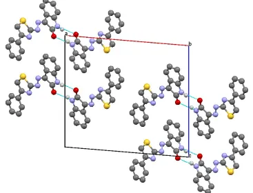
phenyl ring and the indole ring system is 11.5 (1)Å. In the crystal, pairs of N—H···O hydrogen bonds form inversion
Figure 1
The molecular structure of the title compound with displacement ellipsoids drawn at the 50% probability level. The
dashed line indicates an intramolecular N—H···N bond
Figure 2
Part of the crystal structure with hydrogen bonds indicated as dotted lines
3-[(E)-2-(4-Phenyl-1,3-thiazol-2-yl)hydrazin-1-ylidene]indolin-2-one
Crystal data
C17H12N4OS
Mr = 320.37 Monoclinic, P21/c
Hall symbol: -P 2ybc
a = 17.7108 (8) Å
b = 5.1411 (2) Å
c = 15.9065 (6) Å
β = 94.706 (3)°
V = 1443.45 (10) Å3
[image:4.610.121.488.288.568.2]supporting information
sup-3
Acta Cryst. (2014). E70, o1177–o1178F(000) = 664
Dx = 1.474 Mg m−3
Mo Kα radiation, λ = 0.71073 Å
µ = 0.23 mm−1
T = 296 K Block, colourless 0.35 × 0.31 × 0.25 mm
Data collection
Bruker SMART CCD area-detector diffractometer
Graphite monochromator
φ and ω scans
Absorption correction: multi-scan (SADABS; Sheldrick, 1996)
Tmin = 0.887, Tmax = 0.934
11530 measured reflections
3142 independent reflections 2124 reflections with I > 2σ(I)
Rint = 0.039
θmax = 27.1°, θmin = 2.3°
h = −22→22
k = −6→6
l = −20→20
Refinement
Refinement on F2
Least-squares matrix: full
R[F2 > 2σ(F2)] = 0.046
wR(F2) = 0.109
S = 1.09 3142 reflections 208 parameters 0 restraints
Primary atom site location: structure-invariant direct methods
Secondary atom site location: difference Fourier map
Hydrogen site location: inferred from neighbouring sites
H-atom parameters constrained
w = 1/[σ2(F
o2) + (0.0451P)2 + 0.0098P]
where P = (Fo2 + 2Fc2)/3
(Δ/σ)max < 0.001
Δρmax = 0.20 e Å−3
Δρmin = −0.25 e Å−3
Special details
Geometry. All e.s.d.'s (except the e.s.d. in the dihedral angle between two l.s. planes) are estimated using the full covariance matrix. The cell e.s.d.'s are taken into account individually in the estimation of e.s.d.'s in distances, angles and torsion angles; correlations between e.s.d.'s in cell parameters are only used when they are defined by crystal symmetry. An approximate (isotropic) treatment of cell e.s.d.'s is used for estimating e.s.d.'s involving l.s. planes.
Refinement. Refinement of F2 against ALL reflections. The weighted R-factor wR and goodness of fit S are based on F2,
conventional R-factors R are based on F, with F set to zero for negative F2. The threshold expression of F2 > σ(F2) is used
only for calculating R-factors(gt) etc. and is not relevant to the choice of reflections for refinement. R-factors based on F2
are statistically about twice as large as those based on F, and R- factors based on ALL data will be even larger.
Fractional atomic coordinates and isotropic or equivalent isotropic displacement parameters (Å2)
x y z Uiso*/Ueq
H3 0.4433 1.6113 0.7629 0.049* C4 0.37150 (12) 1.3150 (4) 0.73568 (12) 0.0415 (5) H4A 0.3512 1.3113 0.7877 0.05* C5 0.34649 (11) 1.1406 (4) 0.67384 (12) 0.0376 (5) H5 0.3098 1.0188 0.6849 0.045* C6 0.37525 (10) 1.1438 (4) 0.59528 (11) 0.0304 (5) C7 0.34960 (10) 0.9552 (4) 0.52935 (11) 0.0309 (5) C8 0.38282 (11) 0.9102 (4) 0.45680 (11) 0.0367 (5) H8 0.4247 1.0007 0.4406 0.044* C9 0.27677 (11) 0.6406 (4) 0.47676 (11) 0.0314 (5) C10 0.16106 (11) 0.1315 (4) 0.39588 (11) 0.0319 (5) C11 0.14763 (10) −0.0560 (4) 0.32827 (11) 0.0317 (5) C12 0.18154 (11) −0.1039 (4) 0.25430 (12) 0.0409 (5) H12 0.222 −0.003 0.2397 0.049* C13 0.15406 (12) −0.3045 (4) 0.20274 (12) 0.0451 (6) H13 0.1762 −0.3386 0.1528 0.054* C14 0.09393 (12) −0.4556 (4) 0.22454 (12) 0.0422 (6) H14 0.0768 −0.5912 0.1893 0.051* C15 0.05887 (11) −0.4091 (4) 0.29754 (12) 0.0392 (5) H15 0.0183 −0.51 0.312 0.047* C16 0.08638 (10) −0.2078 (4) 0.34775 (11) 0.0319 (5) C17 0.10069 (11) 0.0896 (4) 0.45404 (12) 0.0362 (5)
Atomic displacement parameters (Å2)
U11 U22 U33 U12 U13 U23
supporting information
sup-5
Acta Cryst. (2014). E70, o1177–o1178C17 0.0322 (11) 0.0398 (14) 0.0368 (11) −0.0016 (11) 0.0040 (9) −0.0008 (10)
Geometric parameters (Å, º)
S1—C8 1.711 (2) C4—C5 1.377 (3) S1—C9 1.7203 (19) C4—H4A 0.93 O1—C17 1.240 (2) C5—C6 1.388 (2) N1—C9 1.299 (2) C5—H5 0.93 N1—C7 1.389 (2) C6—C7 1.472 (3) N2—N3 1.341 (2) C7—C8 1.358 (2) N2—C9 1.374 (2) C8—H8 0.93 N2—H2 0.86 C10—C11 1.449 (3) N3—C10 1.294 (2) C10—C17 1.486 (3) N4—C17 1.352 (2) C11—C12 1.386 (2) N4—C16 1.411 (2) C11—C16 1.392 (3) N4—H4 0.86 C12—C13 1.381 (3) C1—C2 1.378 (3) C12—H12 0.93 C1—C6 1.391 (3) C13—C14 1.385 (3) C1—H1 0.93 C13—H13 0.93 C2—C3 1.373 (3) C14—C15 1.382 (3) C2—H2A 0.93 C14—H14 0.93 C3—C4 1.373 (3) C15—C16 1.372 (3) C3—H3 0.93 C15—H15 0.93
C5—C6—C1 117.73 (18) C15—C16—C11 122.65 (18) C5—C6—C7 121.26 (18) C15—C16—N4 128.34 (18) C1—C6—C7 121.00 (17) C11—C16—N4 109.01 (17) C8—C7—N1 114.38 (17) O1—C17—N4 126.73 (19) C8—C7—C6 126.00 (18) O1—C17—C10 126.84 (19) N1—C7—C6 119.59 (16) N4—C17—C10 106.42 (17)
C9—N2—N3—C10 −179.69 (17) N2—N3—C10—C17 0.7 (3) C6—C1—C2—C3 −0.6 (3) N3—C10—C11—C12 −3.6 (4) C1—C2—C3—C4 0.2 (3) C17—C10—C11—C12 176.1 (2) C2—C3—C4—C5 0.4 (3) N3—C10—C11—C16 177.11 (18) C3—C4—C5—C6 −0.7 (3) C17—C10—C11—C16 −3.2 (2) C4—C5—C6—C1 0.3 (3) C16—C11—C12—C13 −1.0 (3) C4—C5—C6—C7 179.04 (18) C10—C11—C12—C13 179.7 (2) C2—C1—C6—C5 0.3 (3) C11—C12—C13—C14 −0.2 (3) C2—C1—C6—C7 −178.37 (17) C12—C13—C14—C15 0.9 (3) C9—N1—C7—C8 0.9 (2) C13—C14—C15—C16 −0.3 (3) C9—N1—C7—C6 −177.30 (17) C14—C15—C16—C11 −0.9 (3) C5—C6—C7—C8 −167.50 (19) C14—C15—C16—N4 178.32 (18) C1—C6—C7—C8 11.2 (3) C12—C11—C16—C15 1.6 (3) C5—C6—C7—N1 10.4 (3) C10—C11—C16—C15 −178.92 (17) C1—C6—C7—N1 −170.89 (17) C12—C11—C16—N4 −177.75 (17) N1—C7—C8—S1 −0.5 (2) C10—C11—C16—N4 1.7 (2) C6—C7—C8—S1 177.49 (15) C17—N4—C16—C15 −178.68 (18) C9—S1—C8—C7 0.06 (16) C17—N4—C16—C11 0.7 (2) C7—N1—C9—N2 179.57 (17) C16—N4—C17—O1 176.05 (19) C7—N1—C9—S1 −0.8 (2) C16—N4—C17—C10 −2.7 (2) N3—N2—C9—N1 −178.05 (17) N3—C10—C17—O1 4.6 (3) N3—N2—C9—S1 2.4 (2) C11—C10—C17—O1 −175.10 (19) C8—S1—C9—N1 0.48 (16) N3—C10—C17—N4 −176.72 (18) C8—S1—C9—N2 −179.92 (16) C11—C10—C17—N4 3.6 (2) N2—N3—C10—C11 −179.72 (16)
Hydrogen-bond geometry (Å, º)
D—H···A D—H H···A D···A D—H···A
N2—H2···O1 0.86 2.12 2.771 (2) 133 N4—H4···O1i 0.86 2.11 2.922 (2) 158