Crystal structure of (3
E
)-3-(2,4-dinitro-phenoxymethyl)-4-phenylbut-3-en-2-one
Ignez Caracelli,a* Stella H. Maganhi,aPaulo J. S. Moran,b Bruno R. S. de Paula,bFelix N. Dellingcand Edward R. T. Tiekinkd
aBioMat-Departmento de Fı´sica, Universidade Federal de Sa
˜o Carlos, 13565-905 Sa˜o Carlos, SP, Brazil,bInstituto de Quı´mica, Universidade Estadual de Campinas, CP 6154, 13083-970 Campinas, SP, Brazil,cDepartmento de Qı´mica, Universidade Federal de Sa˜o Carlos, 13565-905 Sa˜o Carlos, SP, Brazil, anddDepartment of Chemistry, University of Malaya, 50603 Kuala Lumpur, Malaysia. *Correspondence e-mail: [email protected]
Received 15 August 2014; accepted 19 August 2014
Edited by P. C. Healy, Griffith University, Australia
In the title compound, C17H14N2O6, the conformation about the C C double bond [1.345 (2) A˚ ] is E, with the ketone moiety almost coplanar [C—C—C—C torsion angle = 9.5 (2)]
along with the phenyl ring [C—C—C—C = 5.9 (2)]. The
aromatic rings are almost perpendicular to each other [dihedral angle = 86.66 (7)]. The 4-nitro moiety is
approxi-mately coplanar with the benzene ring to which it is attached [O—N—C—C = 4.2 (2)], whereas the one in the ortho
position is twisted [O—N—C—C = 138.28 (13)]. The
mol-ecules associateviaC—H O interactions, involving both O atoms from the 2-nitro group, to form a helical supramolecular chain along [010]. Nitro–nitro N O interactions [2.8461 (19) A˚ ] connect the chains into layers that stack along [001].
Keywords:crystal structure; hydrogen bonding; N O interactions.
CCDC reference:1020026
1. Related literature
For background to biotransformations mediated by Sacchar-omyces cerevisiae, see: Rodrigueset al.(2004); de Paulaet al.
(2013). For a related structure, see: Zukerman-Schpectoret al.
(2014). For interactions between nitro groups, see: Daszkie-wicz (2013).
2. Experimental
2.1. Crystal data
C17H14N2O6
Mr= 342.30
Monoclinic,P21=c a= 12.8459 (6) A˚
b= 7.6983 (4) A˚
c= 19.4283 (8) A˚
= 122.254 (2)
V= 1624.82 (14) A˚3
Z= 4
MoKradiation
= 0.11 mm1
T= 290 K
0.660.450.28 mm
2.2. Data collection
Bruker Kappa APEXII CCD diffractometer
Absorption correction: multi-scan (SADABS; Sheldrick, 1996)
Tmin= 0.699,Tmax= 0.745
10435 measured reflections 2957 independent reflections 2630 reflections withI> 2(I)
Rint= 0.019
2.3. Refinement
R[F2> 2(F2)] = 0.037
wR(F2) = 0.101
S= 1.04 2957 reflections
228 parameters
H-atom parameters constrained
max= 0.20 e A˚
3
min=0.21 e A˚
3
Table 1
Hydrogen-bond geometry (A˚ ,).
D—H A D—H H A D A D—H A
C6—H6 O5i
0.93 2.41 3.1375 (18) 135 C16—H16 O6ii
0.93 2.58 3.292 (3) 134
Symmetry codes: (i)x;yþ1;z; (ii)xþ1;yþ1 2;zþ
1 2.
Data collection: APEX2 (Bruker, 2009); cell refinement: SAINT (Bruker, 2009); data reduction:SAINT; program(s) used to solve structure:SIR97(Altomare et al., 1999); program(s) used to refine structure: SHELXL97 (Sheldrick, 2008); molecular graphics: ORTEP-3 for Windows(Farrugia, 2012),QMol(Gans & Shalloway, 2001) and DIAMOND (Brandenburg, 2006); software used to prepare material for publication:MarvinSketch (ChemAxon, 2010) andpublCIF(Westrip, 2010).
Acknowledgements
We thank Professor Regina H. A. Santos from IQSC–USP for the data collection. The Brazilian agencies CNPq (306121/ 2013-1 to IC), CAPES (808/2009 to IC) and FAPESP (2012/ 22524-9 to SHM) are acknowledged for financial support.
data reports
Acta Cryst.(2014).E70, o1051–o1052 doi:10.1107/S1600536814018819 Caracelliet al.
o1051
Supporting information for this paper is available from the IUCr electronic archives (Reference: HG5406).
References
Altomare, A., Burla, M. C., Camalli, M., Cascarano, G. L., Giacovazzo, C., Guagliardi, A., Moliterni, A. G. G., Polidori, G. & Spagna, R. (1999).J. Appl. Cryst.32, 115–119.
Brandenburg, K. (2006).DIAMOND. Crystal Impact GbR, Bonn, Germany. Bruker (2009).APEX2andSAINT. Bruker AXS Inc., Madison, Wisconsin,
USA.
Daszkiewicz, M. (2013).CrystEngComm,15, 10427–10430. Farrugia, L. J. (2012).J. Appl. Cryst.45, 849–854.
Gans, J. & Shalloway, D. (2001).J. Mol. Graph. Model.19, 557–559. Paula, B. R. S. de, Zampieri, D. S., Rodrigues, J. A. R. & Moran, P. J. S. (2013).
Tetrahedron Asymmetry,24, 973–981.
Rodrigues, J. A. R., Moran, P. J. S., Conceica˜o, G. J. A. & Fardelone, L. C. (2004).Food Technol. Biotechnol.42, 295–303.
Sheldrick, G. M. (1996).SADABS. University of Go¨ttingen, Germany. Sheldrick, G. M. (2008).Acta Cryst.A64, 112–122.
Westrip, S. P. (2010).J. Appl. Cryst.43, 920–925.
supporting information
sup-1
Acta Cryst. (2014). E70, o1051–o1052
supporting information
Acta Cryst. (2014). E70, o1051–o1052 [doi:10.1107/S1600536814018819]
Crystal structure of (3
E
)-3-(2,4-dinitrophenoxymethyl)-4-phenylbut-3-en-2-one
Ignez Caracelli, Stella H. Maganhi, Paulo J. S. Moran, Bruno R. S. de Paula, Felix N. Delling and
Edward R. T. Tiekink
S1. Structural commentary
In the context of the study of biotransformation reactions mediated by Saccharomyces cerevisiae, such as the
bio-reduction of α-haloketones and enones (Rodrigues et al., 2004), the title compound,
(3E)-3-(2,4-dinitrophenoxy-methyl)-4-phenylbut-3-en-2- one, (I), as well as its 4-nitrophenylmethyl analogue, i.e.
(3E)-3-(4-nitrophenoxymethyl)-4-phenylbut-3-en-2-one, (II), (Zukerman-Schpector et al. 2014), were synthesised to be used as substrates in order to
compare their behaviour with that of the 3-halomethyl-4-phenyl-3-buten-2-ones analogues (de Paula et al., 2013). Herein,
the crystal structure determination and spectroscopic details of (I) are described.
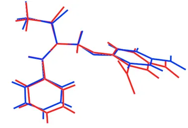
In (I), the conformation about the C═C double bond [1.345 (2) Å] is E. The ketone moiety almost co-planar [C11–C8–
C9–C10 torsion angle = 9.5 (2) °] with the double bond but the phenyl ring is twisted [C8–C11–C12–C17 =
-150.23 (16)°]. The aromatic rings are almost perpendicular to each other [dihedral angle = 86.66 (7)°]. The p-nitro
moiety is approximately co-planar with the benzene ring to which it is attached [O3–N1–C4–C5 = -175.29 (14) °] while
the the one in the o-position is twisted [O5–N2–C2–C3 = -40.76 (17)°].
Fig. 2 shows and overlay diagram of (I) and the inverted molecule of (II) (Zukerman-Schpector et al. 2014). Clearly, the
two molecules adopt very similar conformations.
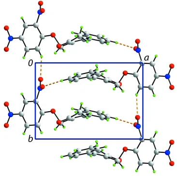
In the crystal packing, molecules associate via C—H···O interactions, whereby the O atoms of the o-nitro group interact
with H derived from a benzene or a phenyl ring, leading to a supramolecular helical chain along [0 1 0]; Fig. 2. There also
N···O5 interactions [2.8461 (19) Å], i.e. nitro···nitro interactions (Daszkiewicz, 2013), that link the chains into a layer.
Layers stack along [0 0 1] without specific intermolecular interactions between them, Fig. 3.
S2. Synthesis and crystallization
Potassium carbonate (232 mg, 2.4 mmol) and 2,4-dinitrophenol (368 mg, 2 mmol) were added to a solution of
4-nitro-phenol (1.53 g, 11 mmol) and 3-bromomethyl-4-phenyl-3-buten-2-one (478 mg, 2 mmol) in dry acetone (4 mL). The
reaction mixture was stirred for 16 hours and the filtered. The solvent was evaporated and the residue was purified by
column chromatography (hexane/EtOAc, 80:20) to afford the product as a colourless solid in 75% yield. The product was
recrystallized by slow evaporation of a 2:1 dichloromethane/hexane mixture. M.pt: 442.2–442.8 K. 1H NMR (CD2Cl2,
400 MHz): δ 2.52 (3H, s), 5.12 (2H, s), 7.41 (d, 1H, J = 9.2 Hz), 7.43-7.56 (5H, m), 7.99 (1H, s), 8.42 (dd, 1H, J = 2.8,
9.2 Hz), 8.72 (d, 1H, J = 2.8 Hz). 13C NMR (CD2Cl2, 150 MHz): δ 26.2, 64.7, 116.1, 122.3, 129.5, 129.7, 130.2, 130.8,
134.5, 134.6, 139.8, 140.9, 148.1, 157.0, 198.4.
S3. Refinement
Carbon-bound H-atoms were placed in calculated positions (C—H 0.93 to 0.97 Å) and were included in the refinement in
Figure 1
The molecular structure of the title showing the atom-labelling scheme and displacement ellipsoids at the 50% probability
level.
Figure 2
Overlay diagram of (I), red image, with inverted (II), blue image, drawn so that the C═C—C(phenyl) atoms are
[image:4.610.106.485.389.648.2]supporting information
sup-3
[image:5.610.121.487.71.435.2]Acta Cryst. (2014). E70, o1051–o1052
Figure 3
Figure 4
A view of unit-cell contents in projection down the b axis. The C—H···O and N···O contacts are shown as orange and
blue dashed lines, respectively.
(3E)-3-(2,4-Dinitrophenoxymethyl)-4-phenylbut-3-en-2-one
Crystal data
C17H14N2O6 Mr = 342.30
Monoclinic, P21/c Hall symbol: -P 2ybc a = 12.8459 (6) Å b = 7.6983 (4) Å c = 19.4283 (8) Å β = 122.254 (2)° V = 1624.82 (14) Å3 Z = 4
F(000) = 712 Dx = 1.399 Mg m−3
Mo Kα radiation, λ = 0.71073 Å Cell parameters from 6161 reflections θ = 2.9–25.3°
µ = 0.11 mm−1 T = 290 K
Irregular, colourless 0.66 × 0.45 × 0.28 mm
Data collection
Bruker Kappa APEXII CCD diffractometer
Radiation source: fine-focus sealed tube Graphite monochromator
ω and φ scans
supporting information
sup-5
Acta Cryst. (2014). E70, o1051–o1052 2630 reflections with I > 2σ(I) Rint = 0.019
θmax = 25.3°, θmin = 1.9°
h = −10→15 k = −7→9 l = −23→19
Refinement
Refinement on F2 Least-squares matrix: full R[F2 > 2σ(F2)] = 0.037 wR(F2) = 0.101 S = 1.04 2957 reflections 228 parameters 0 restraints
Primary atom site location: structure-invariant direct methods
Secondary atom site location: difference Fourier map
Hydrogen site location: inferred from neighbouring sites
H-atom parameters constrained w = 1/[σ2(Fo2) + (0.047P)2 + 0.4196P]
where P = (Fo2 + 2Fc2)/3 (Δ/σ)max < 0.001
Δρmax = 0.20 e Å−3 Δρmin = −0.21 e Å−3
Extinction correction: SHELXL97 (Sheldrick, 2008), Fc*=kFc[1+0.001xFc2λ3/sin(2θ)]-1/4 Extinction coefficient: 0.0323 (19)
Special details
Geometry. All s.u.'s (except the s.u. in the dihedral angle between two l.s. planes) are estimated using the full covariance matrix. The cell s.u.'s are taken into account individually in the estimation of s.u.'s in distances, angles and torsion angles; correlations between s.u.'s in cell parameters are only used when they are defined by crystal symmetry. An approximate (isotropic) treatment of cell s.u.'s is used for estimating s.u.'s involving l.s. planes.
Refinement. Refinement of F2 against ALL reflections. The weighted R-factor wR and goodness of fit S are based on F2, conventional R-factors R are based on F, with F set to zero for negative F2. The threshold expression of F2 > 2σ(F2) is used only for calculating R-factors(gt) etc. and is not relevant to the choice of reflections for refinement. R-factors based on F2 are statistically about twice as large as those based on F, and R- factors based on ALL data will be even larger.
Fractional atomic coordinates and isotropic or equivalent isotropic displacement parameters (Å2)
x y z Uiso*/Ueq
C13 0.57329 (14) 0.1286 (2) 0.19522 (9) 0.0502 (4) H13 0.6557 0.1012 0.2312 0.060* C14 0.52360 (15) 0.1174 (2) 0.11257 (10) 0.0568 (4) H14 0.5729 0.0830 0.0933 0.068* C15 0.40169 (16) 0.1568 (2) 0.05828 (10) 0.0596 (4) H15 0.3690 0.1496 0.0026 0.071* C16 0.32823 (15) 0.2069 (2) 0.08653 (10) 0.0613 (4) H16 0.2458 0.2337 0.0500 0.074* C17 0.37725 (14) 0.2172 (2) 0.16927 (10) 0.0539 (4) H17 0.3268 0.2492 0.1880 0.065* N1 1.22919 (11) 0.1784 (2) 0.33327 (8) 0.0572 (4) N2 0.95589 (10) −0.16711 (15) 0.38003 (7) 0.0443 (3) O1 0.83551 (8) 0.13359 (12) 0.37729 (6) 0.0459 (3) O2 0.78088 (11) 0.27977 (19) 0.51669 (7) 0.0716 (4) O3 1.26869 (11) 0.0420 (2) 0.32347 (8) 0.0767 (4) O4 1.26880 (12) 0.32170 (19) 0.33128 (9) 0.0825 (4) O5 0.95090 (13) −0.28306 (15) 0.33557 (8) 0.0716 (4) O6 0.93475 (11) −0.18635 (15) 0.43354 (8) 0.0673 (4)
Atomic displacement parameters (Å2)
U11 U22 U33 U12 U13 U23
supporting information
sup-7
Acta Cryst. (2014). E70, o1051–o1052 Geometric parameters (Å, º)
C1—O1 1.3405 (15) C10—H10A 0.9600 C1—C6 1.3947 (18) C10—H10B 0.9600 C1—C2 1.3993 (18) C10—H10C 0.9600 C2—C3 1.3773 (17) C11—C12 1.464 (2) C2—N2 1.4610 (16) C11—H11 0.9300 C3—C4 1.375 (2) C12—C17 1.394 (2) C3—H3 0.9300 C12—C13 1.3945 (19) C4—C5 1.380 (2) C13—C14 1.378 (2) C4—N1 1.4638 (17) C13—H13 0.9300 C5—C6 1.379 (2) C14—C15 1.376 (2) C5—H5 0.9300 C14—H14 0.9300 C6—H6 0.9300 C15—C16 1.376 (2) C7—O1 1.4565 (16) C15—H15 0.9300 C7—C8 1.4959 (19) C16—C17 1.381 (2) C7—H7A 0.9700 C16—H16 0.9300 C7—H7B 0.9700 C17—H17 0.9300 C8—C11 1.345 (2) N1—O4 1.2242 (18) C8—C9 1.487 (2) N1—O3 1.2242 (19) C9—O2 1.2176 (19) N2—O6 1.2156 (16) C9—C10 1.502 (2) N2—O5 1.2198 (16)
C9—C8—C7 115.26 (13) O4—N1—C4 117.88 (15) O2—C9—C8 120.24 (14) O3—N1—C4 118.49 (13) O2—C9—C10 120.09 (14) O6—N2—O5 124.41 (13) C8—C9—C10 119.67 (14) O6—N2—C2 118.65 (12) C9—C10—H10A 109.5 O5—N2—C2 116.90 (12) C9—C10—H10B 109.5 C1—O1—C7 117.86 (10)
O1—C1—C2—C3 173.75 (11) C8—C11—C12—C17 −150.23 (16) C6—C1—C2—C3 −3.9 (2) C8—C11—C12—C13 33.5 (2) O1—C1—C2—N2 −5.24 (18) C17—C12—C13—C14 1.2 (2) C6—C1—C2—N2 177.08 (12) C11—C12—C13—C14 177.53 (15) C1—C2—C3—C4 3.50 (19) C12—C13—C14—C15 −0.2 (3) N2—C2—C3—C4 −177.48 (11) C13—C14—C15—C16 −0.4 (3) C2—C3—C4—C5 −0.6 (2) C14—C15—C16—C17 −0.1 (3) C2—C3—C4—N1 179.98 (11) C15—C16—C17—C12 1.1 (3) C3—C4—C5—C6 −1.8 (2) C13—C12—C17—C16 −1.7 (2) N1—C4—C5—C6 177.66 (13) C11—C12—C17—C16 −178.17 (15) C4—C5—C6—C1 1.3 (2) C3—C4—N1—O4 −176.08 (13) O1—C1—C6—C5 −176.05 (13) C5—C4—N1—O4 4.5 (2) C2—C1—C6—C5 1.5 (2) C3—C4—N1—O3 4.2 (2) O1—C7—C8—C11 −94.16 (16) C5—C4—N1—O3 −175.29 (14) O1—C7—C8—C9 88.75 (14) C3—C2—N2—O6 137.34 (13) C11—C8—C9—O2 −171.36 (15) C1—C2—N2—O6 −43.62 (17) C7—C8—C9—O2 5.9 (2) C3—C2—N2—O5 −40.76 (17) C11—C8—C9—C10 9.5 (2) C1—C2—N2—O5 138.28 (13) C7—C8—C9—C10 −173.26 (13) C6—C1—O1—C7 5.4 (2) C9—C8—C11—C12 −178.44 (14) C2—C1—O1—C7 −172.16 (12) C7—C8—C11—C12 4.6 (2) C8—C7—O1—C1 178.57 (11)
Hydrogen-bond geometry (Å, º)
D—H···A D—H H···A D···A D—H···A
C6—H6···O5i 0.93 2.41 3.1375 (18) 135 C16—H16···O6ii 0.93 2.58 3.292 (3) 134