2
000-Acetonaphthone
Ibukun O. Shotonwa and Rene´ T. Boere´*
Department of Chemistry and Biochemistry, University of Lethbridge, Lethbridge, AB, Canada T1K3M4
Correspondence e-mail: [email protected]
Received 15 September 2012; accepted 17 September 2012
Key indicators: single-crystal X-ray study;T= 173 K; mean(C–C) = 0.001 A˚;
Rfactor = 0.040;wRfactor = 0.118; data-to-parameter ratio = 17.6.
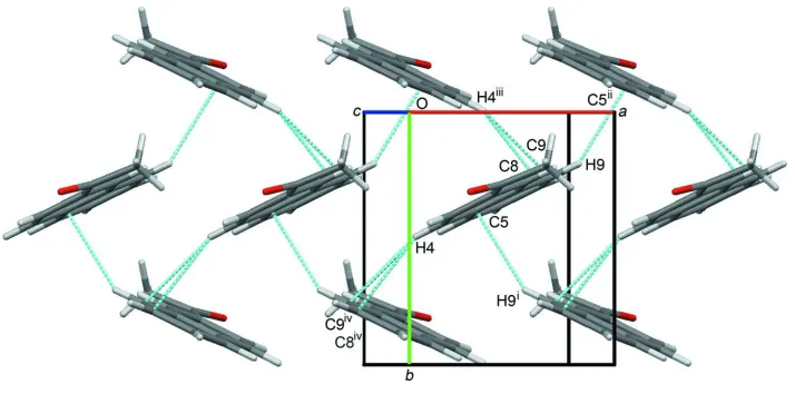
In the structure of the title compound [systematic name: 1-(naphthalen-2-yl)ethanone], C12H10O, the acetyl group is approximately coplanar with the naphthalene ring with a Car—Car—C O torsion angle of 5.8 (2). In the crystal, the molecules are packed in a classic herringbone arrangement typical for aromatic polycycles such as pentacene. They are also linked by weak end-to-end C—H O interactions along theacdiagonal.
Related literature
For synthesis details, see: Bassilios & Salem (1952). For related structures, see: Kemperman et al. (2000); Mattheus et al. (2001); Miyake et al. (1998). For a description of the Cambridge Structural Database, see: Allen (2002).
Experimental
Crystal data
C12H10O
Mr= 170.20 Monoclinic,P21=n
a= 5.9875 (5) A˚
b= 7.4025 (7) A˚
c= 20.2778 (18) A˚
= 93.747 (1)
V= 896.84 (14) A˚3
Z= 4
MoKradiation
= 0.08 mm1
T= 173 K
0.30.250.2 mm
Data collection
Bruker APEXII CCD area-detector diffractometer
Absorption correction: multi-scan (SADABS; Bruker, 2008)
Tmin= 0.701,Tmax= 0.746
12546 measured reflections 2089 independent reflections 1840 reflections withI> 2(I)
Rint= 0.019
Refinement
R[F2> 2(F2)] = 0.040
wR(F2) = 0.118
S= 1.08 2089 reflections
119 parameters
H-atom parameters constrained
max= 0.25 e A˚
3
min=0.23 e A˚
3
Table 1
Hydrogen-bond geometry (A˚ ,).
D—H A D—H H A D A D—H A
C8—H8 O1i
0.95 2.65 3.324 (1) 129
Symmetry code: (i)xþ1 2;yþ
1 2;zþ
1 2.
Data collection:APEX2(Bruker, 2008); cell refinement: SAINT-Plus(Bruker, 2008); data reduction:SAINT-Plus; program(s) used to solve structure: SHELXS97(Sheldrick, 2008); program(s) used to refine structure: SHELXTL (Sheldrick, 2008); molecular graphics: Mercury(Macraeet al., 2008); software used to prepare material for publication:publCIF(Westrip, 2010).
The Natural Sciences and Engineering Research Council of Canada (NSERC) is gratefully acknowledged for a Discovery Grant. The diffractometer was purchased with the help of NSERC and the University of Lethbridge.
Supplementary data and figures for this paper are available from the IUCr electronic archives (Reference: HG5250).
References
Allen, F. H. (2002).Acta Cryst.B58, 380–388.
Bassilios, H. F. & Salem, A. Y. (1952).Bull. Soc. Chim. Fr.pp. 586–592. Bruker (2008). APEX2, SAINT-Plus and SADABS. Bruker AXS Inc.,
Madison Wisconsin, USA.
Kemperman, G. J., de Gelder, R., Dommerholt, F. J., Raemakers-Franken, P. C., Klunder, A. J. H. & Zwanenburg, B. (2000).J. Chem. Soc. Perkin Trans. 2, pp. 1425–1429.
Macrae, C. F., Bruno, I. J., Chisholm, J. A., Edgington, P. R., McCabe, P., Pidcock, E., Rodriguez-Monge, L., Taylor, R., van de Streek, J. & Wood, P. A. (2008).J. Appl. Cryst.41, 466–470.
Mattheus, C. C., Dros, A. B., Baas, J., Meetsma, A., de Boer, J. L. & Palstra, T. T. M. (2001).Acta Cryst.C57, 939–941.
Miyake, Y., Hirose, J., Hasegawa, Y., Sada, K. & Miyata, M. (1998).Chem. Commun.pp. 111–112.
Sheldrick, G. M. (2008).Acta Cryst.A64, 112–122. Westrip, S. P. (2010).J. Appl. Cryst.43, 920–925.
Acta Crystallographica Section E Structure Reports
Online
supporting information
Acta Cryst. (2012). E68, o3112 [doi:10.1107/S1600536812039554]
2
′
-Acetonaphthone
Ibukun O. Shotonwa and Ren
é
T. Boer
é
S1. Comment
2′-Acetonaphthone, (I), is an important example of an aromatic ketone that can be prepared by a classical Friedel-Crafts
acylation reaction (Bassilios & Salem, 1952) and is commercially available from many suppliers. A search of the
Cambridge Structural Database (Allen, 2002; WebCSD August 2012) returned only two previous crystal structures for
(I), in both of which this molecule functions as a guest within an organic host framework. In the structure reported by
Kemperman et al. (2000; refcode MEGXUR), (I) is found as a disordered inclusion compound along with four water
molecules in a clathrate formed by two cephradine molecules. The cages formed by this cephalosporin antibiotic were
shown to be quite flexible and fit guests of differing size, in part by also incorporating varying numbers of
hydrogen-bonded water molecules. This adaptability of the host lattice has been described as permitting "induced fitting" of guest
molecule(s). The cephradine host molecules fully surround their guests and keep individual molecules of (I) separated by
the b axis distance of 7.1965 (3) Å. In the second example (refcode: NECPUG), (I) forms into π-stacks which fill
channels that run along the c axis of a lattice formed from the modified bile acid derivative 3-epiursodeoxycholic acid
(Miyake et al., 1998). The average separation of molecules of (I) along these channels is 3.51 Å, just 0.1 Å greater than
the sums of the van der Waals radii of two carbon atoms. Both of these structures for (I) have very poor precision in the
interatomic distances with mean s.u. of 0.01 Å.
We have therefore determined the crystal structure at 173 K of pure (I). Fig. 1 displays the molecular structure as found
in the crystal lattice. The acetyl group is approximately co-planar with the naphthalene ring and the carbonyl oxygen is
anti to the ring with the torsion angle C1-C2-C11-O1 174.8 (1)°. By comparison, in NECPUG the oxygen atom is in the
syn position. The disorder in MEGXUR precludes a definitive conformational assignment, but the major component
appears to have the oxygen anti as in (I). It is instructive to compare the bond distances determined for pure (I) with those
determined in the host lattices. The high-accuracy structure reported here may also be used to define rigid templates as an
aid in refining future inclusion compounds of (I).
In contrast to the host–guest complexes MEGXUR, which has isolated molecules of (I), and NECPUG with π-stacked
(I), the crystal packing of pure (I) is of the herringbone 2-D edge-to-face type (Figure 2). This arrangement of crystal
packing is reminiscent to that found in pentacene as determined at 90 K (Mattheus et al., 2001). Unlike pentacene,
molecules of (I) are also linked by weak end-to-end by C8-H8···O1 intermolecular interactions (Table 1).
S2. Experimental
A sample of (I) was prepared by the method of Bassilios and Salem (1952).
S3. Refinement
Hydrogen atoms attached to carbon were treated as riding, with C—H = 0.98 Å and Uiso(H) = 1.5Ueq(C) for methyl and C
Figure 1
Molecular structure of (I) drawn with displacement elipsoids at the 50% probability level and showing the atom
numbering scheme.
Figure 2
An extended packing diagram viewed down the c* direction, showing the "herringbone" edge-to-face packing
arrangements. Only atoms involved in short contacts to neighbouring atoms are labelled [Sym. codes: (i) -x + 3/2, y + 1/2,
-z + 1/2; (ii) -x + 3/2, y - 1/2, -z + 1/2; (iii) -x + 1/2, y - 1/2, -z + 1/2; (iv) -x + 1/2, y + 1/2, -z + 1/2]. The O1···H8—C8
H-bonds are not shown but are oriented along the ac diagonal (approximately perpendicular to the page).
1-(Naphthalen-2-yl)ethanone
Crystal data
C12H10O
Mr = 170.20 Monoclinic, P21/n
Hall symbol: -P 2yn
a = 5.9875 (5) Å
b = 7.4025 (7) Å
c = 20.2778 (18) Å
β = 93.747 (1)°
V = 896.84 (14) Å3
Z = 4
F(000) = 360
Dx = 1.261 Mg m−3
Melting point: 326.7 K
Mo Kα radiation, λ = 0.71073 Å Cell parameters from 7818 reflections
[image:3.610.128.485.310.487.2]µ = 0.08 mm−1
T = 173 K
Block, colourless 0.3 × 0.25 × 0.2 mm
Data collection
Bruker APEXII CCD area-detector diffractometer
Radiation source: fine-focus sealed tube, Bruker D8
Graphite monochromator
Detector resolution: 66.06 pixels mm-1
φ and ω scans
Absorption correction: multi-scan (SADABS; Bruker, 2008)
Tmin = 0.701, Tmax = 0.746
12546 measured reflections 2089 independent reflections 1840 reflections with I > 2σ(I)
Rint = 0.019
θmax = 27.6°, θmin = 2.0°
h = −7→7
k = −9→9
l = −26→26
Refinement
Refinement on F2
Least-squares matrix: full
R[F2 > 2σ(F2)] = 0.040
wR(F2) = 0.118
S = 1.08 2089 reflections 119 parameters 0 restraints
Primary atom site location: structure-invariant direct methods
Secondary atom site location: difference Fourier map
Hydrogen site location: inferred from neighbouring sites
H-atom parameters constrained
w = 1/[σ2(F
o2) + (0.0634P)2 + 0.1624P]
where P = (Fo2 + 2Fc2)/3
(Δ/σ)max < 0.001
Δρmax = 0.25 e Å−3
Δρmin = −0.23 e Å−3
Special details
Experimental. A crystal coated in Paratone (TM) oil was mounted on the end of a thin glass capillary and cooled in the gas stream of the diffractometer Kryoflex device.
Geometry. All e.s.d.'s (except the e.s.d. in the dihedral angle between two l.s. planes) are estimated using the full covariance matrix. The cell e.s.d.'s are taken into account individually in the estimation of e.s.d.'s in distances, angles and torsion angles; correlations between e.s.d.'s in cell parameters are only used when they are defined by crystal symmetry. An approximate (isotropic) treatment of cell e.s.d.'s is used for estimating e.s.d.'s involving l.s. planes.
Refinement. Refinement of F2 against ALL reflections. The weighted R-factor wR and goodness of fit S are based on F2,
conventional R-factors R are based on F, with F set to zero for negative F2. The threshold expression of F2 > σ(F2) is used
only for calculating R-factors(gt) etc. and is not relevant to the choice of reflections for refinement. R-factors based on F2
are statistically about twice as large as those based on F, and R- factors based on ALL data will be even larger.
Fractional atomic coordinates and isotropic or equivalent isotropic displacement parameters (Å2)
x y z Uiso*/Ueq
O1 0.29572 (15) 0.31555 (14) −0.00905 (4) 0.0539 (3) C1 0.60105 (15) 0.27553 (12) 0.14913 (4) 0.0256 (2)
H1 0.7348 0.2220 0.1356 0.031*
C2 0.43438 (16) 0.32203 (13) 0.10228 (4) 0.0274 (2) C3 0.23586 (16) 0.40517 (13) 0.12238 (5) 0.0301 (2)
H3 0.1214 0.4389 0.0901 0.036*
C4 0.20800 (15) 0.43702 (13) 0.18753 (5) 0.0284 (2)
H4 0.0745 0.4931 0.2001 0.034*
C5 0.37603 (15) 0.38738 (12) 0.23698 (4) 0.0246 (2) C6 0.35241 (17) 0.41832 (13) 0.30521 (5) 0.0298 (2)
C7 0.51965 (17) 0.36962 (14) 0.35120 (5) 0.0330 (2)
H7 0.5016 0.3906 0.3968 0.040*
C8 0.71788 (17) 0.28884 (14) 0.33168 (5) 0.0325 (2)
H8 0.8322 0.2554 0.3641 0.039*
C9 0.74625 (15) 0.25840 (13) 0.26613 (5) 0.0280 (2)
H9 0.8809 0.2048 0.2533 0.034*
C10 0.57636 (15) 0.30626 (12) 0.21724 (4) 0.0237 (2) C11 0.45374 (18) 0.28604 (15) 0.03043 (5) 0.0351 (3) C12 0.6697 (2) 0.21278 (19) 0.00735 (5) 0.0450 (3)
H12A 0.6551 0.1955 −0.0407 0.067*
H12B 0.7035 0.0967 0.0290 0.067*
H12C 0.7910 0.2984 0.0187 0.067*
Atomic displacement parameters (Å2)
U11 U22 U33 U12 U13 U23
O1 0.0509 (5) 0.0794 (7) 0.0299 (4) 0.0071 (5) −0.0076 (4) −0.0073 (4) C1 0.0253 (4) 0.0241 (4) 0.0279 (5) 0.0005 (3) 0.0048 (3) −0.0005 (3) C2 0.0296 (5) 0.0268 (5) 0.0257 (5) −0.0027 (4) 0.0024 (4) 0.0000 (3) C3 0.0264 (5) 0.0310 (5) 0.0323 (5) 0.0004 (4) −0.0022 (4) 0.0037 (4) C4 0.0238 (4) 0.0264 (5) 0.0355 (5) 0.0024 (3) 0.0041 (4) 0.0015 (4) C5 0.0246 (4) 0.0206 (4) 0.0291 (5) −0.0024 (3) 0.0050 (3) −0.0001 (3) C6 0.0311 (5) 0.0280 (5) 0.0313 (5) −0.0035 (4) 0.0090 (4) −0.0030 (4) C7 0.0386 (5) 0.0357 (5) 0.0254 (4) −0.0101 (4) 0.0062 (4) −0.0031 (4) C8 0.0319 (5) 0.0355 (5) 0.0293 (5) −0.0068 (4) −0.0035 (4) 0.0047 (4) C9 0.0250 (4) 0.0277 (5) 0.0312 (5) −0.0004 (3) 0.0013 (4) 0.0032 (4) C10 0.0237 (4) 0.0207 (4) 0.0268 (4) −0.0017 (3) 0.0027 (3) 0.0014 (3) C11 0.0404 (6) 0.0376 (6) 0.0270 (5) −0.0032 (4) 0.0007 (4) −0.0017 (4) C12 0.0473 (6) 0.0594 (8) 0.0290 (5) 0.0020 (5) 0.0086 (4) −0.0075 (5)
Geometric parameters (Å, º)
O1—C11 1.2185 (13) C6—C7 1.3712 (14)
C1—C2 1.3759 (13) C6—H6 0.9500
C1—C10 1.4169 (12) C7—C8 1.4086 (15)
C1—H1 0.9500 C7—H7 0.9500
C2—C3 1.4216 (13) C8—C9 1.3697 (14)
C2—C11 1.4931 (13) C8—H8 0.9500
C3—C4 1.3629 (14) C9—C10 1.4181 (12)
C3—H3 0.9500 C9—H9 0.9500
C4—C5 1.4215 (13) C11—C12 1.5046 (16)
C4—H4 0.9500 C12—H12A 0.9800
C5—C6 1.4186 (13) C12—H12B 0.9800
C5—C10 1.4220 (12) C12—H12C 0.9800
C2—C1—C10 121.04 (8) C8—C7—H7 119.6
C2—C1—H1 119.5 C9—C8—C7 120.18 (9)
C1—C2—C3 119.51 (8) C7—C8—H8 119.9
C1—C2—C11 122.01 (9) C8—C9—C10 120.56 (9)
C3—C2—C11 118.47 (9) C8—C9—H9 119.7
C4—C3—C2 120.69 (8) C10—C9—H9 119.7
C4—C3—H3 119.7 C1—C10—C9 121.70 (8)
C2—C3—H3 119.7 C1—C10—C5 119.06 (8)
C3—C4—C5 120.88 (8) C9—C10—C5 119.24 (8)
C3—C4—H4 119.6 O1—C11—C2 120.18 (10)
C5—C4—H4 119.6 O1—C11—C12 120.42 (10)
C6—C5—C4 122.35 (8) C2—C11—C12 119.40 (9)
C6—C5—C10 118.84 (8) C11—C12—H12A 109.5
C4—C5—C10 118.80 (8) C11—C12—H12B 109.5
C7—C6—C5 120.39 (9) H12A—C12—H12B 109.5
C7—C6—H6 119.8 C11—C12—H12C 109.5
C5—C6—H6 119.8 H12A—C12—H12C 109.5
C6—C7—C8 120.78 (9) H12B—C12—H12C 109.5
C6—C7—H7 119.6
C10—C1—C2—C3 1.13 (14) C2—C1—C10—C9 179.74 (8)
C10—C1—C2—C11 −178.17 (8) C2—C1—C10—C5 −0.33 (14)
C1—C2—C3—C4 −0.85 (14) C8—C9—C10—C1 −179.72 (8)
C11—C2—C3—C4 178.47 (9) C8—C9—C10—C5 0.34 (14)
C2—C3—C4—C5 −0.25 (15) C6—C5—C10—C1 −179.82 (8)
C3—C4—C5—C6 −179.92 (9) C4—C5—C10—C1 −0.75 (13)
C3—C4—C5—C10 1.04 (14) C6—C5—C10—C9 0.11 (13)
C4—C5—C6—C7 −179.44 (8) C4—C5—C10—C9 179.19 (8)
C10—C5—C6—C7 −0.40 (14) C1—C2—C11—O1 174.03 (10)
C5—C6—C7—C8 0.25 (15) C3—C2—C11—O1 −5.28 (16)
C6—C7—C8—C9 0.22 (15) C1—C2—C11—C12 −5.85 (16)
C7—C8—C9—C10 −0.51 (15) C3—C2—C11—C12 174.85 (10)
Hydrogen-bond geometry (Å, º)
D—H···A D—H H···A D···A D—H···A
C8—H8···O1i 0.95 2.65 3.324 (1) 129