5-(Prop-2-yn-1-yl)-5
H
-dibenzo[
b
,
f
]-azepine: orthorhombic polymorph
M. M. M. Abdoh,aS. Madan Kumar,bK. S. Vinay Kumar,c B. C. Manjunath,bM. P. Sadashivacand N. K. Lokanathb*
aDepartment of Physics, Faculty of Science, An Najah National University, Nabtus West Bank, Palestine,bDepartment of Studies in Physics, Manasagangotri, University of Mysore, Mysore 570 006, India, andcDepartment of Studies in Chemistry, Manasagangotri, University of Mysore, Mysore 570 006, India
Correspondence e-mail: [email protected]
Received 22 November 2012; accepted 28 November 2012
Key indicators: single-crystal X-ray study;T= 103 K; mean(C–C) = 0.003 A˚;
Rfactor = 0.026;wRfactor = 0.067; data-to-parameter ratio = 7.3.
In the title orthorhombic polymorph (space group Iba2), C17H13N, the dihedral angle between the benzene rings is
55.99 (10) and the azepine ring adopts a boat conformation. In the crystal, molecules are linked by C—H contacts. The previously-reported polymorph [Yousuf et al. (2012). Acta Cryst. E68, o1101] crystallizes in the monoclinic system (space groupP21/c) with two molecules in the asymmetric unit.
Related literature
For the previously-reported monoclinic polymorph, see: Yousufet al. (2012). For biochemical background, see: Sada-shivaet al.(2005).
Experimental
Crystal data
C17H13N Mr= 231.28
a= 16.2444 (6) A˚
b= 21.1700 (6) A˚
c= 7.2399 (2) A˚
V= 2489.76 (13) A˚3
MoKradiation
= 0.07 mm1
T= 103 K
0.350.300.25 mm
Data collection
Oxford Diffraction Xcalibur Eos diffractometer
10081 measured reflections
1199 independent reflections 1132 reflections withI> 2(I)
Rint= 0.027
Refinement
R[F2> 2(F2)] = 0.026
wR(F2) = 0.067
S= 1.09 1199 reflections 164 parameters
1 restraint
H-atom parameters constrained
max= 0.08 e A˚3
min=0.10 e A˚
[image:1.610.314.565.308.346.2]3
Table 1
Hydrogen-bond geometry (A˚ ,).
Cg1 and Cg2 are the centroids of the C2/C3/C12–C15 and C6-C11 rings, respectively.
D—H A D—H H A D A D—H A
C10—H10 Cg2i 0.95 2.75 3.659 (2) 160 C18—H18 Cg1ii
0.95 2.58 3.512 (2) 167
Symmetry codes: (i)x;y;z1
2; (ii)x;y;zþ1.
Data collection: CrysAlis PRO(Oxford Diffraction, 2009); cell refinement: CrysAlis PRO; data reduction: CrysAlis PRO; program(s) used to solve structure: SHELXS97 (Sheldrick, 2008); program(s) used to refine structure:SHELXL97(Sheldrick, 2008); molecular graphics: Mercury (Macraeet al., 2006); software used to prepare material for publication:SHELXL97.
SMK thanks UGC–BRS and University of Mysore for the award of a fellowship. MPS gratefully acknowledges financial support (grant No. 37–456/2009[SR]) from the University Grants Commission, New Delhi, India.
Supplementary data and figures for this paper are available from the IUCr electronic archives (Reference: HB6998).
References
Macrae, C. F., Edgington, P. R., McCabe, P., Pidcock, E., Shields, G. P., Taylor, R., Towler, M. & van de Streek, J. (2006).J. Appl. Cryst.39, 453–457. Oxford Diffraction (2009).CrysAlis PRO.Oxford Diffraction Ltd, Yarnton,
Oxfordshire, England.
Sadashiva, M. P., Mallesha, H., KarunakaraMurthy, K. & Rangappa, K. S. (2005).Bioorg. Med. Chem. Lett.15, 1811–1814.
Sheldrick, G. M. (2008).Acta Cryst.A64, 112–122.
Yousuf, S., Khan, M., Fazal, S., Butt, M. & Basha, F. Z. (2012).Acta Cryst.E68, o1101.
Structure Reports
Online
supporting information
Acta Cryst. (2013). E69, o17 [https://doi.org/10.1107/S1600536812048908]
5-(Prop-2-yn-1-yl)-5
H
-dibenzo[
b
,
f
]azepine: orthorhombic polymorph
M. M. M. Abdoh, S. Madan Kumar, K. S. Vinay Kumar, B. C. Manjunath, M. P. Sadashiva and N.
K. Lokanath
S1. Comment
As part of our studies of new derivatives useful for tailoring of biologically active 5-membered heterocyclic rings such as
3,5-disubstituted isoxazole (Sadashiva et al., 2005), we now describe the title compound.
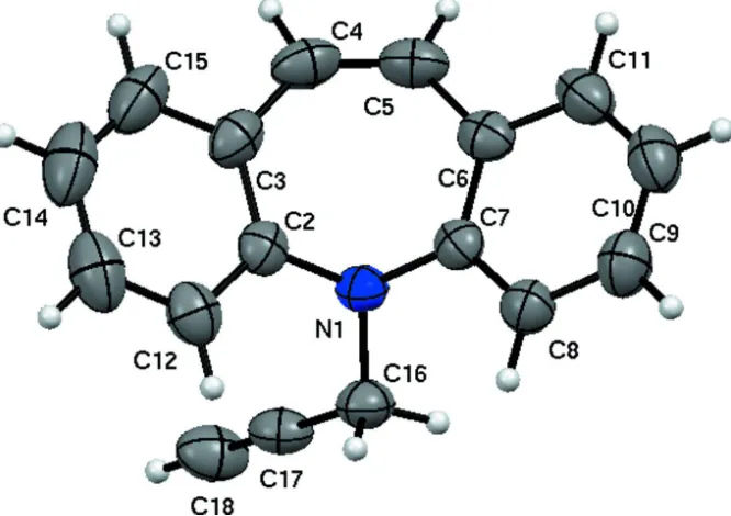
In the title molecule, C17 H13 N (Fig. 1.), benzene rings fused to azepine rings are nearly planar and its geometry is
similar to 5-(Prop-2-ynyl)-5H-dibenzo[b,f]azepine (Yousuf et al., 2012). The dihedral angle between the benzene rings is
55.99 (10)° which is almost equal and large compared to the two molecules repectively for the first polymorph.
Seven-membered azepine ring adopts a boat conformation as indicated by the puckering parameters Q2 = 0.7126 (18) Å,
Q3 = 0.2154 (17) Å, φ2 = 177.93 (15) °, φ3 = 178.1 (5) °, and the total puckering amplitude QT = 0.7444 (17) Å. The title
molecule adopts butterfly shape which may be essential for inhibition pocket which is similar to the reported polymorph
(Yousuf et al., 2012).
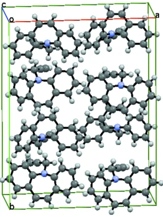
The packing of the title molecules is as shown (Fig. 2.) and features short C—H···π contacts (Table 1).
S2. Experimental
5H-dibenzo[b,f]azepine (1, 0.0026 mol) was taken in a mixture of toluene and water in the ratio 1:1,was added sodium
hydroxide (0.026 mol) followed by tetra-n-butylammonium bromide (TBAB) (0.00286 mol) at room temperature. After
15 minutes, propargyl bromide was added (0.00286 mol) at room temperature. Then, the resulting reaction mixture was
heated at 60°C for 5 h. After completion of reaction (monitored by TLC), the reaction mixture was diluted with water (50
ml). The aqueous layer was extracted with ethyl acetate (3 × 20 ml), the combined ethyl acetate layer was washed with
0.1 N hydrochloric acid (2 × 25 ml), followed by brine solution (2 × 25 ml). Then, the organic layer was dried over
anhydrous sodium sulfate, filtered and concentrated under reduced pressure to afford crude 2, which was purified by
column chromatography over silica gel (60–120 mesh) using Hexane: Ethyl acetate mixture in 9.5:0.5 ratios as eluent.
The pure compound 2 was crystallized in ethyl acetate and hexane to obtain yellow blocks.
1H NMR (DMSO-d6, 300 MHz): δ 7.33 (t, J=7.2 Hz, 2H), 7.13 (t, J=8.7 Hz, 4H), 7.02 (t, J=7.2 Hz, 2H), 6.75 (s, 2H),
4.51 (d, J=1.8 Hz, 2H), 3.08 (s, 1H).
MS (M++1): 232. Melting point (°C): 90 (Uncorrected)
S3. Refinement
In the absence of significant anomalous dispersion effects Friedel pairs were merged. All the hydrogen atoms of the
compound are fixed geometrically (C—H= 0.93–0.97 Å) and allowed to ride on their parent atoms. The poorly fitted
Figure 1
Figure 2
Packing diagram of molecule, viewed along the crystallographic a axis. Dashed lines indicates short contacts.
5-(Prop-2-yn-1-yl)-5H-dibenzo[b,f]azepine
Crystal data
C17H13N
Mr = 231.28
Orthorhombic, Iba2
Hall symbol: I 2 -2c
a = 16.2444 (6) Å
b = 21.1700 (6) Å
c = 7.2399 (2) Å
V = 2489.76 (13) Å3
Z = 8
F(000) = 976
Dx = 1.234 Mg m−3
Mo Kα radiation, λ = 0.71073 Å
Cell parameters from 1199 reflections
θ = 3.2–25.0°
µ = 0.07 mm−1
T = 103 K
Block, yellow
Oxford Diffraction Xcalibur Eos diffractometer
Radiation source: fine-focus sealed tube Graphite monochromator
Detector resolution: 16.0839 pixels mm-1
ω scans
10081 measured reflections
1199 independent reflections 1132 reflections with I > 2σ(I)
Rint = 0.027
θmax = 25.0°, θmin = 3.2°
h = −19→19
k = −25→25
l = −8→8
Refinement
Refinement on F2
Least-squares matrix: full
R[F2 > 2σ(F2)] = 0.026
wR(F2) = 0.067
S = 1.09
1199 reflections 164 parameters 1 restraint
Primary atom site location: structure-invariant direct methods
Secondary atom site location: difference Fourier map
Hydrogen site location: inferred from neighbouring sites
H-atom parameters constrained
w = 1/[σ2(F
o2) + (0.0354P)2 + 0.3725P]
where P = (Fo2 + 2Fc2)/3 (Δ/σ)max < 0.001
Δρmax = 0.08 e Å−3
Δρmin = −0.10 e Å−3
Extinction correction: SHELXL97 (Sheldrick,
2008), FC*=KFC[1+0.001XFC2Λ3/SIN(2Θ)]-1/4
Extinction coefficient: 0.0046 (6)
Special details
Geometry. Bond distances, angles etc. have been calculated using the rounded fractional coordinates. All su's are estimated from the variances of the (full) variance-covariance matrix. The cell e.s.d.'s are taken into account in the estimation of distances, angles and torsion angles
Refinement. Refinement on F2 for ALL reflections except those flagged by the user for potential systematic errors.
Weighted R-factors wR and all goodnesses of fit S are based on F2, conventional R-factors R are based on F, with F set to
zero for negative F2. The observed criterion of F2 > σ(F2) is used only for calculating -R-factor-obs etc. and is not relevant
to the choice of reflections for refinement. R-factors based on F2 are statistically about twice as large as those based on F,
and R-factors based on ALL data will be even larger.
Fractional atomic coordinates and isotropic or equivalent isotropic displacement parameters (Å2)
x y z Uiso*/Ueq
N1 0.23053 (8) 0.36797 (6) 0.6618 (2) 0.0409 (4)
C2 0.31311 (10) 0.36894 (7) 0.5957 (3) 0.0414 (5)
C3 0.34432 (10) 0.42627 (8) 0.5292 (3) 0.0492 (6)
C4 0.29519 (12) 0.48379 (8) 0.5170 (3) 0.0560 (6)
C5 0.21579 (11) 0.48775 (7) 0.4806 (3) 0.0538 (6)
C6 0.15967 (10) 0.43569 (8) 0.4423 (3) 0.0450 (5)
C7 0.16937 (9) 0.37583 (7) 0.5219 (2) 0.0399 (5)
C8 0.11790 (10) 0.32691 (8) 0.4686 (3) 0.0483 (6)
C9 0.05556 (11) 0.33719 (9) 0.3419 (3) 0.0592 (7)
C10 0.04355 (11) 0.39633 (10) 0.2688 (3) 0.0635 (7)
C11 0.09515 (11) 0.44467 (9) 0.3192 (3) 0.0571 (6)
C12 0.36307 (11) 0.31589 (9) 0.5992 (3) 0.0532 (6)
C13 0.44347 (12) 0.31917 (11) 0.5374 (3) 0.0705 (8)
C14 0.47460 (12) 0.37508 (13) 0.4711 (4) 0.0791 (9)
C17 0.26199 (11) 0.33371 (8) 0.9703 (3) 0.0474 (6)
C18 0.30303 (14) 0.34250 (9) 1.1020 (3) 0.0616 (7)
H4 0.32340 0.52250 0.53740 0.0670*
H5 0.19270 0.52900 0.47920 0.0650*
H8 0.12550 0.28600 0.51950 0.0580*
H9 0.02100 0.30320 0.30540 0.0710*
H10 −0.00010 0.40370 0.18410 0.0760*
H11 0.08650 0.48550 0.26830 0.0690*
H12 0.34190 0.27700 0.64430 0.0640*
H13 0.47730 0.28260 0.54070 0.0850*
H14 0.52970 0.37720 0.42790 0.0950*
H15 0.44760 0.46630 0.42260 0.0830*
H16A 0.15230 0.32680 0.84240 0.0590*
H16B 0.22030 0.27930 0.76240 0.0590*
H18 0.33610 0.34960 1.20820 0.0740*
Atomic displacement parameters (Å2)
U11 U22 U33 U12 U13 U23
N1 0.0432 (7) 0.0429 (7) 0.0367 (8) −0.0037 (6) 0.0005 (6) 0.0061 (6)
C2 0.0424 (8) 0.0505 (9) 0.0314 (8) −0.0034 (8) −0.0036 (8) −0.0045 (7)
C3 0.0494 (9) 0.0594 (10) 0.0389 (10) −0.0153 (8) −0.0019 (8) −0.0043 (8)
C4 0.0762 (12) 0.0439 (10) 0.0479 (11) −0.0187 (8) 0.0020 (10) −0.0015 (8)
C5 0.0747 (11) 0.0393 (9) 0.0474 (11) 0.0003 (8) 0.0066 (11) 0.0039 (8)
C6 0.0506 (9) 0.0464 (8) 0.0380 (9) 0.0061 (7) 0.0079 (8) 0.0023 (8)
C7 0.0385 (8) 0.0449 (8) 0.0362 (9) 0.0031 (7) 0.0050 (7) −0.0001 (7)
C8 0.0464 (9) 0.0486 (9) 0.0498 (11) −0.0009 (8) −0.0006 (9) −0.0001 (8)
C9 0.0498 (10) 0.0693 (12) 0.0584 (13) −0.0044 (9) −0.0072 (10) −0.0051 (10)
C10 0.0506 (10) 0.0866 (14) 0.0533 (13) 0.0091 (11) −0.0072 (10) 0.0044 (12)
C11 0.0574 (11) 0.0628 (11) 0.0511 (11) 0.0143 (9) 0.0018 (10) 0.0122 (9)
C12 0.0539 (10) 0.0610 (11) 0.0448 (11) 0.0056 (9) −0.0074 (9) −0.0068 (9)
C13 0.0560 (11) 0.1000 (17) 0.0554 (13) 0.0212 (12) −0.0066 (11) −0.0157 (13)
C14 0.0453 (11) 0.131 (2) 0.0610 (14) −0.0082 (12) 0.0035 (11) −0.0112 (15)
C15 0.0540 (11) 0.0919 (15) 0.0615 (13) −0.0257 (11) 0.0031 (11) −0.0036 (13)
C16 0.0545 (10) 0.0483 (9) 0.0446 (11) −0.0065 (8) −0.0006 (9) 0.0095 (8)
C17 0.0632 (11) 0.0407 (8) 0.0384 (10) −0.0004 (8) 0.0064 (10) 0.0056 (8)
C18 0.0885 (15) 0.0558 (11) 0.0404 (11) −0.0061 (11) −0.0077 (11) 0.0014 (9)
Geometric parameters (Å, º)
N1—C2 1.424 (2) C14—C15 1.369 (3)
N1—C7 1.429 (2) C16—C17 1.454 (3)
N1—C16 1.463 (2) C17—C18 1.178 (3)
C2—C3 1.401 (2) C4—H4 0.9500
C2—C12 1.386 (2) C5—H5 0.9500
C3—C4 1.459 (2) C8—H8 0.9500
C3—C15 1.393 (3) C9—H9 0.9500
C6—C7 1.401 (2) C12—H12 0.9500
C6—C11 1.389 (3) C13—H13 0.9500
C7—C8 1.386 (2) C14—H14 0.9500
C8—C9 1.384 (3) C15—H15 0.9500
C9—C10 1.373 (3) C16—H16A 0.9900
C10—C11 1.372 (3) C16—H16B 0.9900
C12—C13 1.382 (3) C18—H18 0.9500
C13—C14 1.374 (4)
C2—N1—C7 114.53 (14) C3—C4—H4 117.00
C2—N1—C16 117.15 (13) C5—C4—H4 117.00
C7—N1—C16 116.10 (13) C4—C5—H5 117.00
N1—C2—C3 117.96 (14) C6—C5—H5 116.00
N1—C2—C12 122.26 (15) C7—C8—H8 120.00
C3—C2—C12 119.77 (16) C9—C8—H8 120.00
C2—C3—C4 123.10 (16) C8—C9—H9 120.00
C2—C3—C15 118.11 (17) C10—C9—H9 120.00
C4—C3—C15 118.76 (17) C9—C10—H10 120.00
C3—C4—C5 126.88 (16) C11—C10—H10 120.00
C4—C5—C6 127.00 (15) C6—C11—H11 119.00
C5—C6—C7 122.38 (16) C10—C11—H11 119.00
C5—C6—C11 119.39 (16) C2—C12—H12 120.00
C7—C6—C11 118.21 (15) C13—C12—H12 120.00
N1—C7—C6 118.38 (14) C12—C13—H13 120.00
N1—C7—C8 122.01 (14) C14—C13—H13 120.00
C6—C7—C8 119.58 (15) C13—C14—H14 120.00
C7—C8—C9 120.57 (16) C15—C14—H14 120.00
C8—C9—C10 120.19 (17) C3—C15—H15 119.00
C9—C10—C11 119.39 (18) C14—C15—H15 119.00
C6—C11—C10 121.97 (18) N1—C16—H16A 109.00
C2—C12—C13 120.44 (18) N1—C16—H16B 109.00
C12—C13—C14 120.3 (2) C17—C16—H16A 109.00
C13—C14—C15 119.5 (2) C17—C16—H16B 109.00
C3—C15—C14 121.9 (2) H16A—C16—H16B 108.00
N1—C16—C17 110.96 (14) C17—C18—H18 180.00
C16—C17—C18 179.8 (2)
C7—N1—C2—C3 69.0 (2) C4—C3—C15—C14 −178.4 (2)
C7—N1—C2—C12 −112.4 (2) C3—C4—C5—C6 −0.8 (4)
C16—N1—C2—C3 −149.95 (18) C4—C5—C6—C7 32.0 (3)
C16—N1—C2—C12 28.6 (3) C4—C5—C6—C11 −146.4 (2)
C2—N1—C7—C6 −72.36 (18) C5—C6—C7—N1 7.6 (3)
C2—N1—C7—C8 110.12 (17) C5—C6—C7—C8 −174.84 (18)
C16—N1—C7—C6 146.21 (16) C11—C6—C7—N1 −174.02 (16)
C16—N1—C7—C8 −31.3 (2) C11—C6—C7—C8 3.6 (3)
C2—N1—C16—C17 58.9 (2) C5—C6—C11—C10 175.77 (19)
N1—C2—C3—C4 −2.9 (3) N1—C7—C8—C9 175.51 (16)
N1—C2—C3—C15 178.7 (2) C6—C7—C8—C9 −2.0 (3)
C12—C2—C3—C4 178.5 (2) C7—C8—C9—C10 −0.6 (3)
C12—C2—C3—C15 0.1 (3) C8—C9—C10—C11 1.6 (3)
N1—C2—C12—C13 −178.59 (19) C9—C10—C11—C6 0.1 (3)
C3—C2—C12—C13 0.0 (3) C2—C12—C13—C14 −0.3 (3)
C2—C3—C4—C5 −33.6 (4) C12—C13—C14—C15 0.5 (4)
C15—C3—C4—C5 144.8 (2) C13—C14—C15—C3 −0.4 (4)
C2—C3—C15—C14 0.1 (4)
Hydrogen-bond geometry (Å, º)
Cg1 and Cg2 are the centroids of the C2/C3/C12–C15 and C6-C11 rings, respectively.
D—H···A D—H H···A D···A D—H···A
C10—H10···Cg2i 0.95 2.75 3.659 (2) 160
C18—H18···Cg1ii 0.95 2.58 3.512 (2) 167