The role of TET-mediated DNA hydroxymethylation in prostate cancer
E. Smeets, A.G. Lynch, S. Prekovic, T. Van den Broeck, L. Moris, C. Helsen, S. Joniau, F. Claessens, C.E. Massie
PII: S0303-7207(17)30465-3 DOI: 10.1016/j.mce.2017.08.021 Reference: MCE 10058
To appear in: Molecular and Cellular Endocrinology
Received Date: 22 February 2017 Revised Date: 30 June 2017 Accepted Date: 31 August 2017
Please cite this article as: Smeets, E., Lynch, A.G., Prekovic, S., Van den Broeck, T., Moris, L., Helsen, C., Joniau, S., Claessens, F., Massie, C.E., The role of TET-mediated DNA hydroxymethylation in prostate cancer, Molecular and Cellular Endocrinology (2017), doi: 10.1016/j.mce.2017.08.021.
M
AN
US
CR
IP
T
AC
CE
PT
ED
1
The role of TET-mediated DNA hydroxymethylation in prostate cancer
Smeets E1*, Lynch AG2, Prekovic S1, Van den Broeck T1,3, Moris L1,3, Helsen C1, Joniau S3, Claessens F1, Massie CE2
1
Molecular Endocrinology Laboratory, Department of Cellular and Molecular Medicine, KU Leuven, Leuven, Belgium.
2
Cancer Research UK Cambridge Institute, University of Cambridge, Cambridge, UK.
3
Department of Urology, University Hospitals Leuven, Campus Gasthuisberg, Leuven, Belgium. * Corresponding author: elien.smeets@kuleuven.be
Keywords: prostate cancer, epigenetics, TET, DNA hydroxymethylation, 5hmC
Ten-eleven translocation (TET) proteins are recently characterized dioxygenases that regulate
demethylation by oxidizing 5-methylcytosine to 5-hydroxymethylcytosine and further derivatives.
The recent finding that 5hmC is also a stable and independent epigenetic modification indicates
that these proteins play an important role in diverse physiological and pathological processes
such as neural and tumor development. Both the genomic distribution of (hydroxy)methylation
and the expression and activity of TET proteins are dysregulated in a wide range of cancers
including prostate cancer. Up to now it is still unknown how changes in TET and 5(h)mC profiles
are related to the pathogenesis of prostate cancer. In this review, we explore recent advances in
the current understanding of how TET expression and function are regulated in development
and cancer. Furthermore, we look at the impact on 5hmC in prostate cancer and the potential
underlying mechanisms. Finally, we tried to summarize the latest techniques for detecting and
quantifying global and locus-specific 5hmC levels of genomic DNA.
1 Introduction
Recent large-scale genome sequencing studies have classified primary prostate cancer (PCa)
into distinct molecular subtypes, defined by specific genetic alterations (Cancer Genome Atlas
Research, 2015). Gene-fusions involving the ETS family of transcription factors occur in over
50% of primary prostate cancers, with the next most common subtype defined by mutations in
the SPOP gene (~10% cases) (Cancer Genome Atlas Research, 2015). In addition to these
genetic alterations there are recurrent epigenetic changes that occur in over 80% cases and that
show less intra-tumor heterogeneity compared to genetic changes (Bastian, Yegnasubramanian,
Palapattu et al., 2004,Cooper, Eeles, Wedge et al., 2015,Massie, Spiteri, Ross-Adams et al.,
M
AN
US
CR
IP
T
AC
CE
PT
ED
2
changes that vary between tumor foci and change progressively from early to late stage disease
(Brocks, Assenov, Minner et al., 2014,Yegnasubramanian, Haffner, Zhang et al., 2008),
highlighting the ongoing epigenetic evolution in prostate tumors.
The epigenetic changes observed in prostate tumors may be shaped by extrinsic factors (e.g.
metabolic stress (Sciacovelli, Goncalves, Johnson et al., 2016,Thienpont, Steinbacher, Zhao et
al., 2016)) and cell-intrinsic factors (e.g. genetic alterations impinging on epigenetic regulators
(Cancer Genome Atlas Research, 2015,Spans, Van den Broeck, Smeets et al., 2016)). For
example, a rare molecular subtype of prostate cancer is defined by recurrent point mutations in
the IDH1 gene, resulting in increased production of the oncometabolite 2-hydroxyglutarate
(2-HG) that inhibits several 2-oxoglutarate (2-OG) dependent deoxygenases including enzymes
involved in the metabolism of DNA methylation.
In mammalian cells, DNA methylation occurs on the 5’ position of cytosine bases, predominantly
at CpG dinucleotides, and is mediated by DNA methyltransferases (DNMT1, DNMT3a/b) using
S-adenosylmethionine (SAM) as a methyl-donor (Chiang, Gordon, Tal et al., 1996,Gold, Hurwitz
and Anders, 1963). Cytosine methylation (5mC) at transcriptional regulatory elements is
associated with reduced gene expression and tissue-specific 5mC profiles are implicated in
canalization of cellular differentiation (Razin and Riggs, 1980,Roadmap Epigenomics, Kundaje,
Meuleman et al., 2015). 5mC oxidation results in 5-hydroxymethylcytosine (5hmC), a base that
was first identified as a constituent of bacteriophage DNA in 1952 (Wyatt and Cohen, 1952) and
was subsequently found in mammalian tissues in 1972 (Penn, Suwalski, O'Riley et al., 1972). It
was not until 2009 when the ten-eleven translocation methylcytosine dioxygenase (TET) family
of proteins were implicated in the oxidation of 5mC to 5hmC that interest in this base began to
grow (Tahiliani, Koh, Shen et al., 2009). Initial excitement over 5hmC was due to the implication
of this modification in the active demethylation of 5mC during epigenetic reprogramming (He, Li,
Li et al., 2011,Ito, Shen, Dai et al., 2011), however much recent attention has focused on 5hmC
as a stable epigenetic mark involved in transcriptional regulation and lineage specification
(Bachman, Uribe-Lewis, Yang et al., 2014,Koh, Yabuuchi, Rao et al., 2011,Zhang, Su, Jeong et
al., 2016).
The functional impact of 5hmC therefore appears to be context-dependent and locus-specific. In
this review we explore the mechanisms regulating the expression and function of the TET family
and the impact on 5hmC in development and cancer, before focusing on a specific summary of
M
AN
US
CR
IP
T
AC
CE
PT
ED
3
2 The TET gene family
The ten-eleven translocation family of proteins (TET1-3) was initially identified because of the
involvement of TET1 as a fusion partner of the histone methyltransferase (MLL) gene in acute
myeloid leukemia (Lorsbach, Moore, Mathew et al., 2003,Ono, Taki, Taketani et al., 2002). The
TET protein family members are mammalian homologs of the trypanosome base J binding
proteins JBP1 and JBP2, which were found to oxidize the 5-methyl group of thymine (Iyer,
Tahiliani, Rao et al., 2009,Tahiliani et al., 2009). Although 5-hydroxymethylcytosine (5hmC) had
previously been detected in mammalian genomes, the functional importance of this modification
was not further investigated until Tahiliani and colleagues discovered that TET proteins are
dioxygenases that participate in the DNA demethylation pathway by oxidizing 5-methylcytosine
(5mC) to 5-hydroxymethylcytosine (Tahiliani et al., 2009). 5hmC is a substrate for deamination
into 5-hydroxymethyluridine (5hmU) by activation-induced deaminase (AID) and apolipoprotein B
mRNA-editing enzyme complex (APOBEC), followed by base excision repair (BER) (Guo, Su,
Zhong et al., 2011). It can also be converted to formylcytosine (5fC) and finally to
5-carboxylcytosine (5caC) with subsequent base-excision repair to yield unmethylated cytosines
(He et al., 2011,Ito et al., 2011). Decarboxylation of 5caC by unknown enzymes or removal of
the base by thymine DNA glycosylase or other DNA repair enzymes further completes the
demethylation pathway (He et al., 2011,Ito et al., 2011,Tahiliani et al., 2009). For these
reactions, TET enzymes require 2-oxoglutarate (2-OG or α-ketoglutarate) and Fe(II) as
cofactors, rendering their activity acutely dependent on central metabolism and oxygen levels.
The TET family members (TET1-3) all have the same catalytic activity but vary in structural
domains (Figure 1). TET proteins harbor a core catalytic oxygenase domain with a
double-stranded β-helix (DSβH) containing metal-binding residues which are characteristic for 2-OG
and Fe(II)-dependent oxidation (Loenarz and Schofield, 2011). This core domain is typically
preceded by a cysteine-rich domain that seems to be required for enzymatic activity (Iyer et al.,
2009,Tahiliani et al., 2009). TET proteins catalyze demethylation by binding Fe(II) through
conserved His-His-Asp residues. This generates a reactive enzyme-bound Fe(IV)-oxo
intermediate by using molecular oxygen from 2-OG, which reacts with 5mC to yield 5hmC.
At their N-terminus, TET1 and TET3 contain a CXXC zinc finger domain, a motif that is also
found in DNMT1 and is required for direct DNA-binding of DNMT1 (Pradhan, Esteve, Chin et al.,
2008,Xu, Wu, Tan et al., 2011,Zhang, Zhang, Clark et al., 2010). This domain has been reported
to recognize unmodified, methylated and hydroxymethylated cytosines and binds preferentially
M
AN
US
CR
IP
T
AC
CE
PT
ED
4
al., 2013,Xu et al., 2011,Zhang et al., 2010). TET2 does not contain a CXXC-domain but
associates with the IDAX protein which contains a CXXC-domain (Ko, An, Bandukwala et al.,
2013). Divergent sequence specificity has been reported for CXXC-domain containing proteins
(Frauer, Rottach, Meilinger et al., 2011,Risner et al., 2013), and TET deletion mutants lacking
CXXC-domains seem to retain activity to hydroxylate genomic 5mC (Frauer et al., 2011,Liu,
Wang, Deng et al., 2013). Together, these data suggest that the recruitment of TET enzymes to
specific genomic loci may occur through direct CXXC-domain DNA binding and also indirectly as
part of protein-complexes (Nakagawa, Lv, Nakagawa et al., 2015,Yildirim, Li, Hung et al., 2011).
The exact combination of direct binding and protein-interaction partners may in part contribute to
[image:5.595.83.504.294.517.2]the differential recruitment of TET proteins in particular cellular contexts.
Figure 1: Schematic representation of mutations in TET family in PCa. TET1-3 missense, nonsense
substitutions and insertions/deletions are represented in a simplified protein structure showing the DNA
binding domain (DBD) and catalytic domain (CD). The dots represent how frequent the mutations occur in
different samples. The scheme contains both primary PCa samples and metastatic samples (indicated
with an arrow). This representation is based on data retrieved from cBioPortal (http://www.cbioportal.org/), COSMIC (http://cancer.sanger.ac.uk/cosmic) and (Spans et al., 2016). One sample has been excluded for its hyper mutated genotype (TCGA-XK-AAIW; 6517 nonsynonymous mutations among which 4 mutations
M
AN
US
CR
IP
T
AC
CE
PT
ED
5
3 5hmC and the TET enzymes
3.1 Role in transcriptional regulation
One of the best studied covalent DNA modifications is 5mC, which is most prevalent in CpG
islands (70-80% of CpGs are methylated). The accumulation of 5mC at gene promoters and
enhancers is associated with gene repression (Lee, Morton, Epstein et al., 1994,Nelson, Lee,
Nguyen et al., 1997). DNA demethylation occurs in different biological contexts. Passive
demethylation refers to the loss of 5mC in a replication-dependent manner during cell division. A
process for active demethylation was predicted from studies in early development (Mayer,
Niveleau, Walter et al., 2000,Santos, Hendrich, Reik et al., 2002), but this demethylation process
remained unknown for decades. The characterization of TET, thymine-DNA glycosylase (TDG)
and AID/APOBEC activity defined a pathway for 5mC demethylation (Guo, Li, Liang et al.,
2014,He et al., 2011,Morgan, Dean, Coker et al., 2004,Tahiliani et al., 2009), and at least
initially, 5hmC was considered as an intermediate of the demethylation pathway (Ito, D'Alessio,
Taranova et al., 2010,Williams, Christensen and Helin, 2011). Recent studies have confirmed
that in addition to 5hmC being an intermediate step in demethylation, that 5hmC is a stable DNA
modification that is present at higher levels in tissues with low replication rates (Bachman et al.,
2014), and that conversely 5hmC is decreased in rapidly proliferating tissues and in cancer (Jin,
Jiang, Qiu et al., 2011,Lian, Xu, Ceol et al., 2012). Not only are TET proteins involved in the
demethylation pathway, but they have also been shown to physically bind to DNA to prevent
DNA methylation on specific loci (Wu and Zhang, 2011). Recent publication suggests that TET
proteins are not only able to hydroxymethylate DNA, but also RNA (Delatte, Wang, Ngoc et al.,
2016). Therefore, it is believed that TET enzymes have a direct influence on gene transcription.
Indeed, TET1 DNA binding and associated 5hmC markers are found in 5’ gene regulatory
regions and are associated with active gene transcription (Stroud, Feng, Morey Kinney et al.,
2011,Williams, Christensen, Pedersen et al., 2011,Wu and Zhang, 2011). Recent studies have
shown that 5hmC regulates gene transcription at least in part by directing the dynamic
organization and remodeling of chromatin structures. For example, 5hmC can inhibit the binding
of the methyl-CpG binding domain of MeCP2, MBDs and RING/zinc-finger domain-containing
proteins to DNA (Jin, Kadam and Pfeifer, 2010,Valinluck, Tsai, Rogstad et al., 2004). These
chromatin readers repress transcription at loci marked with DNA methylation and therefore, it is
suggested that the 5hmC marks can counteract 5mC-mediated transcriptional repression (Tan
M
AN
US
CR
IP
T
AC
CE
PT
ED
6
An example of the transcriptional activating role of the TET proteins is the enrichment of TET1
and 5hmC at regulatory regions of pluripotency factors such as Nanog in mouse embryonic stem
cells (mESC) (Ito et al., 2010). Knockdown of Tet1 or Tet2 in this system is associated with
lowered expression levels of these factors by promoter hypermethylation (Ficz, Branco,
Seisenberger et al., 2011,Ito et al., 2010). In addition, a transcriptionally repressive role for TET
proteins has been proposed at Polycomb target gene promoters where TET1 contributes to
repressive chromatin complexes in ESCs (Williams et al., 2011,Wu, D'Alessio, Ito et al., 2011).
Based on these findings, 5hmC can be considered as a stable epigenetic marker in its own right,
in addition to being an intermediate of the demethylation pathway. Clearly, the emerging
transcriptional activating and repressing roles of the TET proteins at key genomic loci imply that
they play an important function in normal development, pluripotency and tumor suppression.
3.2 Role in development
During the early stages of development there is a role for TET proteins in epigenetic
reprogramming. Immediately after fertilization, while the paternal and maternal pronuclei move
towards each other, maternal TET3 catalyzes demethylation in the paternal pronucleus (Gu,
Guo, Yang et al., 2011,Iqbal, Jin, Pfeifer et al., 2011,Wossidlo, Nakamura, Lepikhov et al., 2011)
and a partial demethylation in the maternal pronucleus (Guo et al., 2014). Even in primordial
germ cells (PGCs), which migrate into the gonad region at embryonic day 7 and develop into
oocytes and sperm cells, genome-wide demethylation as well as chromatin remodeling is
observed (Hajkova, Ancelin, Waldmann et al., 2008). This demethylation step is carried out
through a replication-dependent mechanism initiated by TET1 and TET2 (Hackett, Sengupta,
Zylicz et al., 2013). Expression patterns of TET enzymes suggest functional specificity: Tet1 and
Tet2 are expressed in PGCs, whereas Tet3 is highly expressed in oocytes (Gu et al., 2011,Hajkova, 2010,Surani and Hajkova, 2010). However, normal development of PGCs is not
ablated by the knockout of individual Tet genes (Dawlaty, Ganz, Powell et al., 2011,Gu et al.,
2011,Ko, Bandukwala, An et al., 2011), suggesting a level of functional redundancy at least
between Tet1 and Tet2.
TET1 also plays an essential role in regulating pluripotency and differentiation in mouse
embryonic stem cells (ESCs). Both TET1 and TET2 are abundantly expressed in mESCs,
accompanied by high 5hmC levels (Ito et al., 2010,Koh et al., 2011,Ruzov, Tsenkina, Serio et
al., 2011). It was recently shown that TET1 can functionally replace transcription factors such as
OCT4 and SOX2 during somatic cell reprogramming to successfully generate induced
M
AN
US
CR
IP
T
AC
CE
PT
ED
7
Tet2 are rapidly downregulated upon differentiation (Ito et al., 2010,Koh et al., 2011,Tahiliani et al., 2009), whereas Tet3 levels rise. Further illustration of selectivity of TET activities came from
Ito and colleagues who reported that KO of Tet1 induced spontaneous differentiation of mESCs
into trophoblasts in vitro, whereas this was not the case for KO of other Tet family members
(Dawlaty et al., 2011,Ito et al., 2010). Tet1 KO in mESCs showed 35% reduction in 5hmC levels
and subtle changes in gene expression (Dawlaty et al., 2011). Surprisingly, the homozygous KO
of Tet1 or Tet2 did not seem to affect normal development in mice (Dawlaty et al., 2011,Li, Cai,
Cai et al., 2011,Moran-Crusio, Reavie, Shih et al., 2011,Quivoron, Couronne, Della Valle et al.,
2011). No difference in gene expression was found in Tet3 KO compared to wild type controls
(Shen, Inoue, He et al., 2014), suggesting compensation from other TET family members. Most
double Tet1/Tet2 deficient mice survive to adulthood, but some die late in embryogenesis or
shortly after birth (Dawlaty, Breiling, Le et al., 2013). In contrast, knockout of Tet3 led to neonatal
lethality (Guo et al., 2011), and Tet1-3 triple deficient mESCs show major defects in
differentiation and reprogramming (Dawlaty, Breiling, Le et al., 2014,Hu, Zhang, Mao et al.,
2014). This stresses the important involvement of TET3 during embryogenesis and illustrates
the partial redundancy between TET proteins.
Postnatally, levels of 5hmC are very tissue- and locus dependent. The highest 5hmC levels have
been found in the central nervous system (CNS) (Globisch, Munzel, Muller et al.,
2010,Szwagierczak, Bultmann, Schmidt et al., 2010). Hydroxymethylation levels in the
cerebellum and hippocampus are higher in adult mice compared to newborns (Szulwach, Li, Li
et al., 2011). In general, 5hmC is even higher in terminally differentiated cells than in progenitor
cells in the CNS (Haffner, Chaux, Meeker et al., 2011,Haffner, Pellakuru, Ghosh et al., 2013,Orr,
Haffner, Nelson et al., 2012). In these cells, the local levels of 5hmC were found to be inversely
correlated with MECP2 binding at regions with differential 5-hydroxymethylation (Szulwach et al.,
2011).
As previously mentioned, Tet1 and Tet2 KO mice are viable and fertile (Dawlaty et al., 2011,Li et
al., 2011,Moran-Crusio et al., 2011), and Tet3 KO leads to neonatal lethality (Gu et al., 2011).
However, Tet1 KO mice do display reduced body mass and smaller litter size (Dawlaty et al.,
2011). More detailed analyses of Tet2 KO mice revealed that TET2 activity is critical for normal
hematopoietic lineage differentiation (Madzo, Vasanthakumar and Godley, 2013,Madzo, Liu,
Rodriguez et al., 2014). TET2 modulates the balance between self-renewal and differentiation in
hematopoietic stem cells. Moreover, mutations in TET2 have been found in multiple types of
leukemia (Ko, Huang, Jankowska et al., 2010,Li et al., 2011,Moran-Crusio et al., 2011,Quivoron
M
AN
US
CR
IP
T
AC
CE
PT
ED
8
leukemia (Albano, Anelli, Zagaria et al., 2011,Chou, Chou, Liu et al., 2011,Ko et al., 2011,Li et
al., 2011,Moran-Crusio et al., 2011,Quivoron et al., 2011,Weissmann, Alpermann, Grossmann et
al., 2012).
3.3 Role in malignancies
The identification of the TET1 gene as a recurrent translocation partner in acute myeloid
leukemia was a first hint to its role in the oncogenic process. Based on rodent models, TET1 can
be considered a tumor suppressor of hematopoietic malignancy and more specifically B cell
lymphoma (Cimmino, Dawlaty, Ndiaye-Lobry et al., 2015). TET2 mutations were found in up to
30% of patients with myeloid malignancies such as chronic myelomonocytic leukemia, acute
myeloid leukemia (AML) and myelodysplastic syndromes (Albano et al., 2011,Chou et al.,
2011,Ko et al., 2010,Madzo et al., 2013,Weissmann et al., 2012). TET2 mutations have also
been reported in solid tumors including clear-cell renal cell carcinoma, prostate cancer (PCa),
and breast cancer (Wahab, Mullally, Hedvat et al., 2009,Chung, Schatoff and
Abdel-Wahab, 2012,Delhommeau, Dupont, Della Valle et al., 2009,Ko et al., 2010,Nickerson, Im,
Misner et al., 2013,Sato, Yoshizato, Shiraishi et al., 2013,Takahashi, 2013,Wu and Ling, 2014).
In myeloid malignancies, mutations in TET2 are mainly loss-of-function whereas in solid tumors
missense mutations are clustered in the catalytic domain of the gene, suggesting that disruption
of TET2 dioxygenase activity provides a selective advantage in multiple tumor types (Ko et al.,
2010). This notion is corroborated by the observation that TET2 mutations in AML are
predominantly associated with genomic hypermethylation (Ko et al., 2010,Madzo et al., 2013).
Recent studies have shown that the overall 5hmC level in genomic DNA is substantially
decreased in many solid tumors, which has been linked to reduced expression levels of the TET
family members (Jin et al., 2011,Kudo, Tateishi, Yamamoto et al., 2012,Lian et al., 2012,Yang,
Liu, Bai et al., 2013). There is an inverse correlation between TET expression and robust tumor
growth and metastasis in colorectal cancer (Gambichler, Sand and Skrygan, 2013,Hsu, Peng,
Kang et al., 2012,Lian et al., 2012). Furthermore, lower TET levels are associated with poor
prognosis of patients with early breast cancer and prostate cancer (Spans, Van den Broeck,
Smeets et al., 2016,Yang, Yu, Hong et al., 2015). Studies in prostate cancer have reported
reduced 5hmC levels in tumor versus normal tissue and suggested this as a potential biomarker
for malignant transformation (Haffner et al., 2011,Yang et al., 2013). More recently, 5hmC levels
were shown to be significantly reduced in ETS-related gene (ERG) negative PCa compared to
normal prostate tissue samples, and this change has been proposed as a potential prognostic
M
AN
US
CR
IP
T
AC
CE
PT
ED
9
al., 2015). These data are suggestive for a tumor suppressive role of TET proteins in prostate
cancer.
4 Regulation of TET genes
4.1 Transcriptional control
The TET enzymes are tightly regulated at many levels of gene expression. Here, most relevant
pathways in prostate cancer will be discussed. Promoter studies revealed two alternative
promoters for mouse Tet1 as well as Tet2, resulting in different isoforms (Neri, Incarnato,
Krepelova et al., 2015,Sohni, Bartoccetti, Khoueiry et al., 2015,Zhang, Xia, Wang et al., 2016).
The Tet1a isoform contains a CXXC DNA-binding domain and is regulated by ESC-cell specific
transcription factors including SOX2 and KLF4, while the Tet1b isoform lacks the CXXC-domain
and is transcribed from an alternative promotor in differentiated cells independently of
pluripotency transcription factors (Neri et al., 2015,Sohni et al., 2015). In human cells only one
TET1 isoform has been characterized, however the human TET1 gene includes an enhancer region that is bound by the pluripotency factors OCT4, SOX2 and NANOG (Neri et al., 2015).
Other transcription factors known to regulate TET expression are the high-mobility group
AT-hook 2 (HMGA2) and hypoxia inducible factor (HIF)-1a (Song, Poliseno, Song et al., 2013,Sun,
Song, Huang et al., 2013). Indeed, several groups reported that all three members of the TET
family can be directly regulated by HIF-1a through binding in the proximal promoter region of
TET genes (Mariani, Vasanthakumar, Madzo et al., 2014,Thienpont et al., 2016,Wu, Chen, Nieh et al., 2015). Furthermore, regulation can be controlled by epigenetic mechanisms such as
hypermethylation of the TET promoter (Chim, Wan, Fung et al., 2010,Cimmino et al., 2015).
4.2 Post-translational control
The activity of TET enzymes is also tighly regulated at the protein level. Major influence on TET
activity include central metabolism and oxygen availability. TET enzymes are oxygen-dependent
dioxygenases and recently tumor hypoxia was shown to reduce TET activity and DNA
demethylation (Thienpont et al., 2016). Furthermore, tricarboxylic acid (TCA) cycle intermediate
metabolites have been implicated in TET regulation. Isocitrate dehydrogenase (IDH) proteins
convert isocitrate to 2-OG using NADP+/NADPH as cofactors (Figure 2). Since 2-OG is an
essential cofactor for dioxygenases such as TET proteins, IDH enzymes are important
regulators of TET activity. Other rate-limiting steps up- and downstream of 2-OG in central
metabolism also have the potential to influence TET activity, including oxygen levels, TCA cycle
M
AN
US
CR
IP
T
AC
CE
PT
ED
10
participating in the TCA cycle such as succinate dehydrogenase (SDH) and fumarate hydratase
(FH) have been described in cancer too (Adam, Yang, Soga et al., 2014,Xiao, Yang, Xu et al.,
2012). Loss-of function mutations in these enzymes result in accumulation of succinate or
fumarate respectively, impairing the activity of 2-OG-dependent dioxygenases such as TET,
reducing DNA demethylation (Sciacovelli et al., 2016).
Figure 2: Crosstalk between TET and key enzymes involved in metabolism and glycosylation.
Several enzymes involved in the TCA cycle such as isocitrate dehydrogenase (IDH), succinate
dehydrogenase (SDH) and fumarate hydratase (FH) have been reported to influence TET activity in
cancer. 2-OG is a product of the TCA cycle and an essential co-factor of TET, thus mutations in IDH as
well as glutamine deficiency affect its hydroxymethylating capacity. High levels of zinc inhibit aconitase
[image:11.595.76.506.217.620.2]M
AN
US
CR
IP
T
AC
CE
PT
ED
11 which glycosylates serine and threonine residues of diverse proteins including TET. TET also enables
GlcNAcylation by recruitment of OGT to the chromatin.
O-Linked N-Acetylglucosamine (GlcNAc) Transferase (OGT) glycosylates hydroxyl groups of
specific serine and threonine residues in many different nuclear and cytoplasmic proteins,
thereby controlling their transcription, activity, stability and localization (Ozcan, Andrali and
Cantrell, 2010). There is a direct interaction between phosphorylated TET proteins and OGT
(Chen, Chen, Bian et al., 2013,Deplus, Delatte, Schwinn et al., 2013,Ito, Katsura, Shimada et al.,
2014,Zhang, Liu, Gao et al., 2014), and TET-OGT complexes mainly co-localize on H3K4me3
positive, CpG-rich promoters at sites of active gene transcription (Chen et al., 2013,Deplus et al.,
2013,Vella, Scelfo, Jammula et al., 2013). This interaction results in the O-GlcNAc modification
of TETs, thereby decreasing available phosphorylation sites and increasing protein stability and
promoting DNA demethylation (Bauer, Gobel, Nagaraj et al., 2015,Shi, Kim, Lu et al., 2013,Vella
et al., 2013). In addition, TET proteins are essential for efficient recruitment of OGT to chromatin
in order to facilitate the modification of histones (Chen et al., 2013,Deplus et al., 2013,Vella et
al., 2013). Increased glucose consumption also leads to a higher production of
UDP-N-acetylglucosamine (UDP-GlcNAc), which is further used by OGT to glycosylate various proteins.
Dysregulation of OGT-TET interaction and co-localization might therefore affect the stability of
TET itself, but also of many other target genes such as tumor suppressor genes and oncogenes
(p53, c-Myc) (Itkonen, Minner, Guldvik et al., 2013,Yang, Kim, Nam et al., 2006).
Additional post-translational modifications have been reported to influence TET activity including
poly (ADP-ribosyl)ation (PARylation) which is catalyzed by enzymes of the poly(ADP-ribose)
polymerase (PARP) family. PARPs use NAD+ as a substrate producing negatively charged
polymers of ADP-ribose which are then hydrolyzed by the poly(ADP-ribose) glycohydrolase
(PARG) (Schreiber, Dantzer, Ame et al., 2006). PARylation participates in highly diverse cellular
processes such as DNA damage response, transcription and apoptosis (Beck, Robert,
Reina-San-Martin et al., 2014,Krishnakumar and Kraus, 2010,Schreiber et al., 2006). A direct
protein-protein interaction between TET1 and PARP1 has been reported associated with PARylation
and enhanced TET1 activity (Ciccarone, Valentini, Zampieri et al., 2015). Recently, TET1 gene
expression has also been found to be positively regulated by PARylation through
hypomethylation of CpG islands in the TET promoter region (Ciccarone, Valentini, Bacalini et al.,
M
AN
US
CR
IP
T
AC
CE
PT
ED
12
Lastly, an important nutritional factor has been implicated in TET regulation at the protein level.
Vitamin C (or L-ascorbic acid) is an essential antioxidant in mammals which is required for
collagen, catecholamine and carnitine biosynthesis (Banhegyi, Benedetti, Margittai et al.,
2014,Englard and Seifter, 1986). It is also an essential cofactor of Fe(II)- and 2-OG-dependent
dioxygenases (such as TET enzymes), acting as an electron donor, adjusting the redox state of
enzymes and reducing Fe(III) to Fe(II). In mouse ES-cells, vitamin C enhances TET activity and
therefore promotes demethylation, leading to global increase of 5hmC levels (Blaschke, Ebata,
Karimi et al., 2013). This activation is possibly mediated by a direct mechanism whereby vitamin
C directly binds to the catalytic domain of TET (Yin, Mao, Zhao et al., 2013).
5 5hmC landscapes in cancer
Hydroxymethylation is inversely associated with cell proliferation as 5hmC levels are decreased
in rapidly proliferating tissues and cancer (Jin et al., 2011,Lian et al., 2012). When staining
different cancer types such as prostate cancer for 5hmC and proliferation marker Ki67, cells that
lost 5hmC present with high levels of Ki67 (Jin et al., 2011). It is believed that 5hmC levels play
an important role in gene regulation, but the genomic distribution of 5hmC marks in normal and
cancer tissues is still largely unknown.
A global decrease of 5hmC was first observed in hematological malignancies (Coutinho,
Monte-Mor, Vianna et al., 2015,Huang and Rao, 2014,Ko, An, Pastor et al., 2015) and later also in solid
cancers including breast cancer, colon cancer, melanoma and prostate cancer (Haffner et al.,
2011,Lian et al., 2012,Murata, Baba, Ishimoto et al., 2015,Spans et al., 2016,Udali, Guarini,
Moruzzi et al., 2015). In melanoma, hypo-5hmC was determined at the gene level for RAC3,
IGF1R and TIMP2 (Lian et al., 2012). However, while the general trend in tumors is global 5hmC decrease, some cancers such as pancreatic cancer are characterized by increased 5hmC levels
(Bhattacharyya, Yu, Suzuki et al., 2013,Huang, Jiang, Li et al., 2013,Navarro, Yin, Ono et al.,
2014).
Technological improvements and detailed studies have revealed more complex landscapes
where 5hmC patterns at individual genomic regions can differ considerably. While tissue and
cellular studies have shown that 5hmC is globally decreased in cancer, high-resolution
genome-wide studies have shown local enrichment of 5hmC at promoters, enhancers and gene bodies of
actively expressed genes (Stroud et al., 2011,Xu et al., 2011). A broad redistribution of 5hmC
M
AN
US
CR
IP
T
AC
CE
PT
ED
13
(Bhattacharyya et al., 2013,Lian et al., 2012,Thomson, Hunter, Lempiainen et al., 2013). This
redistribution generally includes a decrease in 5hmC around transcriptional start sites (TSS) and
increased levels in gene bodies, although the overall level is lower in cancer cells compared to
normal cells.
To date, few studies have studied 5hmC genome-wide in cancer tissues and further work is
needed to map tissue-specific and cancer type-specific 5hmC in order to reveal the diversity of
5hmC changes and to identify molecular signatures.
6 TET and 5hmC in prostate cancer
The clinical behavior of primary as well as metastatic prostate cancer is highly variable with
substantial intra- and interpatient heterogeneity and differential outcome. Current risk
stratifications of primary tumors combine clinical and pathological parameters including Gleason
score, PSA and clinical staging (D'Amico, Whittington, Malkowicz et al., 1998). These risk
stratifications may be refined by the inclusion of genetic and epigenetic profiling, such as point
mutations (SPOP, TP53, PTEN, and FOXA1), copy-number alterations, gene fusions
(TMPRSS2-ERG) and (hydroxy-)methylation landscapes (Barbieri, Baca, Lawrence et al.,
2012,Massie, Mills and Lynch, 2016,Taylor, Schultz, Hieronymus et al., 2010,Tomlins, Rhodes,
Perner et al., 2005,Wang, Shankar, Dhanasekaran et al., 2011).
The Cancer Genome Atlas (TCGA) Research Network recently proposed a new molecular
taxonomy of primary prostate cancer based on data from 333 primary prostate tumors (Cancer
Genome Atlas Research, 2015). They found that 74% of all prostate tumors can be subdivided
in one of seven molecular classes, based on fusions involving ERG (46%), ETV1 (8%), ETV4
(4%) or FLI1 (1%) and on mutations in SPOP (11%), FOXA1 (3%) or IDH1 (1%). All of these
mutated genes either encode transcription factors or are related to gene expression and
epigenetics. Within these different subclasses, epigenetic profiling revealed a diversity of 5mC
changes that was not clearly separated by molecular subtypes but did correlate with gene
silencing. In addition, approximately one in four prostate tumors could not be classified. Either
these unclassified tumors belong to undiscovered subclasses, or there is missing information
linking them to any of the existing classes.
6.1 Genetic alterations of TET genes in prostate cancer
Genetic alterations are infrequent events in prostate cancer. Mutations that have been found in
localized prostate cancer for all three TET genes, showing a similar distribution (Figure 1).
M
AN
US
CR
IP
T
AC
CE
PT
ED
14
having detrimental effects on TET enzymatic activity. Mutations in the TET genes were also
detected in a large cohort of castration-resistant prostate cancer patients (Robinson, Van Allen,
Wu et al., 2015). Again, most of the mutations detected in metastatic PCa are located within the
catalytic domain (included in Figure 1).
Spans and colleagues identified a high risk patient with a TET1 mutation in its catalytic domain
showing distinct hydroxymethylation patterns (Spans et al., 2016). In this study, 15% of the
high-risk prostate cancer samples presented with a loss of at least one TET1 copy and 39 out of 40
samples (97.5%) showed decreased TET1 levels, suggesting other mechanisms of TET1
repression in prostate cancer. Further studies are needed to functionally characterize these
mutations and the consequences of reduced TET1 expression in prostate cancer. If validated,
we propose that these tumors could be added to the IDH1 molecular group of prostate cancers,
since IDH1 mutants are also known to impair TET1 activity and tumors with IDH1 mutations
present with altered 5mC profiles (Xu, Yang, Liu et al., 2011).
6.2 TET transcription in prostate cancer
The mechanisms of transcriptional regulation of the TET genes in prostate cancer are still largely
unknown. However, many signals are expected to converge on the TET genes. The expression
levels of the TET gene regulating transcription factors OCT4, SOX2 and MYC are frequently
altered in prostate cancer (Amini, Fathi, Mobalegi et al., 2014,Gu, Yuan, Wills et al., 2007,Koh,
Bieberich, Dang et al., 2010,Sotomayor, Godoy, Smith et al., 2009,Ugolkov, Eisengart, Luan et
al., 2011,Yu, Cates, Morrissey et al., 2014), suggesting that there may commonly be alterations
on the TET transcription levels. The transcriptional repressor HMGA2 is also selectively
activated during prostate development and its overexpression in prostate cells promotes
formation of prostate intraepithelial neoplasia (PIN) lesions in xenografts (Zong, Huang,
Sankarasharma et al., 2012). In breast cancer cell lines, it was shown that knockdown of
HMGA2 induces TET1 expression which is protective against tumor growth and development of metastases (Sun et al., 2013). Thus, overexpression of HMGA2 in prostate cancer cells might
also downregulate TET expression.
TET expression levels are also controlled at the epigenetic level by the DNA-methyltransferases (DNMTs). DNMT1 acts mainly as a maintenance methyltransferase and DNMT3a/b
predominantly acts as de novo 5mC transferases. DNMT protein levels and activities were both
found to be elevated in prostate cancer cells and tissues compared to normal controls (Gravina,
Ranieri, Muzi et al., 2013,Patra, Patra, Zhao et al., 2002). De novo 5mC by DNMT3a and
M
AN
US
CR
IP
T
AC
CE
PT
ED
15
activity appears to be more related to the early stages of malignancy (Gravina et al., 2013).
TET1 promoter hypermethylation has recently been reported in prostate cancer as well as in multiple other cancers (Chim et al., 2010,Cimmino et al., 2015,Kim, Pierscianek, Mittelbronn et
al., 2011,Li, Li, Mao et al., 2016). Surprisingly, more detailed investigation of the TET1 promoter
region has shown that the observed hypermethylation in prostate cancer is not correlated with its
expression (Figure 3). In contrast, reduced TET1 expression levels were found to be correlated
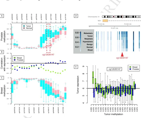
[image:16.595.75.536.226.613.2]with hypermethylation throughout the TET1 promoter region in breast cancer.
Figure 3: Methylation profile of TET1 promoter. [A] Depicting methylation profiles for (equally-spaced)
probes in the promoter and first intron of TET1 for TCGA prostate samples (cg12630147 is
highlighted)(Cancer Genome Atlas Research, 2015). The pattern is the same as [C] seen in Breast
samples from TCGA. [D] Aggregated public data show that the difference in methylation seen in TCGA
between Tumor and Normal tissue of the first-intron regulatory region of TET1 is part of a larger trend.
Depicted are average levels (Beta values) for Blood (from PCa patients), ‘Normal' prostate tissue (from
M
AN
US
CR
IP
T
AC
CE
PT
ED
16 and Metastasis. For a number of probes in the first intron, including cg12630147 (highlighted), there is a
general trend of greater methylation with malignancy. This figure is based on data retrieved from: (Aryee,
Liu, Engelmann et al., 2013,Brocks et al., 2014,Cancer Genome Atlas Research, 2015,Massie et al.,
2015,Naeem, Wong, Chatterton et al., 2014,Paziewska, Dabrowska, Goryca et al., 2014) [B] The
correlation profiles of each methylation probe and TET1 gene expression from TCGA are presented,
showing that methylation of the first intron does not regulate TET1 expression in prostate cancer as it
does in breast. [E] The patterns of association between methylation and expression are highlighted for
probe cg12630147. TCGA data were obtained from the Wanderer tool.
Recent studies have proposed an interaction between the androgen receptor (AR) and TET
regulation. Dhiman and colleagues reported a role for the AR in the regulation of DNA
methylation patterns in gene regulatory regions (Dhiman, Attwood, Campbell et al., 2015).
MicroRNAs such as the miR-29 family are induced upon androgen stimulation and repress TET2
in a high risk prostate cancer model (Takayama, Misawa, Suzuki et al., 2015) (Figure 4).
Moreover, TET2 transcription can be directly repressed by binding of activated AR to the distal
enhancer region of TET2 (Takayama et al., 2015). Furthermore, the AR itself can also be
silenced by DNA methylation in a subset of androgen-deprivation treated prostate cancers,
promoting the development of the disease towards an androgen-independent phenotype
(Jarrard, Kinoshita, Shi et al., 1998,Kinoshita, Shi, Sandefur et al., 2000,Yamanaka, Watanabe,
Yamada et al., 2003).Therefore, it is possible to predict a model whereby a balanced network of
links between AR signaling and TET activity are disrupted at different stages of tumorigenesis to
drive first epigenetic changes and subsequently evolution of resistance to therapy.
Hypoxia has been recognized as an important factor in promoting tumor malignancy (Harris,
2002,Ljungkvist, Bussink, Kaanders et al., 2006,Semenza, 1999). Preclinical data have shown
that hypoxia can induce the selection of aggressive cancer which is characterized by reduced
sensitivity to apoptosis and DNA repair and an increased angiogenesis, proliferation and
metastatic potential (Subarsky and Hill, 2003). Hypoxic primary tumors also form a greater risk
of developing progressive disease (Milosevic, Fyles, Hedley et al., 2004), and prognostic
signatures for poor outcome in prostate cancer include tumor hypoxia (Lalonde, Ishkanian,
Sykes et al., 2014). Increased angiogenesis preceded by hypoxia signaling has been reported in
PCa as well (Milosevic, Chung, Parker et al., 2007). Several groups have shown that TET is
M
AN
US
CR
IP
T
AC
CE
PT
ED
17
2014,Thienpont et al., 2016,Wu et al., 2015) in addition to the direct effects of oxygen availability
on TET enzyme activity. In PCa, there might also be an additional role for androgens in hypoxia
control through the transcriptional regulation of angiogenesis factors, providing another potential
link between PCa biology and TET regulation (Boddy, Fox, Han et al., 2005,Fernandez, Reece,
Ley et al., 2015,Horii, Suzuki, Kondo et al., 2007,Woodward, Wachsberger, Burd et al., 2005).
6.3 TET protein activity in prostate cancer
In addition to the transcriptional regulation of TET genes, the activity and stability of these
enzymes is regulated at the protein level (as outlined in section 4.2). In PCa, androgen signaling
regulates both the expression of TET genes and the recruitment of TET proteins to methylated
DNA. In prostate epithelial cell lines, stimulation of the AR has been reported to induce dynamic
patterns of 5mC in the vicinity of the androgen responsive elements (AREs) at AR target genes
resulting in selective access of regulatory factors to these regions (Dhiman et al., 2015).
Strikingly, TET1 and TDG seem to be co-recruited to these regions upon AR stimulation,
indicating an important role for local demethylation in the transcriptional regulation of AR target
genes. However, these findings requires further validation and investigation of whether the AR
itself is responsible for the recruitment of TET and TDG to the target genes. A recent publication
even shows that TET2 and the AR can form a complex to regulate the expression of
androgen-responsive genes (Nickerson, Das, Im et al., 2016).
Furthermore, there is a clear link between metabolic alterations in PCa and TET protein activity.
As oxygen and 2-OG dependent enzymes, TET proteins are acutely regulated by the
microenvironment and cellular metabolism. For example prostate tumors frequently present with
regions of hypoxia, and a recent study showed that TET activity is reduced in hypoxic conditions
which was associated with hypermethylation of many gene promoters (Thienpont et al., 2016).
This provides one explanation for the early and recurrent 5mC alterations observed in PCa
tumors.
Mutations in TCA enzymes IDH1 (cytosolic) and IDH2 (mitochondrial) are frequently identified in
acute myeloid leukemia, glioma, chondrosarcoma, enchondroma and thyroid cancer patients,
and have also been found in 1% of patients with localized PCa (Chan, Milosevic and Bristow,
2007,Kang, Kim, Oh et al., 2009). A recent TCGA study defined PCa cases with IDH1 mutations
as a separate molecular subclass (Cancer Genome Atlas Research, 2015). Importantly, mutant
M
AN
US
CR
IP
T
AC
CE
PT
ED
18
the conversion of isocitrate to the oncometabolite 2-hydroxyglutarate (2-HG) instead of 2-OG
(Amary, Bacsi, Maggiani et al., 2011,Dang, White, Gross et al., 2009,Hemerly, Bastos and
Cerutti, 2010,Mardis, Ding, Dooling et al., 2009,Murugan, Bojdani and Xing, 2010,Pansuriya,
van Eijk, d'Adamo et al., 2011,Yang, Ye, Guan et al., 2012). Accumulation of this alternative
metabolite prevents catalytic activity of TET enzymes by competing with 2-OG for binding in the
catalytic site of 2-OG-dependent dioxygenases (Xu et al., 2011). Furthermore, a link was also
proposed between the IDH and hypoxia. 2-OG is required for proper function of
prolylhydroxylases (PHD) which are responsible for hydroxylation and promotion of HIF1α
degradation. When conversion of isocitrate is deflected towards the formation of 2-HG, PHD
won’t be able to degrade HIF1α and favoring angiogenesis (Zhao, Lin, Xu et al., 2009). Given
the spatial heterogeneity of hypoxia in PCa (Boutros, Fraser, Harding et al., 2015) and its
potential to apply selective pressure, foci with mutations in IDH1 might be favored in this
environment. This raises the question whether the 1% of PCa samples defined in the IDH1
subclass of the TCGA classification are representative or if there might be more cases wherein
IDH1 mutations are not sampled.
High levels of zinc in the prostate inhibit the activity of the TCA enzyme aconitase (ACO)
(Costello, Liu, Franklin et al., 1997) and some reports have shown that intracellular zinc
concentrations fall during PCa development (Desouki, Geradts, Milon et al., 2007,Singh,
Desouki, Franklin et al., 2006). The resulting alterations in the TCA cycle upstream of 2-OG
production may impact TET activity as well as the 5hmC dynamics during PCa development.
Future studies will be needed to assess the functional consequences of this prostate-specific
phenotype at different stages of prostate malignancy.
Together, there are many mechanisms acting at different levels to regulate TET activity. In this
section we discussed the majority of these mechanisms applicable for prostate cancer (Figure
4). Future and ongoing research will help to identify new mechanisms and elucidate the role of
M
AN
US
CR
IP
T
AC
CE
PT
ED
19 Figure 4: Overview of the important mechanisms influencing TET transcriptional activity and
activity and stability in prostate cancer. Many different pathways are altered during PCa development
and/or progression. TET activity and stability is dependent on mutations in the TET encoding genes,
transcription, androgen regulation and metabolic state such as hypoxia and glucose metabolism. On the
other hand, TET transcriptional activity can be regulated by methylation of CpG islands in its regulatory
regions, TET mutations, androgen signaling partly via miRNAs, hypoxia, products of the TCA cycle and
altered transcription factor levels and activities such as OCT4 and HMGA2.
7 Methods for epigenetic profiling of prostate samples
In order to better understand the spatial and temporal regulation of TET proteins numerous
methods have been developed to detect and quantify the 5hmC content of genomic DNA.
These methods leverage specific chemical conversions, enzymatic labeling, affinity enrichment
or direct detection (Table 1). Each 5hmC analysis method has specific advantages and
limitations; the appropriate choice of method for a given application depends on factors
M
AN
US
CR
IP
T
AC
CE
PT
ED
20
samples to be analyzed; and the optimal sequence resolution (Skvortsova, Zotenko, Luu et al.,
2017). Careful consideration should be given to the balance between genomic resolution and
analytical complexity. Methods that provide a quantitative signal across a genomic locus can
allow a high-throughput analysis of differences between tumor and normal, or groups of tumors
with different clinical features. In contrast, methods that allow single base resolution are more
costly and have a much higher analytical burden, but can identify differences (and patterns of
differences) that would be missed using locus-averaging methods.
7.1 Chemical conversion of 5hmC
Methods using sodium bisulfite conversion (BS) may be considered the current ‘gold standard’
for the analysis of cytosine modifications (Frommer, McDonald, Millar et al., 1992). BS treatment
results in the deamination of unmethylated cytosines to uracil (that is subsequently converted to
thymine), whereas both 5mC and 5hmC are protected from BS-conversion (and subsequently
read out as cytosines; Table 1). Oxidative bisulfite conversion (oxBS) is specific for 5mC alone
and therefore the subtraction of oxBS from BS signals allows the specific identification of
5hmC-modified sites (Booth, Branco, Ficz et al., 2012). This subtractive analysis can provide digital
genome-wide base-pair resolution analysis of both 5mC and 5hmC, albeit at significant financial
and computational cost. The original methods for BS-seq and oxBS-seq require large amounts
of input material (micrograms of genomic DNA) (Booth et al., 2012,Frommer et al., 1992,Lister,
O'Malley, Tonti-Filippini et al., 2008). However, optimized protocols have been developed that
allow lower amounts of starting material (~100ng) (Miura, Enomoto, Dairiki et al., 2012),opening
the way for BS/oxBS analysis of precious clinical samples. It is currently not practical to use
genome-wide sequencing implementations of these methods in studies with large clinical sample
collections due to prohibitive costs and the absence of standardized analysis pipelines for
quantitative differential analysis. Array hybridization platforms for BS analysis have been widely
used in several large-scale studies (e.g. TCGA and ICGC) including several prostate cancer
cohort studies (Brocks et al., 2014,Cancer Genome Atlas Research, 2015). However, the most
popular platform (Illumina 450k array) is no longer in production and the cost of dual analysis
(BS/oxBS) to differentiate 5mC and 5hmC marked loci on newer array platforms is similar to
sequencing based approaches. Where prior knowledge of informative loci exists, as is the case
for prostate cancer (Cancer Genome Atlas Research, 2015,Spans et al., 2016), a targeted
BS/oxBS-sequencing approach is both practical and affordable for use in large cohort studies
M
AN
US
CR
IP
T
AC
CE
PT
ED
21 7.2 Affinity enrichment of 5hmC
In contrast, affinity enrichment methods offer a practical high-throughput alternative to map
genomic loci marked with 5mC or 5hmC (Table 1). These approaches enrich (or ‘pull-down’)
DNA fragments containing the modification of interest using specific antibodies (MeDIP/hMeDIP)
(Ficz et al., 2011,Pomraning, Smith and Freitag, 2009,Weber, Davies, Wittig et al., 2005) or
base-modification binding proteins (e.g. MDB-seq/JBP1-seq) (Robertson, Dahl, Vagbo et al.,
2011,Serre, Lee and Ting, 2010). The relative enrichment at specific genomic loci provides a
quantitative read-out of cytosine modifications at that locus and allows comparative analysis
between conditions or cell types. For example, hMeDIP has been applied to clinical prostate
cancer samples and has identified extensive heterogeneity in 5hmC profiles between tumor and
normal tissue (Spans et al., 2016). Lower costs and less complex downstream analysis make
affinity enrichment methods a practical option for large-scale studies that aim to identify or
monitor differences between cell types or treatment conditions (Uribe-Lewis, Stark, Carroll et al.,
2015,Yegnasubramanian, Wu, Haffner et al., 2011). The resolution of affinity enrichment
methods is limited by the input DNA fragment length (e.g. 100-400bp) and in most methods the
output provides an average signal across a locus, meaning that focal and low-level differences
may be missed in such analyses. Most affinity-based methods also require relatively large
amounts of input material (micrograms of DNA) to analyze both 5mC and 5hmC genome-wide.
7.3 Enzymatic labeling of 5hmC
Several 5hmC analysis methods have employed enzymatic covalent tagging of 5hmC bases
using T4 bacteriophage β-glucosyltransferase (β-GT) (Bhattacharyya et al., 2013,Pastor, Pape,
Huang et al., 2011,Petterson, Chung, Tan et al., 2014,Song, Szulwach, Fu et al., 2011,Yu, Hon,
Szulwach et al., 2012). The β-GT enzyme labels 5hmC residues with glucose molecules, either
protecting these residues from chemical conversion (TAB-seq) (Yu et al., 2012) or restriction
enzyme digestion (HELP-GT, RRHP) (Bhattacharyya et al., 2013,Petterson et al., 2014).
Alternatively, β-GT can incorporate azide-glucose moieties at 5hmC positions, allowing
subsequent tagging with N-hydroxysuccimide-esters to incorporate either biotin for enrichment
methods (hm-Seal) (Song et al., 2011) or fluorophores for direct single molecule quantification
(Song, Diao, Brunger et al., 2016). Modifications of these methods allow analysis from
nanograms of input DNA (nano-hmC-Seal) (Han, Lu, Shih et al., 2016), allowing 5hmC analysis
in small samples (~1000 cells). Most implementations of β-GT tagging of 5hmC result in
quantitative signals at a locus-wide level and therefore may miss focal and low-level differences
M
AN
US
CR
IP
T
AC
CE
PT
ED
22 7.4 Direct detection of 5hmCDirect analysis of 5hmC would circumvent many of the limitations inherent to the methods
outlined above: inaccuracies from chemical conversion or enzymatic labeling efficiencies;
amplification bias and lack of complete genomic coverage; the analytical burden of indirect and
subtractive, relative analysis. Single molecule sequencing platforms (e.g. Nanopore and PacBio)
offer the capability for direct detection of modified bases in native DNA molecules (Laszlo,
Derrington, Brinkerhoff et al., 2013,Schreiber, Wescoe, Abu-Shumays et al., 2013,Wanunu,
Cohen-Karni, Johnson et al., 2011). The long read lengths of these platforms also allow analysis
of genomic regions that are inaccessible to short read and array-based platforms. For example,
a recent study using single molecule real-time sequencing mapped base modifications within
specific LINE repeats in the human genome (Suzuki, Korlach, Turner et al., 2016). Alternative
single molecule approaches are also being developed, including β-GT fluorophore labeling of
single native DNA molecules and direct detection and quantification (Song et al., 2016). Current
implementations of these single molecule technologies have limited throughput (e.g. number of
reads per sequencing run), making genome-wide analysis difficult and also require large
amounts of starting material. In addition, the analysis methods for these newer technologies are
not standardized and continuing platform improvements make standardization unlikely in the
short-term. However, these methods are constantly improving and with increased throughput are
likely to offer the deepest insights into DNA base modifications (including 5hmC) in the future. A
particular advantage with respect to prostate cancer analysis is the fast sample-to-data
turnaround times, opening the possibility of real-time analysis of patient samples in the clinic to
improve diagnostic workflows, prognostication or treatment selection.
Method Name Description Advantages a Disadvantages b References
7 .1 C h e m ic a l c o n v e rs io n
BS and OxBS-sequencing
Bisulfite conversion (5mC + 5hmC) minus Ox-bisulfite conversion (5mC)
Digital signal, base-pair resolution
DNA fragmentation, input >1µg, BS + OxBS analysis for subtract. analysis
(Booth et al., 2012,Frommer et al., 1992)
Tn5mC-seq Tagmentation library
prep, followed by BS or OxBS
Efficient library prep, low input (10ng)
Requires high quality input DNA, BS/oxBS after tagmentation reduces complexity
(Adey and Shendure, 2012,Wang, Gu, Adey et al., 2013)
PBAT Bisulfite treatment,
followed by library prep
100ng DNA for WGBS, base pair resolution
Would require BS and OxBS analysis for subtractive analysis
(Miura et al., 2012)
A ff in it y e n ri c h m
CMS-pulldown BS conversion of 5hmC
to CMS and anti-CMS antibody pull-down
Quantitative, high-throughput, lower analytical burden
BS conversion and antibody specificity/ efficiency, locus avg.
M
AN
US
CR
IP
T
AC
CE
PT
ED
23hMeDIP-seq 5hmC-specific
antibody pull-down
Direct 5hmC test, quantitative, high-throughput, lower analytical burden
Locus wide signal, resolution limited by fragment length, Ab- specificity (1µg input)
(Ficz et al.,
2011,Williams et al., 2011)
JBP1-pulldown T4 β–GT tagging and
JBP1 pull-down
Reagents generated in-house, HTP method
Fragment resolution, high input (1-100µg DNA)
(Robertson et al., 2011,Robertson, Dahl, Ougland et al., 2012) 7 .3 E n z y m e c o n v e rs io n GLIB, hmC-Seal, nano-hmC-Seal
T4 β-GT biotin-tagging of 5hmC
Quantitative, low input (ng DNA), HTP
Resolution limited by fragment length, locus average signal
(Han et al., 2016,Pastor et al., 2011,Song et al., 2011)
TAB-seq T4 β-GT 5hmC
protection, Tet 5mC oxidation, BS convert.
Base pair resolution, 5hmC readout (not BS / oxBS subtractive)
Dependent on enzymatic and BS conversion efficiency
(Yu et al., 2012)
MspI/HpaII digest +/- β-GT variants: HELP-GT, RRHP, HMST
HpaII / MspI digest +/- β-GT protection of 5hmC
Up to base-pair resolution, sensitive, 100ng-1µg input
Single CpG/fragment, GC-rich loci, relative analysis (5hmC/5mC)
(Bhattacharyya et al., 2013,Khare, Pai, Koncevicius et al., 2012,Petterson et al., 2014) Aba-seq or
Pvu-seal-seq
5hmC-specific restriction digest with β-GT tagging and biotin pulldown
Up to base pair resolution, high sensitivity
Single CpG analyzed per fragment, digest and tagging efficiency
(Sun, Dai, Borgaro et al., 2015)
Thiol, selenol or aldehyde enzyme tagging
M.HhaI/M.SssI tag with R-SH/R-SeH donor, then NHS-ester labeling
Direct 5hmC labeling, flexible tagging workflow
Not extensively validated, ~20% recovery and ~1% FP
(Liutkeviciute, Lukinavicius, Masevicius et al., 2009,Liutkeviciute, Kriukiene, Grigaityte et al., 2011)
7 .4 S in g le m o le c u le a n a ly s is Direct single molecule sequencing
Direct 5hmC detection in single molecule SMRT or nanopore sequencing
Direct detection, digital readout, long reads, low consensus error, rapid output
High input DNA, no standardized analysis, low reads per run for WGS analysis
(Flusberg, Webster, Lee et al.,
2010,Laszlo et al., 2013,Schreiber et al., 2013,Wanunu et al., 2011)
Tagged single molecule sequencing
T4 β-GT biotin tag and SMRT-seq or nanopore seq
Improved signal with bulky Glc.
modification
Dependent on tagging efficiency
(Song, Clark, Lu et al., 2011,Zahid, Zhao, He et al., 2016)
BS and Ox-BS single molecule sequencing
Bisulfite conversion and SMRT-seq
Longer read analysis: captures regions not possible by short-reads
Ox-BS conversion efficiency, DNA fragmentation
(Yang, Sebra, Pullman et al., 2015)
Thiol labeling (BS mediated)
Thiolation of 5hmC in ssDNA with nanopore seq
Improved detection of 5hmC
Efficiency, specificity and throughput not well characterized
[image:24.595.68.566.107.660.2](Lu and He, 2013)
Table 1: Summary of selected published methods for the analysis of 5hmC. Subset: BS-conversion;
affinity enrichment; enzymatic tagging; direct analysis. a Base-pair resolution methods allow the
assessment of individual cytosine residues at each locus analyzed. b Locus average methods provide a
global view of 5mC and 5hmC at a given locus (in most cases the resolution is defined by DNA fragment
M
AN
US
CR
IP
T
AC
CE
PT
ED
24
8 Conclusions
The importance of TET enzymes and 5hmC in development and disease is clear, as are the
multiple levels of regulation of these important epigenetic regulators. By contrast, the role of
these proteins in PCa is only beginning to be understood, but the combination of insights from
studies in other systems and high-throughput analysis methods offer great potential for future
studies. Over the coming years we will no doubt learn more about the role of TET and 5hmC in
PCa biology, and the potential for TET proteins and their targets to act as disease markers and
therapeutic targets to improve patient outcomes.
9 Acknowledgements
Massie C. is funded by an ERC grant (337905) and acknowledges support of the University of
Cambridge, the Cancer Research UK Cambridge Centre and Hutchison Whampoa Limited.
Claessens F. and Joniau S. hold grants from Fonds Wetenschappelijk Onderzoek-Vlaanderen
(GOA9816N, G.0684.12N, G.0830.13N). Van den Broeck T. is supported by a PhD fellowship
from Fonds Wetenschappelijk Onderzoek-Vlaanderen (11ZO616N). This work was also
supported by the KU Leuven (GOA/15/017) and Kom op tegen Kanker.
10 References
Abdel-Wahab, O., Mullally, A., Hedvat, C., Garcia-Manero, G., Patel, J., Wadleigh, M., Malinge, S., Yao, J., Kilpivaara, O., Bhat, R., Huberman, K., Thomas, S., Dolgalev, I., Heguy, A., Paietta, E., Le Beau, M.M., Beran, M., Tallman, M.S., Ebert, B.L., Kantarjian, H.M., Stone, R.M., Gilliland, D.G.,
Crispino, J.D. and Levine, R.L., 2009. Genetic characterization of TET1, TET2, and TET3 alterations in myeloid malignancies, Blood. 114, 144-7.
Adam, J., Yang, M., Soga, T. and Pollard, P.J., 2014. Rare insights into cancer biology, Oncogene. 33, 2547-56.
Adey, A. and Shendure, J., 2012. Ultra-low-input, tagmentation-based whole-genome bisulfite sequencing, Genome Res. 22, 1139-43.
Albano, F., Anelli, L., Zagaria, A., Coccaro, N., Minervini, A., Rossi, A.R. and Specchia, G., 2011. Decreased TET2 gene expression during chronic myeloid leukemia progression, Leuk Res. 35, e220-2. Amary, M.F., Bacsi, K., Maggiani, F., Damato, S., Halai, D., Berisha, F., Pollock, R., O'Donnell, P.,
Grigoriadis, A., Diss, T., Eskandarpour, M., Presneau, N., Hogendoorn, P.C., Futreal, A., Tirabosco, R. and Flanagan, A.M., 2011. IDH1 and IDH2 mutations are frequent events in central