Amino Acid Assisted Hydrothermal Synthesis of In(OH)
3Nanoparticles
Controlled in Size and Shape
Takafumi Sasaki, Masafumi Nakaya, Kiyoshi Kanie
*and Atsushi Muramatsu
*Institute of Multidisciplinary Research for Advanced Materials, Tohoku University, Sendai 980-8577, Japan
Size and shape controlled indium hydroxide (In(OH)3) nanoparticles are readily obtained by amino acid assisted hydrothermal synthesis
from an aqueous system. The shape control is achieved by the utilization of adsorption of amino acid on the growing surfaces of the nanoparticles, and rod- and cubic-shaped In(OH)3 nanoparticles are selectively formed in the presence of glycine and L-aspartic acid,
respectively. Furthermore, by utilization of two-step aging technique in L-aspartic acid system, originally developed by the gel-sol method, the cubic-shaped In(OH)3nanoparticles with narrow size distribution are successfully obtained. [doi:10.2320/matertrans.M2009236]
(Received July 6, 2009; Accepted September 11, 2009; Published November 11, 2009)
Keywords: amino acid, hydrothermal synthesis, indium hydroxide, nanoparticle, size and shape control
1. Introduction
Recent remarkable progress in synthetic chemistry of inorganic nanoparticles enabled us to obtain various types of the inorganic nanoparticles with controlling their shapes and sizes.1–3)In particular, solution based synthetic systems have
large advantages for the precise shape and size control of the inorganic nanoparticles.4–8)The gel-sol method, we have recently developed, is a particle deposition system from solution for the preparation of monodispersed nanoparticles in large quantities.9–21) In this system, once formed gels consist of an intermediate of the desired nanoparticles prevent coagulation of growing nanoparticles. Furthermore, by the formation of the gels, concentration of direct precursors of the solid particles in the solution is kept low state among the particle growth. These two points play critical roles to obtain monodispersed nanoparticles. For example, by using the gel-sol method, monodispersed hematite, -Fe2O3, nano- and fine-particles are produced
from ferric hydroxide gel through a two-step phase trans-formation from Fe(OH)3 gel to a solid intermediate,
-FeOOH, of a medium solubility and from -FeOOH to
-Fe2O3.9–16) Among the dissolution-recrystallization
proc-esses, shape control, leading to obtain various shapes of -Fe2O3 nanoparticles, is easily achieved. The shape control
is usually performed by adsorption of shape controllers on the specific crystal planes during the particle growth period. The shape controllers such as anions, organic molecules, and surfactants inhibit the particle growth towards perpendicular to the adsorbed crystal planes. This particle preparation technique is applicable to the preparation of shape-controlled anatase-type TiO2 nanoparticles.17–20) In this case, amines,
carboxylic acids, and amino acids are effective for the introduction of anisotropic shapes.
In the present study, we have succeeded in precise size and morphology control of In(OH)3 nanoparticles in the
presence of amino acids in a hydrothermal reaction system based on the gel-sol technique. The In(OH)3 nanoparticles
can be applied as a precursor of indium tin oxide (ITO)
nanoparticles.22–24) ITO is one of the most representative
transparent conductive oxide materials, and it has been extensively investigated owing to many ITO applications.25)
However, only a few examples have been reported so far for the direct synthesis of ITO nanoparticles with controlling the shapes.8,26)In this regards, preparation of size and shape
controlled In(OH)3 nanoparticles would lead to the efficient
procedure for the preparation of size and shape controlled ITO nanoparticles with highly conductivity.
2. Experimental Procedure
All reagents of the highest commercial quality were purchased from Wako Pure Chemical Industries, Ltd. and were used as received, unless otherwise noted. Water was doubly distilled, deionized, and filtered prior to use. X-ray diffraction (XRD) measurements were carried out on a Rigaku Ultima-IV system using CuK radiation (40 kV, 40 mA) equipped with a D/teX Ultra detector. TEM observations were performed by using a Hitachi H7650 system with an acceleration voltage 100 kV.
The experimental procedure for the preparation of In(OH)3
nanoparticles was as follows. An indium chloride (InCl3)
aqueous solution (1.0 mol L1) was filtered through a 0.2mm PTFE filter prior to use to remove insoluble particulates. A sodium hydroxide (NaOH) aqueous solution (3.0 mol L1) was added dropwise to 3.5 mL of the InCl3 solution under
agitation at 0C to adjust pH from 8 to 10 in a screw-capped
Pyrex bottle so that the white-colored suspensions were formed. After the total volume of the suspensions was adjusted to 7.0 mL with H2O, the suspensions were mixed
under the agitation with 7.0 mL of aqueous amino acid solution (0.10 mol L1) such as glycine (Gly), L-aspartic
acid (Asp), L-serine, and L-lysine. By the addition of NaOH solution (3.0 mol L1), pH of the amino acid solutions was
pre-adjusted to 8–10. Here, the total concentrations of In3þ
and amino acid were 0.25 and 0.050 mol L1, respectively. The resulting suspensions were aged at 100C for 24 h.
Solid precipitates from the aged suspensions were collected by centrifugation at 18,000 rpm for 10 min, and the sediments were washed three times with water followed by centrifug-ing. The resulting precipitates were dried at 60C, and the *Corresponding authors: kanie@tagen.tohoku.ac.jp, mura@tagen.tohoku.
ac.jp
particles in the presence of amino acids
Indium hydroxides suspensions obtained by the mixing of InCl3solution with NaOH one were aged at 100C for 24 h in
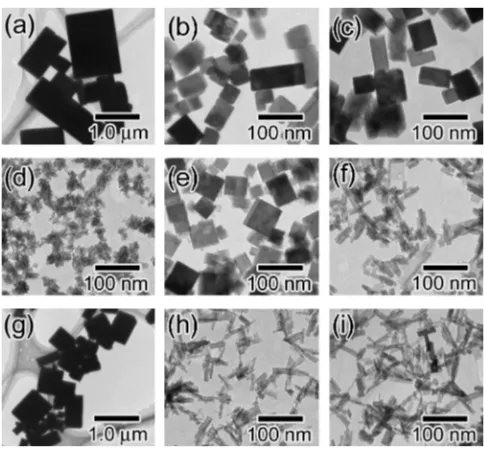
the presence of 0.050 mol L1 Gly or Aspand its absence. Figure 1 shows TEM images of the particles obtained at different initial pH. The products in the absence contained mixture of rectangular- and cubic-shaped particles regardless the initial pH (Fig. 1(a)–(c)), whose structure was identified as In(OH)3(JCPDS No. 076-1464) in a single-phase by XRD
analysis. The mean particle size was drastically decreased from severalmmto ca 100 nm with increasing the initial pH from 8 to 10. In contrast, as shown in Fig. 1(d), particles of irregular shape and rough surfaces with less than 50 nm in the diameter were formed for 0.050 mol L1Aspat initial pH 8.
Cubic-shaped particles with the relatively broad size distri-bution in 40–70 nm were predominantly formed at pH 9 (Fig. 1(e)), in contrast to the formation of the rectangular-shaped particles in the absence of amino acids (Fig. 1(b)). The size of the cubic-shaped particles was decreased to 20– 30 nm by increasing in Asp concentration to 0.25 mol L1. The particles shape became rod-like with the further increase in initial pH to 10 (Fig. 1(f)). XRD measurements showed that the solid particles mainly consisted of In(OH)3 crystal
structure with few undefined solid phase when the initial pH was adjusted to 8 and 10 in the presence of 0.050 mol L1 Asp. In contrast, only the In(OH)3 phase was observed at
pH 9 forAspsystems.
tions, the higher initial pH results in the more number of nuclei due to higher OH concentration: namely, the
as-prepared particle size is the smaller. In fact, the mean particle size was decreased with increasing the initial pH, except for
Asp systems. In contrast, in the case of Asp systems, the particle shape was significantly affected by the initial pH. The aspect ratio of cubic-shaped particles formed in Asp
system at the initial pH 9 is smaller than that of rectangular-shaped particles. This shape control may be due to the suppression to the growth by the specific adsorption ofAsp
on the surface. In fact, Ninhydrin tests of the supernatant solutions after 24 h aging at 100C in the Aspsystems with
the initial pH 9 and 10 indicated 25% and 10% uptake of original Asp feed on the particle surface. This difference seems due to the strong dependence of the adsorption behavior ofAspon pH, because of the relationship between the dissociation state of Asp and surface charge of the particles.27–29) The cubic particles, obtained in the present study, were bounded by {100} planes, determined by high resolution TEM observation. The specific adsorption ofAsp
onto {100} surface of the In(OH)3particles may be stronger
than the other ones. In this regard, the shape control to cube on In(OH)3 nanoparticles must be achieved by the specific
adsorption ofAsp. The detailed formation mechanism is now under the investigation, in particular, on particle morphology change on precise shape control of the In(OH)3nanoparticles.
It can be summarized that we have succeeded in the size-and shape-control of In(OH)3 nanoparticles to form both
cubic- and rod-shapes by changing amino acids and the optimization of the initial pH.
3.2 Effect of aging period on the particle formation
Figure 2 exhibits TEM images of the products formed after different aging times at 100C in amino acids free systems at the initial pH 9. The products formed just after mixing of InCl3 solution with NaOH seemed amorphous
In(OH)3, and no crystalline In(OH)3particles were observed
(Fig. 2(a)). After 30 min, In(OH)3 nanoparticles of
rectan-gular, cubic, and irregular shapes were formed as shown in Fig. 2(b). The particles clearly grew with aging time and finally the mean particle size reached 100 nm after 24 h (Fig. 2(c)). The XRD patterns of the products before aging at 100C indicated amorphous phase because of no crystalline
peak. The crystalline In(OH)3 was confirmed as a single
phase after 30 min. The crystallinity was improved by the extension of aging time. Thus, XRD results were consistent with the TEM observations.
TEM photos in Fig. 3 show time evolution of the products with aging time at 100C in the presence ofAspat the initial pH 9. Figure 3(a) indicates that the initially formed precip-itates before aging at 100C were amorphous like gel. After
[image:2.595.47.289.514.740.2]30 min, the particles with both of irregular and thin plate shapes were observed (Fig. 3(b)). No cubic-shaped In(OH)3
particle was found in the products aged until 3 h (Fig. 3(c)). The cubic-shaped In(OH)3particles were clearly recognized
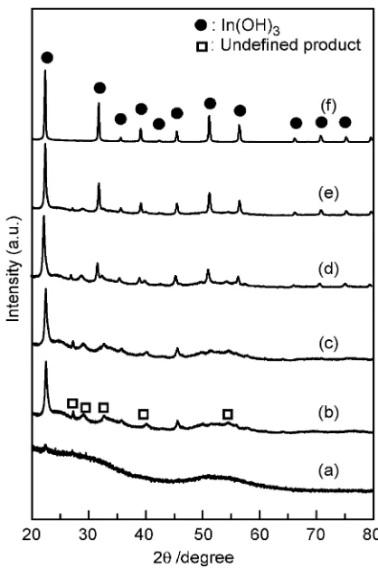
in the products formed after aging for 6 h (Fig. 3(d)). The cubic nanoparticles grew with the aging time, while the other particles, such as thin plate-like and irregular ones, were decreased (Fig. 3(e)). The thin plate-like particles were completely disappeared after 24 h, and no irregular-shaped particles were observed after 72 h (Fig. 3(f)). The XRD patterns of the products at different aging time are shown in Fig. 4. No crystalline phase was found at 0 h (Fig. 4(a)). Peaks of2¼22:2, 35.5, and 45.4 in the XRD patterns of
the products obtained after more than 30 min can be assigned as In(OH)3, but undefined peaks (2¼27:3, 28.8, 32.4, 39.8,
and 54.2) were also found until 8 h aging: They are not
matched with JCPDS data. By the aging, the undefined peaks were decreased, and peaks derived from In(OH)3were
increased. Finally, the undefined peaks were totally disap-peared at 24 h aging, and the In(OH)3was formed in a single
phase. This behavior was consistent with the morphological changes shown in Fig. 3. Thus, it can be identified that the
undefined peaks were derived from the formation of thin plate-like solid, which is, we believe, an intermediate to final product, In(OH)3. Such an intermediate was only observed in
the presence of Asp. Regarding this behavior, the cubic-shaped In(OH)3 nanoparticles were formed through
dissolu-tion of the undefined solid intermediates with thin-plate morphology. In the case of Gly system, In(OH)3 particles
were nucleated and grew through direct deposition without any formation of an intermediate, similar to amino-acid free systems.
3.3 Intermediate-assisted size distribution control of cubic-shaped In(OH)3nanoparticles
As mentioned above, during the generation of In(OH)3
nanoparticles in the presence of Asp, the formation of undefined solids as an intermediate was ascertained. Bene-ficial use of a solid intermediate as a reservoir of precursor complexes to desired product in the gel-sol method, is one of the most powerful tools for the preparation of mono-dispersed fine particles.13) Based on the idea of the gel-sol
method, we focused on the utilization of the undefined solid intermediate for the further size-control to obtain monodis-persed In(OH)3nanoparticles. Initially, we have investigated
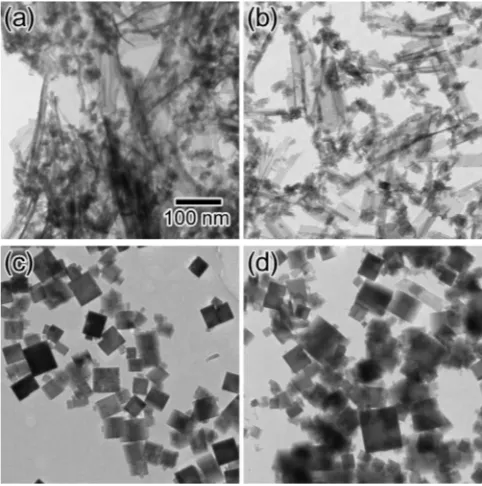
the effect of aging temperatures in Aspsystem to predom-inantly obtain the solid intermediate. The TEM images and the XRD patterns of the products obtained at 50 and 70C for
24 h are shown in Fig. 5(a)–(b) and 6(a)–(b), respectively. The products formed at both temperatures mainly consisted of the thin plate precipitates, which were confirmed as the Fig. 3 TEM images of the solid particles formed by aging for 0 h (a), 0.5 h
(b), 3 h (c), 6 h (d), 8 h (e), and 72 h (f) at 100C in the presence of
0.050 mol L1Asp. The scale bar in (a) is common for all images.
Fig. 4 XRD patterns of the precipitates in samples aged for 0 h (a), 0.5 h (b), 3 h (c), 6 h (d), 8 h (e), and 24 h (f) at 100C in the presence of
0.050 mol L1Asp.
Fig. 2 TEM images of the precipitates in samples aged for 0 h (a), 0.5 h (b), and 24 h (c) at 100C in the absence of amino acids. The scale bar in (a) is
[image:3.595.320.529.69.383.2] [image:3.595.49.290.72.183.2] [image:3.595.48.290.244.467.2]same intermediate as shown in Fig. 3(b). However, peak at 22.2 derived from the formation of In(OH)
3 was also
observed by the aging at 70C (Fig. 6(b)). Thus, we set the
first aging temperature to 50C for 24 h as an optimized
condition to predominantly obtain the solid intermediate. To accelerate In(OH)3 nucleation in a short period through
dissolution of once formed solid intermediate, the aging temperature was rapidly increased to 100C, and aged for
48 h at the temperature. The TEM image and the XRD pattern of the resulting solid particles, obtained by the two-step aging, are shown in Fig. 5(c) and 6(c), respectively. The solid particles can be identified as crystalline In(OH)3 as a
single-phase. As shown in TEM images (Fig. 5(a) and 5(c)), the
tion control of an intermediate has been reported in the preceding studies of the gel-sol method.10)Also in the case of the present report, the nucleation and growth of In(OH)3
nanoparticles might be clearly separated with use of once-formed solid intermediate as reservoir of precursor to them, which can decrease the super-saturation in the system to avoid the spontaneous nucleation during the particle growth. Thus, the amino acid-assisted two-step hydrothermal syn-thesis seems favourable for the preparation of monodispersed In(OH)3 nanoparticles.
4. Conclusion
In summary, shape-controlled highly crystalline In(OH)3
nanoparticles were successfully prepared by amino acid-assisted hydrothermal synthesis. The rod-shaped In(OH)3
nanoparticles with the major axis about 50 nm were mainly formed by usingGlyas a shape controller, while both of rod and cubic-shaped particles were formed in the absence of amino acid. On the other hand, only cubic-shaped In(OH)3
nanoparticles were formed by using Asp and adjusting the initial pH. The size distribution of the cubic-shaped nanoparticles was improved by two-step aging based on the gel-sol technique. The particle deposition system utilizing dissolution-recrystallization process of solid intermediate based on the gel-sol technique would be a quite effective and promising procedure for the preparation of highly mono-dispersed nanoparticles in large quantities.
Acknowledgements
This work was financially supported by METI & NEDO Rare Metal Substitute Materials Development Project.
REFERENCES
1) T. Kasuga, M. Hiramatsu, A. Hosono, T. Sekino and K. Niihara: Langmuir14(1998) 3160–3163.
2) M. Niederberger: Acc. Chem. Res.40(2007) 793–800.
3) T. Teranishi, M. Saruyama and M. Kanehara: Chem. Lett.38(2009) 194–199.
4) L. Gou and C. J. Murphy: Nano Lett.3(2003) 231–234. 5) S. Yang and L. Gao: J. Am. Chem. Soc.128(2006) 9330–9331. 6) J. Zhang, S. Ohara, M. Umetsu, T. Naka, Y. Hatakeyama and T.
Adschiri: Adv. Mater.19(2007) 203–206.
7) O. Durupthy, J. Bill and F. Aldinger: Cryst. Growth Des.7(2007) 2696–2704.
8) R. A. Gilstrap Jr., C. J. Capozzi, C. G. Carson, R. A. Gerhardt and C. J. Summers: Adv. Mater.20(2008) 4163–4166.
9) T. Sugimoto and K. Sakata: J. Colloid Interface Sci.152(1992) 587– 590.
10) T. Sugimoto, K. Sakata and A Muramatsu: J. Colloid Interface Sci.159
(1993) 372–382. Fig. 5 TEM images of the solid particles obtained at different aging
temperature in the presence ofAsp. (a) 50C, 24 h; (b) 70C, 24 h; (c)
50C, 24 h then 100C, 48 h; (d) 100C, 48 h. The scale bar in (a) is
common for all images.
Fig. 6 XRD patterns of the precipitates formed at different aging temper-ature in the presence ofAsp. (a) 50C, 24 h; (b) 70C, 24 h; (c) 50C, 24 h
[image:4.595.48.289.73.316.2] [image:4.595.66.270.380.579.2]11) T. Sugimoto, M. M. Khan and A. Muramatsu: Colloid Surf. A 70
(1993) 167–169.
12) T. Sugimoto, S. Waki, H. Itoh and A. Muramatsu: Colloid Surf. A109
(1996) 155–165.
13) K. Kanie, A. Muramatsu, S. Suzuki and Y. Waseda: Mater. Trans.45
(2004) 968–971.
14) S.-K. Kwon, K. Kimijima, K. Kanie, A. Muramatsu, S. Suzuki and E. Matsubara: Mater. Trans.46(2005) 155–158.
15) T. Sugimoto:Monodispersed Particles, (Elsevier, Amsterdam, 2001). 16) Y. Waseda and A. Muramatsu, eds.:Morphology Control of Materials
and Nanoparticles, (Springer, Berlin, 2003).
17) T. Sugimoto, K. Okada and H. Itoh: J. Dispersion Sci. Technol.19
(1998) 143–161.
18) T. Sugimoto, X. Zhou and A. Muramatsu: J. Colloid Interface Sci.259
(2003) 43–52.
19) T. Sugimoto, X. Zhou and A. Muramatsu: J. Colloid Interface Sci.259
(2003) 53–61.
20) K. Kanie and T. Sugimoto: Chem. Commun.14(2004) 1584–1585.
21) K. Kanie, H. Sakai, J. Tani, H. Takahashi and A. Muramatsu: Mater. Trans.48(2007) 2174–2178.
22) H. Zhu, Y. Wang, N. Wang, Y. Li and J. Yang: Mater. Lett.58(2004) 2631–2634.
23) H. Zhu, K. Yao, Y. Wo, N. Wang and L. Wang: Semicond. Sci. Technol.19(2004) 1020–1023.
24) Q. Tang, W. Zhou, W. Zhang, S. Ou, K. Jiang, W. Yu and Y. Qian: Cryst. Growth Des.5(2005) 147–150.
25) R. B. H. Tahar, T. Ban, Y. Ohya and Y. Takahashi: J. Appl. Phys.83
(1998) 2631–2645.
26) Y. Endo, T. Sasaki, K. Kanie and A. Muramatsu: Chem. Lett.137
(2008) 1278–1279.
27) H. Tanaka, K. Miyajima, M. Nakagaki and S. Shimabayashi: Chem. Pharm. Bull.37(1989) 2897–2901.
28) C. E. Giacomelli, M. J. Avena and C. P. D. Pauli: Langmuir11(1995) 3483–3490.