Conformational changes and the self-assembly of
alpha-synuclein
MASON, Rebecca
Available from Sheffield Hallam University Research Archive (SHURA) at: http://shura.shu.ac.uk/24181/
This document is the author deposited version. You are advised to consult the publisher's version if you wish to cite from it.
Published version
MASON, Rebecca (2018). Conformational changes and the self-assembly of alpha-synuclein. Doctoral, Sheffield Hallam University.
Copyright and re-use policy
Conformational Changes and the Self-assembly of
Alpha-synuclein
Rebecca Jane Mason
A thesis submitted in partial fulfilment of the requirements of
Sheffield Hallam University
for the degree of Doctor of Philosophy
i. Abstract
Parkinson's disease is the second most common neurodegenerative disease, affecting 0.1 - 0.2% of the population. Incidence of this debilitating disorder rises to 1% of the population over the age of 65, posing a substantial socioeconomic burden on the UKs aging population. There are currently no disease altering treatments for PD, in part due to the incomplete knowledge of the disease mechanism.
The misfolding and aggregation of the protein alpha-synuclein is associated with a range of neurological disorders, including Parkinson's disease. Alpha-synuclein has been irrefutably linked to Parkinson's disease through both genetic and pathological data, with increasing evidence suggesting prefibrillar oligomeric forms of the protein are the toxic species. However, the precise molecular mechanisms through which this protein is linked to the disease are currently unknown. Consequently, increasing knowledge of alpha-synuclein is of great importance, as new discoveries will potentially further the development of new therapeutics for the disease.
In this thesis the primary aim was to conduct investigations into the structural and functional aspects of N-terminally acetylated alpha-synuclein and its oligomers through a combination of Electrospray Ionisation – Ion Mobility Spectrometry – Mass Spectrometry, biochemical and cell culture assays. Alpha-synuclein is a known metal binding protein and the copper binding and subsequent conformational changes and aggregation of modified and mutated forms of the protein were investigated. A novel loss of metal binding function was found for the N-terminally acetylated H50Q form of the protein. The conformational effects of N-terminal acetylation on the oligomeric forms of the protein were also investigated. It was demonstrated that this co-translational modification of alpha-synuclein did not affect oligomer formation or function. Low order oligomers were found to be dynamic during the course of aggregation, by the use of isotopically labelled forms of the protein. Lastly the response of SH-SY5Y cells treated with alpha-synuclein oligomers with the ability to seed intracellular aggregation was investigated, demonstrating that this procedure evokes a stress response in these cells, highlighting a potential mode of action.
Together, investigations presented in this thesis have demonstrated the role of a constitutive co-translational modification in ligand binding and oligomer assembly. Furthermore, the cellular stress response to treatment with these oligomers was also determined giving insights into disease progression. Results have highlighted the importance of using the biologically relevant form of a protein when performing in
vitro experiments, validated the cellular effects of previously characterised
ii. Candidates statement
iii. Dissemination: Scientific Publications
Mason R., Paskins A., Dalton C., Smith D., Copper Binding and Subsequent Aggregation of
iv. Dissemination: Conference Presentations
65th ASMS Conference on Mass Spectrometry and Allied Topics 2017 (Indianapolis, USA), Mason, R. Dalton, C. Smith, D. Investigating the dynamics and assembly of alpha-synuclein
amyloid oligomers by ESI-IMS-MS - Poster presentation.
BMSS Annual Meeting 2016 (Eastbourne, UK)/Parkinson's UK Research Conference 2016 (Leeds, UK), Mason, R. Dalton, C. Smith, D. Copper binding and subsequent aggregation of
alpha-synuclein is modulated by N-terminal acetylation and ablated by the H50Q mutation -
Poster presentation.
63rd ASMS Conference on Mass Spectrometry and Allied Topics 2015 (St Louis, USA), Mason, R. Dalton, C. Smith, D. Effect of post-translational modifications on the metal
Table of Contents
i. Abstract ... 2
ii. Candidates statement ... 3
iii. Dissemination: Scientific Publications ... 4
iv. Dissemination: Conference Presentations ... 5
vi. List of Tables ... 12
vii. List of Figures ... 13
viii. Abbreviations ... 16
Chapter 1: General Introduction ... 18
1.1 Parkinson’s disease ... 19
1.1.1 Genetics of Parkinson’s disease... 20
1.1.1.1 SNCA ... 22
1.1.1.2 LRRK2 ... 23
1.1.1.3 DJ-1... 24
1.1.1.4 PINK1/PRKN ... 24
1.1.2 Pathophysiology of Parkinson’s disease ... 25
1.2 Proteins ... 27
1.2.1 Intrinsically disordered proteins ... 30
1.2.2 Protein aggregation ... 30
1.3 Alpha-synuclein ... 32
1.3.1 Physiological role of alpha-synuclein ... 32
1.3.2 Alpha-synuclein structure ... 32
1.3.3 Alpha-synuclein aggregation ... 34
1.4.1 Electrospray Ionisation ... 36
1.4.2 Overview of the Synapt G2 HDMS ... 43
1.4.3 Ion mobility spectrometry ... 46
1.4.3.1 Drift time ion mobility ... 47
1.4.3.2 Travelling wave ion mobility ... 49
1.4.4 ESI-IMS-MS of biological systems ... 53
1.4.4.1 ESI-IMS-MS of alpha-synuclein ... 55
1.4.4.1.1 Alpha-synuclein structure as determined by MS ... 56
1.4.4.1.2 Solution conditions ... 59
1.4.4.1.3 Ligand binding ... 60
1.4.4.1.4 Aggregation... 61
1.5 Thesis overview ... 62
Chapter 2: The effect of N-terminal acetylation and H50Q mutation on copper binding of alpha-synuclein ... 63
2.1 Introduction ... 64
2.1.1 Environmental factors and PD ... 64
2.1.2 Metals in PD... 64
2.1.3 The association between alpha-synuclein and metals ... 65
2.1.3.1 Alpha-synuclein mutations and metals ... 68
2.1.3.2 Alpha-synuclein modifications and metals ... 69
2.2 Aims and Objectives ... 71
2.3 Materials and Methods ... 73
2.3.1 Molecular biology of pET23a-ASYN expression plasmid ... 73
2.3.2 pACYCduet-naa20-naa25 plasmid purification ... 73
2.3.3 Site directed mutagenesis of pET23a-ASYN ... 74
2.3.4 Production of chemically competent cells ... 74
2.3.6 Agarose gel electrophoresis ... 76
2.3.7 Expression of recombinant proteins ... 77
2.3.8 Protein purification ... 77
2.3.9 SDS-PAGE ... 78
2.3.10 Sample preparation for mass spectrometry analysis ... 79
2.3.11 ESI-IMS-MS analysis ... 79
2.3.12 Thioflavin T (ThT) fluorescence ... 80
2.4 Results and discussion ... 82
2.4.1 Site directed mutagenesis of pET23a-ASYN ... 82
2.4.2 Co-transformation of BL21 (DE3) cells ... 83
2.4.3 Expression and purification of alpha-synuclein recombinant proteins ... 85
2.4.4 Mass spectrometry confirmation of successful protein production ... 85
2.4.5 ESI-IMS-MS analysis of recombinant alpha-synuclein reveals a disordered protein with two distinct populations ... 88
2.4.6 Ion mobility analysis of recombinant alpha-synuclein reveals multiple conformations present at each charge state ... 90
2.4.7 Investigating the effect of N-terminal acetylation and H50Q mutation on alpha-synuclein conformation by ESI-IMS-MS ... 93
2.4.8 Copper binding to alpha-synuclein is observed by ESI-MS ... 97
2.4.9 Copper binding to alpha-synuclein results in altered populations of conformations detectable by ESI-IMS-MS ... 100
2.4.10 Copper binding to alpha-synuclein increases the rate of protein aggregation ... 104
2.4.11 Copper binding to alpha-synuclein is altered by N-terminal acetylation and H50Q mutation ... 106
2.4.12 Addressing the effect of N-terminal acetylation and H50Q mutation on copper induced conformational change to alpha-synuclein ... 109
Chapter 3 – Investigating alpha-synuclein aggregation using ESI-MS and ESI-IMS-MS
... 118
3.1 Introduction ... 119
3.2 Aims and Objectives ... 122
3.3 Materials and Methods ... 124
3.3.1 Production of recombinant alpha-synuclein ... 124
3.3.2 Production of 15Nlabelled recombinant alpha-synuclein ... 124
3.3.3 Production of MS compatible Type C alpha-synuclein oligomers ... 124
3.3.4 Aggregation of alpha-synuclein ... 124
3.3.5 Mass spectrometry ... 125
3.3.6 Western blot ... 125
3.3.7 Dot blot procedure ... 125
3.3.8 Thioflavin T (ThT) fluorescence ... 126
3.4 Results and discussion ... 127
3.4.1 Low order oligomeric species of alpha-synuclein are detected in both the unmodified and acetylated protein immediately following reconstitution ... 127
3.4.2 Low order oligomeric species of alpha-synuclein are dynamic ... 130
3.4.3 Structurally comparable species of Type C alpha-synuclein oligomers are present in solutions prepared from both unmodified and acetylated protein ... 133
3.4.4 ESI-MS analysis of alpha-synuclein aggregation ... 136
3.4.5 Aggregates of alpha-synuclein are formed over the time course utilised for ESI-MS experiments ... 141
3.5 Conclusions and Future Work ... 144
Chapter 4 - Validation and further characterisation of the effect of alpha-synuclein oligomers on SH-SY5Y cells ... 147
4.1.1 Oligomeric species of alpha-synuclein have been proposed as the toxic form of the protein
... 148
4.1.1.1 Alpha-synuclein membrane disruption ... 149
4.1.1.2 Alpha-synuclein propagation ... 150
4.1.2 Oxidative stress is associated with PD ... 153
4.1.3 Cell culture models of PD ... 155
4.2 Aims and Objectives ... 157
4.3 Methods ... 158
4.3.1 Production of alpha-synuclein oligomers ... 158
4.3.2 Maintenance and cryopreservation of SH-SY5Y neuroblastoma cell line ... 158
4.3.3 CellTox™ Green Cytotoxicity Assay ... 159
4.3.4 Resazurin reduction assay ... 159
4.3.5 Immunocytochemistry ... 160
4.3.6 Preparation of conditioned media ... 161
4.3.7 Dot blotting... 161
4.3.8 GSH/GSSG-GloTM Assay ... 162
4.3.9 CellROX™ Green assay ... 163
4.3.10 Polysome profiling ... 163
4.3.10.1 Extract preparation ... 163
4.3.10.2 Preparation of sucrose density gradients ... 164
4.3.10.3 Sedimentation of extracts on polysome gradients ... 164
4.3.11 Statistical Analysis ... 165
4.4 Results and discussion ... 166
4.4.1 Type A oligomers produced from both unmodified and N-terminally acetylated alpha-synuclein reduce the viability of SH-SY5Y cells ... 166
4.4.2 Type C oligomers produced from both unmodified and N-terminally acetylated alpha-synuclein cause an increase in cytosolic alpha-alpha-synuclein aggregate formation in SH-SY5Y cells .... 168
4.4.4 Type C oligomers evoke a stress response in SH-SY5Y cells ... 176
4.4.4.1 Treatment of SH-SY5Y cells with Type C oligomers results in an increase in ROS ... 177
4.4.4.2 Treatment of SH-SY5Y cells with Type C oligomers results in an altered GSH/GSSG ratio ... 179
4.4.4.3 Treatment of SH-SY5Y cells with Type C oligomers results in an altered monosome/polysome ratio ... 180
4.5 Conclusions and future work ... 183
Chapter 5 - General discussion ... 189
5.1 H50Q mutation of N-terminally acetylated alpha-synuclein results in a loss of the proteins ability to bind to copper. ... 191
5.2 N-terminal acetylation of alpha-synuclein was found to not alter the formation of oligomeric species or their function. ... 192
5.3 Treatment of SH-SY5Y cells with Type C alpha-synuclein oligomers inducing a stress response. ... 193
5.4 Concluding remarks ... 194
Appendix ... 196
vi. List of Tables
vii. List of Figures
viii. Abbreviations
AA Ammonium acetate ATD Arrival time distributions AFM Atomic force microscopy AD Autosomal dominant AR Autosomal recessive CSF Cerebral spinal fluid CEM Chain ejection model CRM Charged residue model CSD Charge state distribution CSI Charge state ion
Ω Collisional cross sectional area DTIMS Drift time ion mobility spectrometry Parkin E3 ubiquitin-protein ligase parkin ESI Electrospray ionisation
EGCG Epigallocatechin gallate
GWAS Genome wide association studies GSH Glutathione (reduced)
GSSG Glutathione (oxidised)
IDP Intrinsically Disordered Protein IEM Ion evaporation model
MS Mass spectrometry m/z Mass to charge ratio MAO Monoamine oxidase NAT N-acetyltransferase
NAC Non-amyloid-beta component NMR Nuclear magnetic resonance OS Oxidative stress
PD Parkinson's disease
PMSF Phenylmethane sulfonyl fluoride DJ-1 Protein/nucleic acid deglycase DJ-1
Pink1 Phosphate and tensin homolog-induced putative kinase 1 PBS Phosphate buffered saline
RF Radio frequency
ROS Reactive oxygen species SDS Sodium dodecyl sulfate SRIG Stacked ring ion guides
SNPpc Substantia nigra pars compacta ThT Thioflavin T
3D Three dimensional TOF Time of flight
TWIMS Travelling wave ion mobility spectrometry TIC Total Ion Chromatogram
1.1 Parkinson’s disease
Parkinson's disease (PD) is a progressive, neurological condition estimated to affect between 1 and 2 of the population per 1000 in industrialised countries (von Campenhausen et al., 2005), meaning there are approximately 65000 - 130000 individuals in the UK with the condition. The three main symptoms of PD are tremor, slowness of movement (bradykinesia) and rigidity. A wide range of other physical and psychological symptoms are often experienced, including depression, anxiety, balance problems, loss of sense of smell, insomnia, problems with urination and constipation, erectile and sexual dysfunction, swallowing difficulties and memory problems (Schapira, Chaudhuri and Jenner, 2017).
The classical motor symptoms of PD are caused by a loss of dopaminergic neurons in the brain, leading to a reduction in dopamine levels. Motor difficulties arise as dopamine plays a vital role in regulating movement through acting as a neurotransmitter. As PD progresses the severity of symptoms escalate, leading it to be increasingly difficult for sufferers to live independently. The primary risk factor of PD is age, with incidence rising to 1% for the over 60's (de Lau and Breteler, 2006) and 2.6% for those between 85 and 89 (de Rijk et al., 2000). Consequently, PD will pose an increased social and economic burden on society in the UK, as the population ages.
types of medication commonly used are levodopa, dopamine agonists and monoamine oxidase-B inhibitors. Levodopa is absorbed by neurons, where it is converted into dopamine. Initially levodopa can have a significant positive effect on motor symptoms; however, its effects are gradually reduced as more neurons are lost to the disease, leaving fewer to absorb the levodopa. Dopamine agonists act as substitutes for dopamine in the brain and have a similar, but milder, effect compared to levodopa. Dopamine agonists can have a variety of undesirable side effects, such as hallucinations, confusion and the development of compulsive behaviours. Monoamine B inhibitors increase dopamine by blocking the effect of monoamine oxidase-B, an enzyme which breaks down dopamine. This improves symptoms of PD, but to a lesser degree than levodopa. While these treatments can provide relief from some PD symptoms, they lose their effectiveness as the disease progresses, leaving sufferers further debilitated as neurodegeneration continues. Despite decades of research, the cause of the loss of the dopamine producing neurons is still unknown. Therefore, we currently have no treatments to alter the progression of the disease. As such, further research to increase understanding of the cellular and molecular aspects of the aetiology of PD are required in order to develop disease altering treatments.
1.1.1 Genetics of Parkinson’s disease
5-have given insights into the proteins and cellular pathways which may underlie the disease.
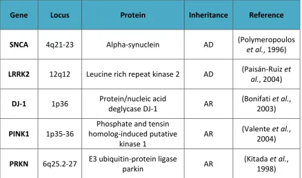
Familial PD can be caused by alterations in several genes, with the most significant monogenic causes being highly penetrant mutations in SNCA, LRRK2, DJ-1, PINK1 and
PRKN, as summarised in Table 1.1. An overarching theme of the function of these genes is that they are linked to either the protein degradation system, which when disrupted leads to an accumulation of potentially toxic proteins, or to mitochondrial quality and function, impairment of which leads to an accumulation of free radicals and oxidative stress (OS).
Gene Locus Protein Inheritance Reference
SNCA 4q21-23 Alpha-synuclein AD (Polymeropoulos
et al., 1996)
LRRK2 12q12 Leucine rich repeat kinase 2 AD (Paisán-Ruı ́z et
al., 2004)
DJ-1 1p36 Protein/nucleic acid
deglycase DJ-1 AR
(Bonifati et al., 2003)
PINK1 1p35-36
Phosphate and tensin homolog-induced putative
kinase 1
AR (Valente et al., 2004)
PRKN 6q25.2-27 E3 ubiquitin-protein ligase
parkin AR
[image:22.595.108.547.382.641.2](Kitada et al., 1998)
The inheritance pattern of PD is dependent on the affected gene, with SNCA and
LRRK2 alterations being inherited in an autosomal dominant (AD) fashion and DJ-1,
PINK1 and PRKN having autosomal recessive (AR) inheritance. A summary of these
genes and their association with PD is provided below.
1.1.1.1 SNCA
The first genetic link to PD occurred in 1996, when Polymeropoulos et al. discovered a locus at 4q21-q23 which was associated with AD PD in a large Italian family (Polymeropoulos et al., 1996). Shortly after, the SNCA gene, encoding the protein alpha-synuclein, was mapped to this locus. Upon sequencing of the SNCA gene a mutation causing the substitution of an alanine at amino acid 53 with a threonine (A53T) was identified in this Italian family, in addition to three unrelated Greek families with high incidence of PD (Polymeropoulos et al., 1997). Subsequently the A53T mutation, along with five other point mutations (A30P, E46K, H50Q, G51D, and A53E), has been identified in many families of varied origin from around the world (Krüger et al., 1998; Zarranz et al., 2004; Lesage et al., 2013; Proukakis et al., 2013; Pasanen et al., 2014). Copy number variation of SNCA have also been found to be associated with PD (La Cognata et al., 2017) displaying a dose-related phenotype, with severity of clinical presentation and disease progression correlating to SNCA dosage (Ross et al., 2008).
alpha-regulation of neurotransmitter release through mediation of vesicular trafficking (Vargas et al., 2014) and the maintenance of synaptic function and plasticity of dopaminergic neurons (Burré, 2015). Alpha-synuclein is the major constituent of Lewy-bodies (LBs), amyloid containing structures which are found in surviving cells in PD patients on post mortem (Spillantini et al., 1997).
1.1.1.2 LRRK2
The first evidence that the LRRK2 gene was associated with PD came in 2002, when a locus at 12p11.2-q13.1 was linked to autosomal dominant Parkinsonism in a Japanese family (Funayama et al., 2002). In 2004 the LRRK2 gene was mapped to this locus (Paisán-Ruı ́z et al., 2004). LRRK2 mutations are the most common known contributor to PD, estimated to account for between 1-5% of sporadic cases and 5-20% of familial cases worldwide (Correia Guedes et al., 2010). Mutations in this gene display variable penetrance, depending largely on ethnicity (Ross et al., 2010), and usually present as mid-to-late onset with slow disease progression. Single nucleotide polymorphisms in
the LRRK2 gene have also been consistently found as one of the main risk factors for
sporadic PD by GWAS (Nalls et al., 2014).
LRRK2 encodes for the highly conserved, multi-domain protein leucine rich repeat
1.1.1.3 DJ-1
In 2003 it was discovered that mutations in DJ-1, encoding protein/nucleic acid deglycase DJ-1 (DJ-1), were associated with early onset, AR PD (Bonifati et al., 2003). DJ-1 has been associated with a number of cellular processes and is most widely accepted as having a role in the protection of neuronal cells from OS (Cookson, 2012). Mutations in the DJ-1 protein cause a loss of its protective properties against OS and DJ-1 knockout increases the vulnerability of cells to oxidative insult (Goldberg et al., 2005).
1.1.1.4 PINK1/PRKN
In 1998 mutations in the PRKN gene were identified in a Japanese family with AR juvenile Parkinsonism (Kitada et al., 1998). Mutations in PRKN have since been identified in patients of many ethnicities, and are the most common cause of familial, early onset PD (Schulte and Gasser, 2011). PRKN encodes the E3 ubiquitin-protein ligase parkin (parkin) which plays a role in the proteasome degradation system through the ubiquitination of proteins, targeting them for degradation. PD associated parkin mutations result in a loss of E3 ligase function and subsequent toxic accumulation of proteins which cannot be degraded by the parkin dependent ubiquitin/proteasome pathway (Shimura et al., 2000).
ubiquitinates proteins on the outer membrane of the mitochondria, triggering autophagy (Pickrell and Youle, 2015).
In addition to the five well characterised PD causative genes described here, polymorphic variants in many additional genes have been linked to PD susceptibility, with GWAS highlighting new associations as more studies are carried out. A recent meta-analysis of GWAS identified 41 risk loci for PD, many of which encoded genes involved in lysosomal biology, mitochondrial biology and autophagy (Chang et al., 2017). Overall, mutations associated with PD mainly appear to disrupt the protein degradation system or affect mitochondrial quality and function. The genetics of PD is complex but has contributed to understanding of the pathogenesis of the disease, which may by extension ultimately be leveraged for therapeutic benefit.
1.1.2 Pathophysiology of Parkinson’s disease
While external examination of the PD brain is typically unremarkable, two major neuropathological findings are most commonly found (Mackenzie, 2001). The first is a loss of pigmented dopaminergic neurons in the substantia nigra pars compacta (SNpc), found upon examination of brain sections, an example of which is shown in Figure 1.1 (a). It is thought that around 50% of nigral neurons are lost before the motor symptoms of PD begin to appear, and on autopsy a loss of over 80% is typically seen (Hartmann, 2004). The significant loss of neuromelanin-pigmented neurons in the SNpc, and subsequent dopamine deficit in the nigrostriatal system, represent the most important hallmark of PD, leading to the major motor symptoms observed in patients.
alpha-synuclein in the form of amyloid fibrils (Spillantini et al., 1997). A variety of other proteins are detectable in LBs, most notably ubiquitin, neurofilament protein, heat shock proteins and tau (Goedert et al., 2012). The presence of ubiquitin is indicative of LBs representing a structural manifestation of a protective cellular response. Classical LBs are spherical, intracytoplasmic neuronal inclusions with a typical diameter between 8 µm and 30 µm. Upon H&E staining they have an eosinophilic hyaline core and a pale peripheral halo, as shown in Figure 1.1 (b).
Figure 1.1: Example images of typical PD pathology. Shown in (a) is horizontal sections
The presence of alpha-synuclein in LBs has warranted significant attention, particularly as the protein is strongly linked to PD through genetic evidence, as described in Section 1.1.1.1. Therefore, both genetic and pathological evidence point towards synuclein playing a key role in the pathogenesis of PD. Research into alpha-synuclein has been an extremely active field over the past 20 years and investigations of this protein are the focus of this thesis.
1.2 Proteins
Proteins constitute the major building blocks of the cell, being fundamental components of almost every function involved in life. Proteins are biological polymers of amino acids linked by an amide bond. Amino acids are small, organic molecules consisting of an amino group (-NH2), a carboxyl group (-COOH), a hydrogen atom and a variable R group, as shown in Figure 1.2. It is this variable R group which defines the properties of an amino acid. Proteins have innumerable cellular functions, and typically adopt a unique structure which determines this function. There are four levels of protein structure, as shown in Figure 1.3.
Figure 1.2: Diagram of an amino acid. The common structure of amino acids,
1.2.1 Intrinsically disordered proteins
Challenging the traditional model that protein structure governs its function, many biologically active proteins possess substantial regions lacking a stable secondary structure, which are termed Intrinsically Disordered Proteins (IDPs). Alpha-synuclein is an example of such a protein. Despite lacking significant secondary structure, IDPs have been shown to be involved in many biological functions (Uversky, 2011) such as cell signalling (Iakoucheva et al., 2002) and transcription (Minezaki et al., 2006), often gaining structure on the binding of ligands. IDPs typically contain a low proportion of hydrophobic and order promoting amino acids, and a high proportion of charged and disorder promoting amino acids (Dyson, 2016; Uversky, 2016), leading to flexible and highly heterogenic conformations. Numerous IDPs have been implicated in disease, such as p53 in cancer, amyloid beta in Alzheimer’s disease and alpha-synuclein in Parkinson’s disease (reviewed by Babu, 2016).
1.2.2 Protein aggregation
Protein aggregation is a common feature in nature, with functional amyloids playing important roles in processes such as structure and protection, cell-cell recognition and epigenetic inheritance (Pham, Kwan and Sunde, 2014). However, the misfolding and subsequent aggregation of proteins is known to be implicated in the pathogenesis of many disease states, termed protein misfolding diseases.
amyloid fibrils share many characteristics, including a common structure, a typical X-ray diffraction pattern and common histological characteristics. The common structure of amyloid fibrils consists of a regular cross beta structure, with beta sheets stacked parallel to the fibril axis. The fibrils are unbranching assemblies, many μm in length and typically 5 - 15 nm wide under electron microscopy (Toyama and Weissman, 2011). A variety of possible protein species can be populated during the aggregation pathway, as described in Figure 1.4.
Figure 1.4: Illustration of the possible species adopted by proteins during the
aggregation process. Image taken from (Invernizzi et al., 2012).
1.3 Alpha-synuclein
1.3.1 Physiological role of alpha-synuclein
Alpha-synuclein is a highly abundant protein in the brain. Accounting for approximately 1% of total protein in neuronal cells (Mizuno et al., 2012), alpha-synuclein is mainly localised to the presynaptic terminals of neurons (Iwai et al., 1995). While the precise physiological function of alpha-synuclein is largely unknown, it is believed to contribute to activity at the synapse (reviewed by Burré, 2015). Specifically, it is thought to be involved in the regulation of neurotransmission through participation in vesicular trafficking and neurotransmitter release (Clayton and George, 1998), and has been demonstrated to interact with various components of the SNARE complex (Burré et al., 2010). In addition to being abundant in the brain, alpha-synuclein is found in many other tissues, including the skin (Rodríguez-Leyva et al., 2014), heart (Iwanaga et al., 1999) and gastrointestinal tract (Shannon et al., 2012), suggesting its function may be more varied than purely the regulation of neurotransmission. Animal and cellular models have implicated a possible involvement of alpha-synuclein in many biological functions, including dopamine biosynthesis (Perez et al., 2002), metal binding (Tavassoly et al., 2014), mitochondrial function (Pozo Devoto and Falzone, 2017) and microtubule organisation (Cartelli et al., 2016).
1.3.2 Alpha-synuclein structure
other groups report an intrinsically disordered structure when the protein is purified under similar conditions (Fauvet et al., 2012). In addition, in-cell nuclear magnetic resonance spectroscopy (NMR) has shown the protein remains in an intrinsically disordered state when introduced to the cell by electroporation (Theillet et al., 2016).
post-translational modification site of the protein; the phosphorylation of the serine residue at position 129. This modification is highly associated with PD, with approximately 90% of the alpha-synuclein found in LBs being phosphorylated at this site compared to approximately 4% which is found to be phosphorylated within the cytosol (Walker et al., 2013).
Figure 1.5: The structure of alpha-synuclein. Alpha-synuclein is a 140-residue protein
comprised of three distinct regions; the highly conserved N-terminus, containing a series of imperfect KTKEGV repeats involved in lipid binding, the central non amyloid- β component (NAC) which is essential for the protein's aggregation, and the largely unfolded, negatively charged C-terminus. Known familial mutations are shown in pink and acetylation/phosphorylation sites are shown in blue.
1.3.3 Alpha-synuclein aggregation
found in LBs in not the normal state of the protein in healthy individuals. However, the exact mechanism of alpha-synuclein aggregation is still uncertain, and currently it is not known whether the aggregation of the protein is a cause or a consequence of PD.
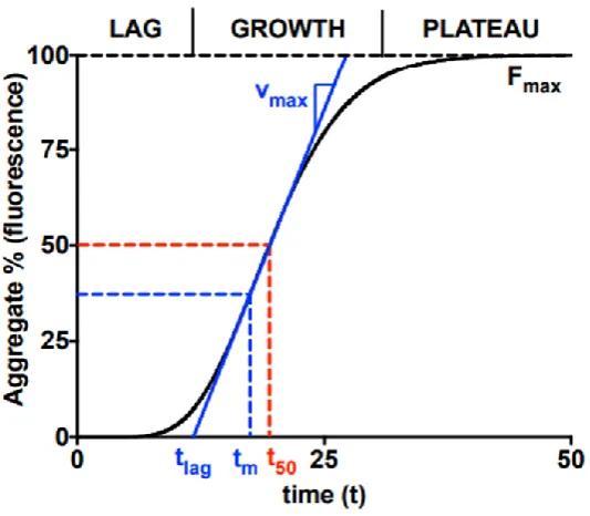
It is thought that alpha-synuclein is involved in PD through misfolding of the usually disordered monomer into highly structured, beta-sheet rich amyloid fibrils via a population of potentially transient toxic soluble oligomers and protofibrils (discussed in detail in Section 4.1.1). The formation of these oligomers is preceded by the collapse of extended forms of the protein into a partially folded intermediate (Uversky, Li and Fink, 2001a). Further fibrillation of alpha-synuclein is a nucleation dependent process (Conway, Harper and Lansbury, 2000), where the disordered monomer first assembles into soluble oligomers during an initial lag phase until a nucleus is formed. Aggregates then form rapidly around this nucleus during an elongation phase, until a thermodynamic equilibrium between fibrillar aggregates and monomer is established. The familial PD mutations have been shown to alter alpha-synuclein oligomerisation and subsequent aggregation (Winner et al., 2011), demonstrating a probable role for alpha-synuclein misfolding and aggregation in PD. Ligand binding has also been demonstrated to effect alpha-synuclein aggregation, with binding of some compounds such as divalent metal cations (Han, Choi and Kim, 2018) and biological polyamines (Krasnoslobodtsev et al., 2012) increasing aggregation, and others such as squalamine (Perni et al., 2017) and curcumin (Herva et al., 2014) decreasing aggregation.
1.4 Mass spectrometry
suited to the study of diverse and transient non-covalent complexes such as amyloid oligomers. Mass spectrometers consist of three fundamental components; an ionisation source, analyser and detector. Samples are introduced into the mass spectrometer via an ionisation source, which generates gas phase ions from the analyte. Ions are then separated by one or more mass analysers, based on their mass (m) to charge (z) ratios (m/z). The separated ions then reach a detector which measures the relative abundance of each ion of a particular m/z, converting it to an electrical signal and presenting this information in the form of a mass spectrum.
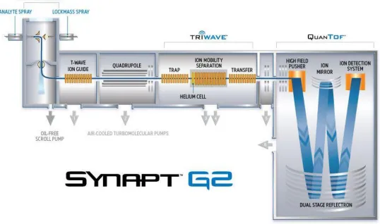
Many different configurations of mass spectrometers are commercially available, with diverse combinations of ionisation source, mass analysers and detectors. MS data presented in this thesis were predominantly obtained on a Waters SynaptTM G2 HDMS instrument (Waters Corporation, Wilmslow, Manchester, UK), using a TriVersa NanoMate electrospray ionisation (ESI) source (Advion, Ithaca, USA). The ESI process is described in detail below.
1.4.1 Electrospray Ionisation
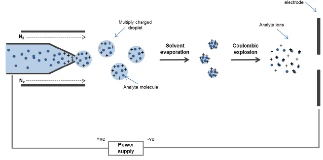
used for protein analysis, and used exclusively in this thesis) a positive voltage is applied to the needle and a negative voltage applied to the counter-electrode in the instrument. This leads to a build-up of protons at the tip of the needle. As a consequence of this strong electric field the sample emerging from the tip is dispersed into an aerosol of highly charged droplets, aided by introducing nitrogen as a nebulising gas flowing around the outside of the capillary. This flow of gas directs the Taylor cone towards the mass spectrometer. As the charged droplet travels towards the sample cone, evaporation of the solvent occurs. This results in a reduction of droplet size, which becomes unstable as it reaches its Raleigh limit, where the surface tension is balanced by Coulombic repulsion. At this stage the droplet undergoes Coulombic explosion, resulting in many smaller, more stable droplets which in turn undergo desolvation and consequently more Coulombic explosion events.
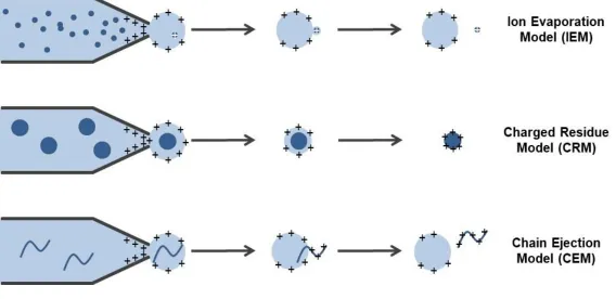
Gas phase ions are formed when the ion is released from the droplet and the charge is transferred onto the analyte (Figure 1.6). It is believed that the exact mechanism through which an analyte is released from the droplet is dependent on the properties of the analyte. Three main models by which ions may be released from the charged droplet have been proposed; the ion evaporation model (IEM), the charged residue model (CRM) and the chain ejection model (CEM). In the IEM, low molecular weight charged species are emitted from nanometer-sized droplets, which have shrunk by evaporation until the field strength at the surface of the droplet is large enough for ions to be expelled (Nguyen and Fenn, 2007). In the CRM a solvent droplet containing a single analyte fully evaporates, with the residual charge being transferred to the analyte inside (Fernandez de la Mora, 2000). The most recently proposed model is the CEM, in which a disordered polymer is partially ejected from the droplet after migrating to the surface of the droplet due to the exposure of hydrophobic residues. This leads to protons attaching to the exposed portion of the ion, followed by further extrusion and ultimate ejection of the extended chain (Konermann et al., 2013). Much of the current evidence suggests that folded proteins formed by ESI from buffered aqueous solution ionize by the CRM, small ions by the IEM, and unfolded, disordered proteins by the CEM (Donor et al., 2017). A schematic illustrating these three models of ion formation by ESI is shown in Figure 1.7.
Based on the CRM, the number of charges that a protein will obtain can be estimated based on the mass of the protein when acquired from aqueous ammonium acetate at neutral pH (Peschke, Blades and Kebarle, 2002). The calculated charge can be estimated by the Rayleigh limit, with the size equivalent to the protein in question as follows.
𝑧𝑅 = 8𝜋/𝑒(𝛾𝜀 0𝑅3)
1 2
Equation 1: Predicted charge calculation, where γ is the surface tension of the aqueous
solution droplet, ε0 is the empirical permittivity of the vacuum, e is the elementary
charge and R is the radius of the droplet.
Making the assumption that the radius of the protein correlates to its mass, and its density is similar to water, a simplified equation can be created as shown below:
𝑧𝑅 = 0.078 𝑀12
Equation 2: Calculated charge of a protein (zR), in which M is the Mass in Da.
1.4.2 Overview of the Synapt G2 HDMS
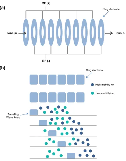
1.4.3 Ion mobility spectrometry
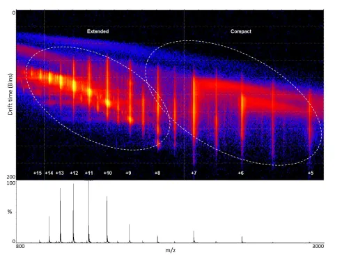
IMS is a widely used technique which separates gas phase ions based on differences in their mobility through a buffer gas under a weak electric field. Mobility is quantified as the time taken for an ion to traverse through a drift tube of a given length. The time an ion will take is dependent on its charge, mass and conformation. IMS provides structural information in the form of calculated rotationally averaged Ω, providing a valuable structural biology tool for characterising co-populated protein conformations under varying conditions. The Ω of an ion is a calculation of the momentum transfer of its average area as it tumbles through a gas, and is typically measured in Angstroms squared (Å2). Ω is related to the chemical structure (mass and size) and three-dimensional conformation (or shape) of an ion. Ω is not an intrinsic property of an analyte, as measured Ω is additionally dependent on the identity of drift gas used, experimental temperature and the electric field used during the measurements (May, Morris and McLean, 2017).
1.4.3.1 Drift time ion mobility
There are two main types of drift tubes commonly used for IMS of proteins, traditional drift time ion mobility (DTIMS) and travelling wave ion mobility (TWIMS). DTIMS measures the time taken for an ion to traverse through a drift tube filled with a buffer gas (typically nitrogen or helium), in the presence of a uniform electric field. Ω can be directly measured from DTIMS using the Mason-Schamp equation (Mason and Schamp, 1958), described below.
𝐾 = 𝑣𝐷 𝐸
Equation 3
The mobility of a gas phase ion (K) is defined by its drift velocity (vD) divided by the electric field (E). As the drift velocity (vD) is related to the drift time (tD) taken to traverse the length (L) of the drift tube, the mobility of an ion can be experimentally determined from its observed drift time as shown in Equation 4:
𝐾 = 𝑣𝐷 𝐸 =
𝐿 𝑡𝐷𝐸
Equation 4
𝐾0 = 𝐾 × 273.2 𝑇 ×
𝑃 760
𝐾 0 = 𝐿 𝑡𝐷𝐸 ×
273.2 𝑇 ×
𝑃 760
Equation 5
Once the mobility of an ion (K) has been established, Ω can then be calculated using the following equation, where z is the number of charges on the analyte ion, e is the charge on an electron (1.6022 × 10⁻¹⁹ C), kb is the Boltzmann constant, T is the temperature and N is the number density of the buffer gas and [𝑚1
𝐼+
1
𝑚𝑁] is the
reciprocal of the reduced mass between the analyte ion (mI) and the buffer gas (mN):
𝐾 = (18𝜋)
1 2
16
𝑧𝑒
(𝑘𝑏𝑇) 1 2
[1 𝑚𝐼+
1 𝑚𝑁]
1 2 1 𝑁 1 Ω Equation 6
Substituting the definition for K in terms of K0 and rearranging the equation to make Ω the subject describes the proportionality between Ω and average drift time (tD):
Ω = (18𝜋)
1 2
16
𝑧𝑒
(𝑘𝑏𝑇) 1 2
[ 1 𝑚𝐼+
1 𝑚𝑁]
1 2760 𝑃 𝑇 273.2 1 𝑁
𝑡𝐷𝐸 𝐿
From this equation the Ω of an ion can be determined by recording its drift time through a buffer gas filled conventional ion mobility cell at a range of electric fields.
1.4.3.2 Travelling wave ion mobility
In order to calculate Ω from TWIMS, correction factors must be incorporated into the conventional ion mobility equations developed for linear drift tubes to take into account the non-linear effects of the TWIMS instruments. These correction factors can be determined empirically through experimental calibration once acquisition parameters have been optimised. In the following equation, A is the correction factor for the electric field parameters (E and L in conventional ion mobility equations) and B is a compensatory factor for the non-linear effect of the TWIM instrumentation:
Ω = (18𝜋)
1 2
16
𝑧𝑒
(𝑘𝑏𝑇) 1 2
[ 1 𝑚𝐼 +
1 𝑚𝑁]
1 2760 𝑃 𝑇 273.2 1 𝑁𝐴𝑡𝐷𝐵
Equation 8
Ω can be defined as a charge and mass independent measure, reduced Ω (Ω'), by dividing the above equation by the absolute charge (ze) and reduced mass as shown in equation 9:
Ω′ = (18𝜋)
1 2
16
1
(𝑘𝑏𝑇) 1 2 760 𝑃 𝑇 273.2 1 𝑁𝐴𝑡𝐷
𝐵
Equation 9
These parameters can then be incorporated into one constant, A’ as shown in equation 8. A’ is a correction factor for the temperature, pressure and electric field and B is a correction factor for the non-linear effects of the TWIMS device:
Ω′ = 𝐴′𝑡𝐷𝐵
Ω can therefore be expressed as equation 11:
Ω = 𝑧𝑒 [ 1 𝑚𝐼+
1 𝑚𝑁]
1 2
𝐴′𝑡 𝐷𝐵
= 𝑧𝑒 [ 1 𝑚𝐼+
1 𝑚𝑁]
1 2
Ω′
Equation 11
It is also necessary to compensate for the mass dependent transit time of an ion outside of the mobility region of the instrument, which is achieved by use of equation 12:
𝑡′𝐷 = 𝑡𝐷 − (𝐶√𝑚 𝑧⁄ /1000)
Equation 12
Where t'D is the corrected drift time (msec), tD is the measured drift time (msec) and C is a constant, found in the control software as the enhanced duty cycle delay coefficient.
Figure 1.10: Example calibration curve of denatured standards of known Ω. Ω' versus drift time (msec) for multiply charged ions of cytochrome c, myoglobin and ubiquitin under denaturing solution conditions.
1.4.4 ESI-IMS-MS of biological systems
The study of proteins by IMS-MS was enabled through the development of ESI, allowing intact proteins and protein complexes to be transferred into the gas phase. ESI-MS and ESI-IMS-MS have emerged as useful tools for the study of many biological systems, with applications in many areas, such as the study of protein subunit stoichiometry, protein modifications and ligand binding and protein-metal interactions (reviewed by Sharon and Robinson, 2007).
Ωs were in good agreement with measurements obtained from more traditional analysis techniques such as X-ray crystallography and NMR, or from theoretical values (Scarff et al., 2008; Smith et al., 2009; Leney et al., 2014). Growing evidence supports the retention of solution structures in the gas phase (Ruotolo et al., 2002; Ouyang et al., 2003; Florance et al., 2011), providing experimental design and instrumental conditions have been carefully considered (Breuker and McLafferty, 2008; Chen and Russell, 2015).
Gross structural information of a protein can be inferred from MS alone through assessment of its CSD. Typically, compact proteins will present with few charge states due to limited ionisable sites being present on their surface, while more disordered or unfolded proteins have more exposed ionisable sites, and therefore a wider CSD (Testa
et al., 2013). Gaussian fitting of a protein’s CSD can aid in the investigation of different
2003), studies from the Jarrold group investigating the conformations of bovine pancreatic trypsin inhibitor and cytochrome C (Shelimov et al., 1997), and studies from the Clemmer group investigating the effect of experimental conditions on ubiquitin conformers (Li et al., 1999).
The study of amyloid formation is one biological system which has benefited greatly from the advances in ESI-IMS-MS, as understanding the morphological transitions which occur at early stages of protein aggregation is of critical importance for developing diagnostic and therapeutic strategies for amyloid diseases. This technique has been used extensively in the study of early stages of aggregation of proteins implicated in protein misfolding diseases, providing valuable information on the co-existing, transient conformations which often exist during this process. Key examples include investigations into the Alzheimer's disease associated amyloid-β (Bernstein et al., 2009; Gessel et al., 2012) and tau (Larini et al., 2013), dialysis-related amyloidosis associated β-2-microglobulin (Smith et al., 2011; Woods et al., 2011; Leney et al., 2014) and type 2 diabetes mellitus associated amylin (Bernstein et al., 2009; Young et al., 2014). This body of work has increased knowledge of the process of protein aggregation through structural characterization of intermediate, oligomeric species of aggregate formation, differentiation of isobaric peaks in the mass spectrum and investigation of the binding mode and subsequent conformational changes of enhancers and inhibitors of aggregation.
1.4.4.1 ESI-IMS-MS of alpha-synuclein
it has proved challenging to investigate the conformation of this protein using conventional structural biology tools such as X-ray crystallography or NMR. In an attempt to overcome these challenges, alpha-synuclein has been studied using MS methods by a number of groups. Areas of interest included attempts to characterise the structure of alpha-synuclein, investigations into the effect of solution conditions and ligand binding on alpha-synuclein conformation, and studies of oligomeric species of the protein and its aggregation. Summaries of findings from key studies in these areas are provided below.
1.4.4.1.1 Alpha-synuclein structure as determined by MS
The intrinsic structure of alpha-synuclein has been investigated using MS methods, with varying results. Alpha-synuclein has been extensively analysed in both positive and negative ionisation mode by several groups. Table 1.2 summarises findings from key studies into the structure of alpha-synuclein under native conditions using ESI-MS.
Table 1.2: Summary of key ESI-MS studies of alpha-synuclein
Ionisation mode
Protein
conc. Buffer CSD (mon) CSD (dimer) Reference
above and below this range. A minimum Ω of 1043 Å2 and a maximum Ω of 2742 Å2 was observed, comparable to those observed by Illes Toth et al. (2013). A high level of variance in measured Ω was noted, indicating conformational inconsistency, which they theorised could be due to either unresolved species or interconversion of species within the timescale of the experiment. Phillips et al. (2015) utilised IMS to investigate conformations of alpha-synuclein present following chemical crosslinking, which identified three distinct conformational families, compact (∼1200 Å2), extended (∼1500 Å2) and unfolded (∼2350 Å2). These measurements correlated to those observed in solution and correspond well to IMS findings for the non-crosslinked protein.
Due to alpha-synuclein being a negatively charged protein under physiological conditions, several investigations into this protein have been conducted using negative mode ESI, with results mirroring those seen in positive ionisation mode. An early study by Bernstein et al. (2004) found the alpha-synuclein monomer to have a CSD of -6 ≤ z ≤ -16, with a CSD of dimers from -17 ≤ z ≤ -21, and a study by Natello et al. (2011) bimodal CSD cantered at the -14/-15 and -7/-8 CSIs. Consistent with positive mode studies, these reports indicate the protein is intrinsically disordered.
reports by Phillips et al. (2015) and Beveridge et al. (2015), where spectra obtained from identically prepared alpha-synuclein samples taken under highly similar source conditions displayed significant differences in the relative population of different charge states, as well as differences in the distribution between monomeric and oligomeric CSIs.
1.4.4.1.2 Solution conditions
In addition to looking at the native structure of alpha-synuclein, investigations into the effect of solution conditions on structure have been an active area of research, due to the known link between solution condition and alpha-synuclein aggregation.
The effect of altered pH has been investigated in a number of studies. Bernstein et al.
(2004) found that reducing the pH from neutral to 2.5 resulted in a significant narrowing of CSD, from -6 ≤ z ≤ -16 to -6 ≤ z ≤ -11, suggesting the presence of predominantly compact conformations in acidic conditions. Using IMS the average Ω across all CSIs was calculated to be 2530 Å2 at pH 7, which reduced to 1690 Å2 at pH 2.5, corresponding to a more globular, compact structure. It was also found that the acidic conditions employed altered dimer formation, as the CSD of dimers from -17 ≤ z
The effect of various alcohols has also been investigated, which has further demonstrated the enrichment of particular conformations in response to solution conditions. Frimpong et al. (2010) found an abundance of compact forms upon addition of 10% - 60% alcohol while Natello et al. (2011) found methanol stabilised compact forms and hexafluoro-2-propanol and tetrafluoroethylene promoted partially folded, intermediate forms.
1.4.4.1.3 Ligand binding
Another key area of interest has been research into the effect of various ligands on alpha-synuclein. As with altered solution conditions, it is known that binding of certain ligands can enhance or inhibit alpha-synuclein aggregation, and as such investigations into early conformational changes which occur upon binding to these ligands is of great interest due to the insights it could potentially give into alpha-synuclein pathology.
The binding of many other small molecules has been investigated using IMS-MS, including investigations into spermine (Grabenauer et al., 2008), calcium (Han, Choi and Kim, 2018), dopamine (Illes-Toth, Dalton and Smith, 2013), epigallocatechin gallate (EGCG) (Konijnenberg et al., 2016) and gallic acid (Liu et al., 2014). A common finding of these studies was an increase in compact conformations upon binding of compounds known to increase alpha-synuclein aggregation, such as spermine (Grabenauer et al., 2008), and prevention of the formation of compact conformations by compounds which inhibit aggregation, such as gallic acid (Liu et al., 2014).
1.4.4.1.4 Aggregation
1.5 Thesis overview
Chapter 2: The effect of N-terminal acetylation and
2.1 Introduction
Various metal ions are known to bind to alpha-synuclein and are found in LBs along with aggregated forms of the protein. Characterising the metal binding ability and subsequent conformational changes of alpha-synuclein can give important insights into the association between metal binding and protein aggregation. In this chapter, the effect of the H50Q familial mutation and N-terminal acetylation on the copper binding of alpha-synuclein was investigated.
2.1.1 Environmental factors and PD
Epidemiological studies have linked various environmental factors to increased incidence of PD. Well established factors include exposure to pesticides and herbicides (Elbaz et al., 2009; Tanner et al., 2011), metals (Gorell et al., 1999; Willis et al., 2010), solvents (Goldman et al., 2012) and Polychlorinated Biphenyls (Caudle et al., 2012).
2.1.2 Metals in PD
zinc) in the SNpc and striatum of PD patients has been widely reported (Kozlowski et al., 2012; Dusek et al., 2015; Gardner et al., 2017). However, it remains to be established whether these alterations in metal homeostasis are a cause or consequence of PD (for a review see Rasheed et al., 2017).
2.1.3 The association between alpha-synuclein and metals
Figure 2.1: Illustration of proposed localisation of alpha-synuclein metal binding sites.
Alpha-synuclein has a high affinity for copper, with two main binding sites at the N-terminus. A third, lower affinity copper binding site exists at the C-terminus, which can bind other divalent metals, including Fe(II) Cu(II) and Mn(II). Figure adapted from Carboni and Lingor (2015).
homeostasis, such as transferrin receptor protein 1 and ferritin. A predicted IRE has been identified in the 5'-UTR of SNCA, and it has been demonstrated that alpha-synuclein translation can be regulated by iron (Febbraro et al., 2012).
Figure 2.2: Illustration of IRP regulation of translation and stability of IRE-containing
mRNAs. IRPs bind to IREs located in either the 5′ or 3′ untranslated regions of specific
mRNAs. When iron is limited, IRPs bind with high affinity to 5′ IRE mRNAs and repress translation, and to 3′ IRE mRNAs, stabilize these mRNAs. When iron is abundant, IRPs do not bind IREs, resulting in the translation of 5′ IRE-containing mRNAs and degradation of 3′ IRE-containing mRNAs. Figure taken from Anderson et al. (2012).
and Brown, 2011). Further research has implied that the major catalytically active form of alpha-synuclein is a membrane-associated tetramer (McDowall, Ntai, Hake, et al., 2017a) and the associated ferrireductase activity is also detectable in vivo (McDowall, Ntai, Honeychurch, et al., 2017b). These studies provide substantial evidence for alpha-synuclein being involved in iron homeostasis, and points to a physiological function for alpha-synuclein as a metalloprotein. Therefore a paradoxical situation exists between the ability of alpha-synuclein to undergo metal induced aggregation in
vitro and its potential role as a metal binding protein in vivo.
2.1.3.1 Alpha-synuclein mutations and metals
2.1.3.2 Alpha-synuclein modifications and metals
The diversity of the human proteome is much greater than would be predicted by the genome. A large part of this diversity is due to protein modifications; alterations to proteins which occur co- or post-translationally, allowing a protein to act in multiple ways. In vitro studies of alpha-synuclein have historically used recombinant protein obtained from bacterial overexpression. Such expression produces unmodified alpha-synuclein. However, in vivo alpha-synuclein has been shown to be constitutively acetylated at its N-terminus (Anderson et al., 2006), a common co-translational modification of nascent polypeptides, particularly those which retain their initiating methionine residue such as alpha-synuclein (Bradshaw, 1989).
Figure 2.3: Schematic of N-terminal acetylation. N-acetyltransferase enzymes catalyse the transfer of an N-acetyl group from acetyl-coA to the alpha-amino group of a protein's N-terminus.
2.2 Aims and Objectives
Prior studies investigating wild type N-terminally acetylated alpha-synuclein have successfully used ensemble based spectroscopy techniques to probe structural changes brought about by metal binding (Moriarty et al., 2014). However, changes to individual conformational states of alpha-synuclein have not been investigated. To date, the effect of the familial H50Q mutation on Cu2+ binding of biologically relevant N-terminally acetylated alpha-synuclein has not been investigated. In this chapter, ESI-IMS-MS has been employed to observe the copper binding and subsequent conformational transitions of individual conformational states of unmodified and N-terminally acetylated forms of wild type (WT) and H50Q alpha-synuclein, using conditions suitable to maintain protein-metal complexes in the gas-phase. ESI-IMS-MS has been utilized due to its ability to interrogate dynamic ensembles of the same mass by separating extended and collapsed conformations (Jenner et al., 2011), with changes in solution conformation detected in the gas phase as changes in Ω. This method also allows the binding of ligands to specific conformational states to be determined (Hopper and Oldham, 2009).
The specific aims of this chapter were:
1. To produce N-terminally acetylated and H50Q mutated recombinant forms of alpha-synuclein.
3. To establish whether N-terminal acetylation or H50Q mutation alter the aggregation propensity of alpha-synuclein.
2.3 Materials and Methods
2.3.1 Molecular biology of pET23a-ASYN expression plasmid
A modified pET23a expression plasmid containing the nucleotide sequence for the expression of alpha-synuclein was provided by Dr David Smith (Sheffield Hallam University, UK). Throughout this thesis this plasmid will be referred to as pET23a-ASYN.
2.3.2 pACYCduet-naa20-naa25 plasmid purification
2.3.3 Site directed mutagenesis of pET23a-ASYN
H50Q mutant alpha-synuclein was created by modification of the pET23a-ASYN plasmid via site directed mutagenesis, using a QuikChange® II XL Site-Directed Mutagenesis Kit by Stratagene following manufacturer's instructions.
Briefly, two complimentary oligonucleotides containing the desired mutation, flanked by unmodified nucleotide sequence were designed in house and synthesized by Sigma Aldrich LTD. Primers used were:
Forward: 5' GCGTTGTCCAAGGGGTTGCG 3'
Reverse: 5' CGCAACCCCTTGGACAACGC 3'
Oligonucleotide primers, each complementary to opposite strands of the vector, were extended during temperature cycling by PfuUltra HF DNA polymerase without primer displacement. Extension of the oligonucleotide primers generated a mutated plasmid containing staggered nicks. Following temperature cycling, the product was treated with Dpn I endonuclease to digest the parental DNA template and to select for mutation containing synthesized DNA. The nicked vector DNA incorporating the desired mutations was then transformed into XL10-Gold ultracompetent cells. Mutated plasmids were then isolated using a QIAprep Spin Miniprep Kit, following manufacturer's instructions. Throughout this thesis this mutated plasmid will be referred to as pET23aH50Q-ASYN.
2.3.4 Production of chemically competent cells
glycerol stocks onto an agar plate containing 100 mg/mL ampicillin and incubated overnight at 37 °C/5% CO2. A single colony was used to inoculate a 5 mL starter culture of LB (containing 10 g/mL Tryptone, 10 g/mL NaCl, 5 g/mL yeast extract and 100 mg/mL ampicillin). 500 µL of starter culture was transferred to 50 mL pre warmed LB and cultured at 37 °C until the OD600 reached between 0.5 and 0.7. The culture was then aliquoted into chilled 50 mL falcon tubes and centrifuged at 4000 rpm for 5 minutes at 4 °C. The supernatant was discarded, the pellet resuspended in 30 mL TBFI (30 mM KOAc, 100 mM RbCl, 50 mM MnCl, 10 mM CaCl2, pH 5.8) and incubated on ice for one hour. They were then centrifuged again at 4000 rpm for 5 minutes at 4 °C and the pellet resuspended in 4 mL TBFII (10 mM MOPS, 10 mM RbCl, 75mM CaCl2, 15% Glycerol, pH 7.0). 200 µL aliquots of chemically competent cells were flash frozen in liquid nitrogen and stored at -80 °C.
2.3.5 Transformation of competent cells
100 µL of BL21 (DE3) chemically competent cells were transformed with 1 µL of the appropriate plasmids required to express each protein, as shown in Table 2.1, with the addition of β-mercaptoethanol to a final concentration of 24 mM.
pET23a-ASYN pET23aH50Q-ASYN pNatB
Unmodified, WT
Acetylated WT
Unmodified H50Q
Acetylated H50Q
Reaction mixtures were incubated for 30 minutes on ice. The mixture was then exposed to 45 second heat shock in a 42 °C water bath and returned to ice for 2 minutes. 900 µL of preheated, sterile SOC medium (2% tryptone, 0.5% yeast extract, 10 mM NaCl, 2.5 mM KCl, 10 mM MgCl2, 10 mM MgSO4, and 20 mM glucose) was added and the reactions incubated at 37 °C with aggitation at 200 rpm for one hour to allow the bacteria to express antibiotic resistance. 100 µL of the reaction was spread onto agar plates containing 100 mg/mL ampicillin (for those transformed with the pET23a plasmids only) or 100 mg/mL ampicillin and 25 mg/mL chloramphenicol (for those transformed with both pET23a and pNatB plasmids) to select for bacteria which had been successfully transformed. Agar plates were incubated at 37 °C/5% CO2 overnight.
2.3.6 Agarose gel electrophoresis
2.3.7 Expression of recombinant proteins
Single colonies from agar plates were incubated in 100 mL LB media (containing 100 mg/mL ampicillin or 100 mg/mL ampicillin and 25 mg/mL chloramphenicol as appropriate) overnight at 37 °C with agitation at 200 rpm. Expression was induced by inoculating 15 mL of this overnight culture into 1 L of auto induction media (Formedium, UK) containing 100 mg/mL ampicillin or 100 mg/mL ampicillin and 25 mg/mL chloramphenicol. This was incubated for 18 hours at 28 °C with agitation at 180 rpm. Cells were then harvested by centrifugation at 8,000 rpm, 4 °C for 20 minutes.
2.3.8 Protein purification
lyophilised. Lyophilised protein was dissolved in buffer A and injected onto a HiLoad® 26/600 SuperdexTM 200 Prep Grade size exclusion column (Amersham Biosciences) using 25 mM Tris-HCl, 10 mM NaCl pH 8.0 as running buffer. SDS-PAGE analysis of size exclusion fractions was performed and purified fractions containing only alpha-synuclein were pooled, dialysed at 4 °C against ultrapure water, re-lyophilised and stored at -20 °C for further experiments. Protein concentration was measured by absorbance at 280 nm using an extinction coefficient of 5960 M-1cm-1 on a nanodrop spectrophotometer.
2.3.9 SDS-PAGE
Separating and stacking gels were prepared as shown in Table 2.2. Glass plates were assembled according to manufacturer's instructions. The separating gel was cast and allowed to polymerise, followed by casting of the stacking gel and insertion of a comb to form sample wells.
Separating gel (mL) Stacking gel (mL)
ddH2O 3.4 2.9
40% acrylamide 2.4 0.75
1.5 M Tris-HCL pH 8.8 2 -
0.5 M Tris-HCL pH 6.8 - 1.25
10% SDS 0.08 0.05
10% APS 0.08 0.05
TEMED 0.008 0.005
Table 2.2: Composition of 12% SDS-PAGE gel.
(Bio-rad) and electrophoresed in 1x running buffer at 100 V. To aid molecular weight determination 5 µL protein marker (broad range, P7702S, New England Biolabs) was loaded into one well. On completion of electrophoresis, gels were stained with InstantBlue Protein Stain (Expedeon) and visualised using a LI-COR Odyssey and Odyssey Infrared Imaging software (LI-COR, Nebraska, USA).
2.3.10 Sample preparation for mass spectrometry analysis
Protein samples were prepared for MS analysis by dissolving lyophilized protein and CuCl2 to a final concentration of 20 μM in 50 mM ammonium acetate pH 7.0. Protein to metal ratios of 1:0 and 1:1 were prepared and mixed immediately prior to analysis.
2.3.11 ESI-IMS-MS analysis
independent time of flight (Ruotolo et al., 2008). Arrival time distributions (ATD) were determined using the Mass Lynx v4.1 software (Waters, Manchester, UK). ATD for each charge state at its respective m/z value were extracted from the Driftscope plots available within the MassLynx suite of software. Plots were then fitted with Microsoft Excel using least square regression to a minimum number of Gaussian distributions using multiples of the following equation:
𝑦 = 𝐴 𝑤√𝜋/2𝑒
−(𝑥−𝑥𝑜)2
2.𝑤2
Where A = total area under the curve from the baseline, x0 = center of the peak, and w = width of the peak at half height. This model describes a normal (Gaussian) probability distribution function.
2.3.12 Thioflavin T (ThT) fluorescence
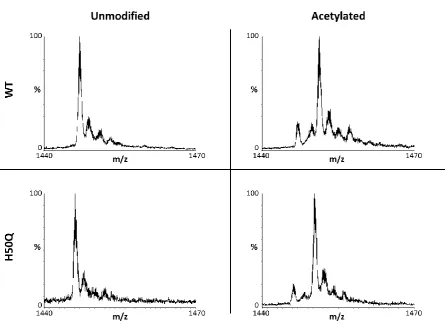
2.4 Results and discussion
In order to determine the effect of protein modification and mutation of a key metal binding residue on alpha-synuclein, recombinant protein was produced with and without N-terminal acetylation, with and wit