
organic papers
o2828
Lehmler and Parkin C12H6Br4O2 doi:10.1107/S160053680502458X Acta Cryst.(2005). E61, o2828–o2830
Acta Crystallographica Section E
Structure Reports Online
ISSN 1600-5368
3,3
000,5,5
000-Tetrabromo-4,4
000-dihydroxybiphenyl
H.-J. Lehmlera* and S. Parkinb
aThe University of Iowa, Department of
Occupational and Environmental Health, 100 Oakdale Campus, 124 IREH, Iowa City, IA 52242-5000, USA, andbUniversity of Kentucky, Department of Chemistry, Lexington, KY 40506-0055, USA
Correspondence e-mail: [email protected]
Key indicators
Single-crystal X-ray study
T= 90 K
Mean(C–C) = 0.005 A˚
Rfactor = 0.022
wRfactor = 0.048
Data-to-parameter ratio = 16.7
For details of how these key indicators were automatically derived from the article, see http://journals.iucr.org/e.
#2005 International Union of Crystallography
Printed in Great Britain – all rights reserved
The dihedral angle between the benzene rings in the title compound, C13H6Cl6O, is 49.06 (8). The molecule has crystallographic twofold rotation symmetry.
Comment
Brominated flame retardants (BFRs), which are used in a wide range of electrical and electronic equipment, are regarded as some of the most highly effective flame retardants used in the plastics industry today (Tange & Drohmann, 2005). Environ-mental studies have detected BFRs, such as polybrominated
diphenylethers (PBDEs) or hexabromocyclododecane
(HBCD), in a wide range of environmental matrixes and in human blood and tissue samples, thus raising human health concerns (Birnbaum & Staskal, 2004; de Boer, 2004; Domingo, 2004). To reduce the environmental risk associated with conventional BFRs, there is an interest in reactive BFRs, which are incorporated into the polymer, thus reducing their potential to leach out of the plastic and enter the environment (Borms & Georlette, 2004). The title compound, (I), is one example of such a reactive BFR.
The toxicity of (I) has been poorly investigated. Similar to other biphenyls of environmental relevance, such as poly-brominated and polychlorinated biphenyls, the three-dimen-sional structure of the title compound will be one important determinant of its mechanisms of toxicity (Kania-Korwelet al., 2004; Lehmler, Parkin & Robertson, 2002; Lehmler, Parkin & Robertson, 2001; Lehmler, Robertson & Parkin, 2001; Lehmler, Robertsonet al., 2002; McKinney & Singh, 1988). In particular, binding to molecular target sites will be determined by the dihedral angle between the two benzene rings. The crystal structure of (I) presented here provides an accurate depiction of its three-dimensional structure, thus adding to our understanding of its interactions with potential target sites in biological systems.
The molecule of (I) has crystallographic twofold rotation symmetry in the solid state. The dihedral angle between the benzene rings is 49.06 (8), which is surprisingly large compared with the calculated value of 38 in an aqueous solution [calculated with MM2 using GB/SA water solvent
continuum as implemented byMACROMODEL5.0 (Stillet al., 1990)]. This is in contrast with other non-ortho-substituted brominated biphenyls, which typically display dihedral angles near or below the calculated angle in solution. For example, the two independent molecules in 4-bromobiphenyl have dihedral angles of 20.4 and 17.8 (Brock, 1980), whereas the two independent molecules in 4,40-dibromobiphenyl have dihedral angles of 38 and 42(Kronebusch et al., 1976). The chloro analog of (I), 3,30,5,50-tetrachloro-4,40 -dihydroxy-biphenyl, is even planar in the crystalline form (McKinney & Singh, 1988). This tendency of non-ortho-substituted biphenyl derivatives to adopt a more planar conformation in the crystal structure is due to stabilizing intermolecular interactions resulting from a stacking arrangement of the benzene rings (McKinney & Singh, 1988).
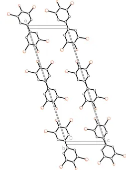
Molecules of the title compound form stacks along the b
axis (Fig. 2). Within these stacks, the distance between the planes (defined by the C atoms in the benzene rings) is 3.526 (3) A˚ . This value is close to the distance of 3.49–3.54 A˚ between 3,30,5,50-tetrachloro-4,40-dihydroxybiphenyl mol-ecules (McKinney & Singh, 1988) and of 3.54 A˚ between the planes in layered aromatic hydrocarbons (Czikkely et al., 1970), thus suggesting the presence ofinteractions between 3,30,5,50-tetrabromo-4,40-dihydroxybiphenyl molecules. Despite these intermolecular interactions, (I) does not adopt a planar conformation in the crystalline form. This observation suggests that, in comparison with related compounds such as 3,30,5,50-tetrachloro-4,40-dihydroxybiphenyl, (I) may interact differently with molecular targets sites and, thus, may have different mechanism(s) of toxicity.
Experimental
The title compound was synthesized by bromination of 4,40
-dihy-droxybiphenyl (5 g) with a slight excess of bromine in warm glacial acetic acid (150 ml). The reaction mixture was allowed to cool to ambient temperature and the crude product was filtered off. Colorless crystals were obtained upon crystallization from ethanol at 277 K.
Crystal data
C12H6Br4O2
Mr= 501.77
Monoclinic,C2
a= 23.4583 (9) A˚
b= 3.8928 (2) A˚
c= 7.5495 (3) A˚
= 108.376 (2) V= 654.26 (5) A˚3
Z= 2
Dx= 2.547 Mg m
3 MoKradiation Cell parameters from 3354
reflections
= 1.0–27.5 = 12.29 mm1
T= 90.0 (2) K Flattened rod, colorless 0.380.150.05 mm
Data collection
Nonius KappaCCD diffractometer
!scans at fixed= 90
Absorption correction: multi-scan (SADABS; Sheldrick, 1997)
Tmin= 0.183,Tmax= 0.541 3668 measured reflections 1403 independent reflections
1341 reflections withI> 2(I)
Rint= 0.037
max= 27.4
h=27!30
k=5!4
l=9!9
Refinement
Refinement onF2
R[F2> 2(F2)] = 0.022
wR(F2) = 0.048
S= 1.07 1403 reflections 84 parameters
H-atom parameters constrained
w= 1/[2(F
o2) + (0.0142P)2] whereP= (Fo2+ 2Fc2)/3
(/)max= 0.005 max= 0.55 e A˚
3 min=0.56 e A˚
3
Extinction correction:SHELXL97
Extinction coefficient: 0.0014 (4) Absolute structure: Flack (1983),
with 554 Friedel pairs Flack parameter: 0.055 (17)
organic papers
Acta Cryst.(2005). E61, o2828–o2830 Lehmler and Parkin C
[image:2.610.46.298.70.225.2]12H6Br4O2
o2829
Figure 2
[image:2.610.307.560.74.415.2]The crystal packing of (I), viewed approximately down the b axis, illustrating the stacking of the molecules along thebaxis. H atoms have been omitted.
Figure 1
H atoms were found in difference Fourier maps and subsequently refined using a riding model, in which the H-atom coordinates were either determined geometrically (Car—H) or placed in the maximum
electron density calculated in a toroid beyond the parent atom (O— H). Bond distances for H were fixed at Car—H = 0.95 A˚ and O—H =
0.84 A˚ , whileUiso(H) values were defined as either 1.2Ueqor 1.5Ueq
of the atom to which they were connected, respectively.
Data collection: COLLECT (Nonius, 1998); cell refinement:
DENZO–SMN (Otwinowski & Minor, 1997); data reduction:
DENZO–SMN; program(s) used to solve structure: SHELXS97
(Sheldrick, 1997); program(s) used to refine structure:SHELXL97
(Sheldrick, 1997); molecular graphics: SHELXTL/PC (Sheldrick, 1994); software used to prepare material for publication:SHELX97-2
(Sheldrick, 1997) and local procedures.
This research was supported by grant No. ES012475 from the National Institute of Environmental Health Sciences, NIH.
References
Birnbaum, L. S. & Staskal, D. F. (2004).Environ. Health Perspect.112, 9–17. Boer, J. de (2004).Environ. Chem.1, 81–85.
Borms, R. & Georlette, P. (2004).Kunstst.-Plast. Eur.94, 256–260.
Brock, C. P. (1980).Acta Cryst.B36, 968–971.
Czikkely, V., Foersterling, H. D. & Kuhn, H. (1970).Chem. Phys. Lett.6, 207– 210.
Domingo, J. L. (2004).J. Chromatogr. A,1054, 321–326. Flack, H. D. (1983).Acta Cryst.A39, 876–881.
Kania-Korwel, I., Parkin, S., Robertson, L. W. & Lehmler, H.-J. (2004).Acta Cryst.E60, o1652–o1653.
Kronebusch, P., Gleason, W. B. & Britton, D. (1976).Cryst. Struct. Commun.5, 839–842.
Lehmler, H.-J., Parkin, S. & Robertson, L. W. (2001).Acta Cryst.E57, o111– o112.
Lehmler, H.-J., Parkin, S. & Robertson, L. W. (2002).Chemosphere,46, 485– 488.
Lehmler, H.-J., Robertson, L. W. & Parkin, S. (2001).Acta Cryst.E57, o590– o591.
Lehmler, H.-J., Robertson, L. W., Parkin, S. & Brock, C. P. (2002).Acta Cryst.
B58, 140–147.
McKinney, J. D. & Singh, P. (1988).Acta Cryst.C44, 558–562. Nonius (1998).COLLECT. Nonius BV, Delft, The Netherlands.
Otwinowski, Z. & Minor, W. (1997). Methods in Enzymology, Vol. 276,
Macromolecular Crystallography, Part A, edited by C. W. Carter & R. M. Sweet, pp. 307–326. New York: Academic Press.
Sheldrick, G. M. (1994).SHELXT/PC. Version 5. Siemens Analytical X-ray Instruments Inc., Madison, Wisconsin, USA.
Sheldrick, G. M. (1997).SADABS,SHELXL97,SHELXS97andSHELX97-2. University of Go¨ttingen, Germany.
Still, W. C., Tempczyk, A., Hawley, R. C. & Hendrickson, T. (1990).J. Am. Chem. Soc.112, 6127–6129.
Tange, L. & Drohmann, D. (2005).Polym. Degrad. Stab.88, 35–40.
organic papers
o2830
Lehmler and Parkin Csupporting information
sup-1
Acta Cryst. (2005). E61, o2828–o2830
supporting information
Acta Cryst. (2005). E61, o2828–o2830 [https://doi.org/10.1107/S160053680502458X]
3,3
′
,5,5
′
-Tetrabromo-4,4
′
-dihydroxybiphenyl
H.-J. Lehmler and S. Parkin
3,3′,5,5′-Tetrabromo-4,4′-dihydroxybiphenyl
Crystal data C12H6Br4O2 Mr = 501.77
Monoclinic, C2
Hall symbol: C 2y
a = 23.4583 (9) Å
b = 3.8928 (2) Å
c = 7.5495 (3) Å
β = 108.376 (2)°
V = 654.26 (5) Å3
Z = 2
F(000) = 468
Dx = 2.547 Mg m−3
Mo Kα radiation, λ = 0.71073 Å
Cell parameters from 3354 reflections
θ = 1.0–27.5°
µ = 12.29 mm−1
T = 90 K
Flattened rod, colourless 0.38 × 0.15 × 0.05 mm
Data collection Nonius KappaCCD
diffractometer
Radiation source: fine-focus sealed tube Graphite monochromator
Detector resolution: 18 pixels mm-1
ω scans at fixed χ = 90°
Absorption correction: multi-scan (SADABS; Sheldrick, 1997) Tmin = 0.183, Tmax = 0.541
3668 measured reflections 1403 independent reflections 1341 reflections with I > 2σ(I) Rint = 0.037
θmax = 27.4°, θmin = 1.8°
h = −27→30
k = −5→4
l = −9→9
Refinement
Refinement on F2
Least-squares matrix: full R[F2 > 2σ(F2)] = 0.022 wR(F2) = 0.048
S = 1.07
1403 reflections 84 parameters 1 restraint
Primary atom site location: structure-invariant direct methods
Secondary atom site location: difference Fourier map
Hydrogen site location: inferred from neighbouring sites
H-atom parameters constrained w = 1/[σ2(F
o2) + (0.0142P)2] where P = (Fo2 + 2Fc2)/3 (Δ/σ)max = 0.005
Δρmax = 0.55 e Å−3 Δρmin = −0.56 e Å−3
Extinction correction: SHELXL97, Fc*=kFc[1+0.001xFc2λ3/sin(2θ)]-1/4 Extinction coefficient: 0.0014 (4)
Absolute structure: Flack (1983), with 554 Freidel pairs
supporting information
sup-2
Acta Cryst. (2005). E61, o2828–o2830
Special details
Geometry. All e.s.d.'s (except the e.s.d. in the dihedral angle between two l.s. planes) are estimated using the full
covariance matrix. The cell e.s.d.'s are taken into account individually in the estimation of e.s.d.'s in distances, angles and torsion angles; correlations between e.s.d.'s in cell parameters are only used when they are defined by crystal symmetry. An approximate (isotropic) treatment of cell e.s.d.'s is used for estimating e.s.d.'s involving l.s. planes.
Refinement. Refinement of F2 against ALL reflections. The weighted R-factor wR and goodness of fit S are based on F2,
conventional R-factors R are based on F, with F set to zero for negative F2. The threshold expression of F2 > σ(F2) is used
only for calculating R-factors(gt) etc. and is not relevant to the choice of reflections for refinement. R-factors based on F2
are statistically about twice as large as those based on F, and R- factors based on ALL data will be even larger.
Fractional atomic coordinates and isotropic or equivalent isotropic displacement parameters (Å2)
x y z Uiso*/Ueq
O1 0.79585 (10) 0.0368 (7) 0.0480 (3) 0.0136 (6)
H1 0.7755 −0.1217 −0.0177 0.020*
Br1 0.800271 (14) −0.27253 (8) −0.32434 (4) 0.01155 (11)
Br2 0.882855 (15) 0.35008 (11) 0.38611 (4) 0.01348 (11)
C1 0.96912 (15) 0.0543 (9) 0.0062 (5) 0.0120 (8)
C2 0.92158 (15) −0.0757 (8) −0.1405 (5) 0.0105 (8)
H2 0.9287 −0.1587 −0.2500 0.013*
C3 0.86460 (15) −0.0841 (9) −0.1271 (4) 0.0095 (8)
C4 0.85181 (14) 0.0389 (9) 0.0304 (5) 0.0089 (7)
C5 0.89905 (15) 0.1718 (9) 0.1743 (4) 0.0113 (8)
C6 0.95698 (14) 0.1774 (9) 0.1646 (4) 0.0101 (8)
H6 0.9888 0.2656 0.2663 0.012*
Atomic displacement parameters (Å2)
U11 U22 U33 U12 U13 U23
O1 0.0103 (12) 0.0165 (16) 0.0151 (13) −0.0001 (11) 0.0053 (10) −0.0023 (11)
Br1 0.00859 (17) 0.01384 (18) 0.01063 (17) −0.00060 (15) 0.00076 (12) −0.00128 (15)
Br2 0.01527 (19) 0.01576 (19) 0.01113 (18) −0.00011 (14) 0.00664 (14) −0.00244 (14)
C1 0.0123 (17) 0.0126 (18) 0.0119 (18) −0.0016 (15) 0.0048 (14) 0.0022 (15)
C2 0.0129 (17) 0.011 (2) 0.0065 (16) −0.0008 (14) 0.0020 (14) −0.0013 (14)
C3 0.0095 (16) 0.008 (2) 0.0077 (16) 0.0019 (14) −0.0016 (13) 0.0005 (13)
C4 0.0055 (16) 0.0096 (19) 0.0129 (16) 0.0023 (14) 0.0048 (13) 0.0037 (13)
C5 0.0161 (17) 0.012 (2) 0.0067 (15) −0.0002 (15) 0.0048 (13) 0.0000 (14)
C6 0.0082 (16) 0.009 (2) 0.0130 (17) −0.0008 (14) 0.0039 (13) −0.0005 (15)
Geometric parameters (Å, º)
O1—C4 1.361 (4) C2—C3 1.372 (5)
O1—H1 0.8400 C2—H2 0.9500
Br1—C3 1.902 (3) C3—C4 1.399 (5)
Br2—C5 1.888 (3) C4—C5 1.385 (5)
C1—C2 1.395 (5) C5—C6 1.384 (4)
C1—C6 1.398 (4) C6—H6 0.9500
supporting information
sup-3
Acta Cryst. (2005). E61, o2828–o2830
C4—O1—H1 109.5 O1—C4—C5 118.9 (3)
C2—C1—C6 118.4 (3) O1—C4—C3 123.6 (3)
C2—C1—C1i 120.2 (4) C5—C4—C3 117.5 (3)
C6—C1—C1i 121.4 (4) C6—C5—C4 121.4 (3)
C3—C2—C1 120.3 (3) C6—C5—Br2 120.1 (3)
C3—C2—H2 119.9 C4—C5—Br2 118.5 (3)
C1—C2—H2 119.9 C5—C6—C1 120.5 (3)
C2—C3—C4 121.9 (3) C5—C6—H6 119.7
C2—C3—Br1 120.2 (2) C1—C6—H6 119.7
C4—C3—Br1 117.9 (3)
C6—C1—C2—C3 −0.6 (5) O1—C4—C5—C6 179.4 (3)
C1i—C1—C2—C3 178.3 (3) C3—C4—C5—C6 −1.2 (5)
C1—C2—C3—C4 0.8 (5) O1—C4—C5—Br2 −1.0 (5)
C1—C2—C3—Br1 −178.3 (3) C3—C4—C5—Br2 178.5 (2)
C2—C3—C4—O1 179.6 (3) C4—C5—C6—C1 1.3 (5)
Br1—C3—C4—O1 −1.4 (5) Br2—C5—C6—C1 −178.3 (3)
C2—C3—C4—C5 0.1 (5) C2—C1—C6—C5 −0.4 (5)
Br1—C3—C4—C5 179.2 (3) C1i—C1—C6—C5 −179.3 (3)
![3 (2,5 Dimethylphenyl) 8 methoxy 2 oxo 1 azaspiro[4 5]dec 3 en 4 yl 3 (2 bromo 4 fluorophenyl)acrylate](data:image/gif;base64,R0lGODlhAQABAIAAAP///wAAACH5BAEAAAAALAAAAAABAAEAAAICRAEAOw==)