Feet are one of the primary points of contact between an animal and its environment. Consequently, they feature morphological adaptations required for survival of an animal in its natural habitat. Larger animals often minimise friction between the feet and the substratum during locomotion with hard-walled hooves and claws. In comparison, smaller animals maximise friction, and a variety of attachment devices has evolved accordingly. A broad diversity of coupling and clamp-like structures that ensure attachment to rough surfaces can be found (e.g. Nachtigall, 1974; Stork, 1980a; Gorb, 2001; Scherge and Gorb, 2001); other devices enable attachment to comparably smooth surfaces. These latter systems usually involve small-scale structural modifications and can be categorised into wet adhesion and dry adhesion systems. The former is present in frogs (e.g. Emerson and Diehl, 1980; Green, 1981; Hanna and Barnes, 1991) and many insects. Flies, for example, make use of an adhesive fluid that is secreted from specialised glands and pores (e.g. Hasenfuss, 1977; Bauchhenß, 1979; Walker et al., 1985; Gorb, 1998). Dry adhesion, on the other hand, is used by geckoes (e.g. Gecko gecko). The great adhesive capacity of these reptiles relies on ultrastructural specialisations of their foot pads (e.g. Ruibal and Ernst, 1965; Hiller, 1968; Williams and Peterson, 1982; Stork, 1983; Autumn et al., 2000).
An analogous ultrastructure is found in spiders. In addition to the tarsal claws, which are present on the tarsus of all spiders, adhesive hairs can be distinguished in many species. These adhesive hairs are either distributed over the entire tarsus, as for example in Lycosid spiders (Rovner, 1978), or concentrated on the pretarsus as a tuft (scopula) lying ventral
to the claws (Hill, 1977), as also found in the jumping spider
Evarcha arcuata (Salticidae), where a scopula is found on
each pretarsus. So far, the effectiveness of these attachment structures has not been analysed. In the present study, the adhesive force (Fa) of the cuticular scopula was analysed via atomic force microscopy. This method permits highly localised measurements of mechanical surface parameters (Binning et al., 1986; Radmacher et al., 1994) and, thus, for the first time, the determination of the adhesive characteristics of the tiny terminal ends (setules) that supply the initial contact area with the substrate (Fig.·1).
Materials and methods Animals
Spiders of the species Evarcha arcuata Clerck (Salticidae) were collected near Saarbrücken (south-western Germany), weighed and kept frozen until scanning electron microscopy (SEM) preparation. Atomic force microscopy (AFM) analyses were carried out on recently captured, untreated individuals.
Scanning electron microscopy
Prior to SEM studies, individuals were dehydrated in ascending acetone concentrations (70%, 80%, 90%, 100%), cleaned with ultrasound, critical-point-dried (CPD 030 Critical Point Dryer; Bal-Tec, Witten/Ruhr, Germany) and sputter-coated with gold (SCD 005 Sputter Coater; Bal-Tec). Specimens were examined in high vacuum using a Zeiss DSM 940A electron microscope at 10–15·kV.
doi:10.1242/jeb.00478
The feet of the jumping spider Evarcha arcuata attach to rough substrates using tarsal claws. On smooth surfaces, however, attachment is achieved by means of a claw tuft, the scopula. All eight feet bear a tarsal scopula, which is equipped with setae, these again being covered by numerous setules. In E. arcuata, an estimated 624·000 setules, with a mean contact area of 1.7×10⁵·nm2, are present. The spider’s entire contact area thus totals 1.06×10¹¹·nm2. Adhesion to the substrate does not depend on the secretion of an adhesive fluid. Analysis via atomic
force microscopy (AFM) shows that a single setule can produce an adhesive force (Fa) of 38.12·nN perpendicular to a surface. Consequently, at a total Faof 2.38×10⁻²·N and a mean body mass of 15.1·mg, a safety factor (SF; Fa/Fm, where Fmis weight) of 160 is achieved. Tenacity (τn; Fa/A, where A is area of contact) amounts to 2.24×10⁵·N·m–2.
Key words: dry adhesion, ultrastructure, scopula, claw tuft, seta, setule, safety factor, atomic force microscopy, scanning electron microscopy, spider, Evarcha arcuata.
Summary
Introduction
Adhesion measurements on the attachment devices of the jumping spider
Evarcha arcuata
A. B. Kesel*, A. Martin and T. Seidl
Department of Zoology, Technical Biology and Bionics, Saarland University, D-66041 Saarbrücken, Germany
*Author for correspondence (e-mail: a.kesel@rz.uni-sb.de)
Atomic force microscopy
In order to carry out adhesion measurements with AFM, untreated individuals captured shortly before use were supinely embedded in 5-min epoxide resin (R&G GmbH, Waldenbuch, Germany). Scopula hairs were kept free of the embedding medium. To ensure that the mechanical properties of the setules were not altered by a covering layer of epoxide, capillary rise of the fluid resin between the setules was avoided by a short waiting period prior to embedding of specimens (resin curing time approximately 1.5·min). Further preparations were not necessary for this measuring technique. Measurements were accomplished under ambient conditions (23°C; 45% air humidity).
A commercial AFM (Topometrix® Explorer; controller software SPMLab 4.01; Santa Clara, CA, USA) was used to measure the adhesive force of the terminal setule contact area. Pointspectroscopy was performed in order to obtain data. In doing so, a highly local contact between certain defined points on the sample and the instrument’s probe was established. According to Hartmann (1991), this application of ATM is especially suitable for determining van der Waals forces of samples. In this case, the probe was an ultrathin silicon–nitride cone, mounted on a cantilever. Prior to the actual probing of the setule surface, the cantilever was lowered towards a glass surface and tapped onto the latter. Due to this dynamic contact, the cone-shaped probe tip was flattened, which allowed us to assume a two-dimensional, flat contact area for the probe tip when interacting with the setules. Probe tip area (3.6×10⁵·nm2) was determined using SEM, as was the successful flattening of the probe tip. The cantilever had a spring constant of 5.95·N·m–1.
At a constant velocity (0.5·m·s–1), the probe was slowly brought into contact with the sample and then retracted, passing through a predetermined traverse path (l; 200–400·nm; maximum error, ±4·nm). Contact was made perpendicular to the ventral surface of the scopula. The traverse path l was registered by a linearised scanner (EX 179807) via strain gauge. Due to the probe’s low driving velocity, load application was taken as quasi-static (Burnham and Kulik, 1999).
During probe–sample contact, the cantilever was deflected, in turn leading to the deflection of a laser beam that was projected on the upper surface of the cantilever. Laser deflection was measured by the change in current of a photodetector. This change in current served as a measurement signal directly related to the traverse path and was recorded by an internal data processor. The current changes were converted to force values based on a previous calibration of the instrument. The calibration of the experimental set-up was accomplished by applying known masses to the cantilever and recording the occurring current alterations. A calculated regression equation obtained in this process served as a calibration curve. The internal AFM measurement error for the registered forces accounted for a maximum of 10%. No further data processing was performed. Data were plotted as a force–distance curve in which the readings of the ‘pull-off’ forces during spontaneous detachment of probe and sample represented the adhesive force Fa between the two (Fig.·4; Radmacher et al., 1994). Control measurements were conducted on glass as well as with the epoxide used (resin curing time approximately 1·h under ambient conditions).
Results
Scanning electron microscopy
In E. arcuata, no additional claws or coupling structures other than the above-mentioned tarsal claws were found on any of the feet. As was to be expected, however, all eight feet were equipped with a tuft-like scopula ventral to the claws (Fig.·1). Fig.·1. (A) Lateral view of the tarsal adhesive apparatus of E.
arcuata, showing both claws (Cl) and the scopula (Sc). (B) Ventral
The scopulae were composed of single setae, which were covered by numerous tiny cuticular processes (setules; Fig.·2). Setule tips on the ventral side of the setae were broadened towards their distal end, forming a flattened triangular surface with a mean area of 1.7×10⁵±0.34×10⁵·nm2 (N=7; Fig.·3). These tips represent the direct points of contact with the substrate. Setule density is clearly lower on the dorsal side of the setae, and single setules are not broadened at their distal end but taper down to a point.
With a mean setule density of 2.1±1.0·setules·µm–2(N=48) or 2.1×10⁶·setules·mm–2 on the setae’s ventral side and an estimated scopula area of 0.037±0.008·mm2(N=4) per scopula, a single foot is provided with roughly 78·000 setules. This gives a total of more than 624·000 possible contact points with a substratum for all eight feet.
Atomic force microscopy
Using the recorded data from pointspectroscopy (Table·1), as well as estimates of areas obtained from the SEM micrographs, the total force of adhesion, Fa, was calculated as follows.
The contact area of the probe tip (3.6×10⁵·nm2) was clearly larger than the surface area of the setule tips (1.7×10⁵·nm2; see Fig.·3). Thus, the possibility arises that the probe tip comes into contact with more than one setule at a time. Such multiple events are easily identified by their ‘stepped’ pull-off character, and curves that contained such multiple pull-off events were not taken into consideration for the further calculation of adhesive forces. Therefore, we assume that the terminal setule Fig.·2. (A) Each seta is covered by numerous setules, which are
tapered on the seta’s dorsal side. (B) On the seta’s ventral side, the setule density is noticeably higher. Here, setules are broadened towards their ends, forming a sail-like shape.
Fig.·3. (A) The terminal setule broadening represents the contact point between the spider and the substrate. (B) A setule density of 1.5·setules·µm–2 (mean setule density: 2.1±1.0·setules·µm–2; N=48)
as well as a mean setule area of 1×10⁵·nm2(±0.34×10⁵·nm2; N=7)
tip represents the relevant adhesive contact area. A mean Faof 38.12±14.6·nN (N=45; Fig.·4) was obtained from the force–distance curves of the AFM measurements. Thus, the mean adhesion was 38.12·nN per setule.
Given an estimated 78·000 setules or contact points per scopula, a single foot is calculated to produce an adhesive force of 2.97×10⁻³·N when contact to the substrate is maximal. Providing that all eight feet or, respectively, all eight scopulae are in full contact with the underlying surface, adhesion perpendicular to the substrate would measure 2.38×10⁻²·N, and the tenacity [τn; the ratio of Fato contact area (A)] would then be 2.24×10⁵·N·m–2.
E. arcuata has a mean body mass of 15.1±1.96·mg (N=8), which corresponds to a weight (Fm) of 1.48×10⁻⁴·N. Consequently, the adhesive force of E. arcuata is 160 times its weight when maximum contact with a surface is achieved.
Discussion
SEM analysis – design and material
As a typical representative of the Dionycha, only two main claws are present on the pretarsus of E. arcuata. A smaller middle hook as well as other claws or hairs with analogous functions to those described in Araneids (Foelix, 1992) are absent. The comparatively simple design of the tarsal claw apparatus can be interpreted as an adaptation to a roaming and hunting lifestyle. Jumping spiders do not build webs and hence do not require specialisations for handling silk threads. Therefore, no bristled hairs or accessory claws have developed. However, an attachment system with such an organisation of setae and setules possesses an enormous adaptability to any given substrate and results in the largest possible contact area between the two. In particular, the setose elements, with setal lengths ranging between 100·µm and 280·µm, setule lengths of 3–4·µm, a setule stem diameter of 0.2·µm and the setule tip surface area of approximately 0.17·µm2 provide the geometrical features required for this high adaptability.
Fine-scale specialisation of tarsal elements to such a high degree is not uncommon in arthropods, yet in E. arcuata it reaches a level that is far more differentiated than so far described in other arthropods, e.g. insects. Setal length in marmelade hoverfly Episyrphus balteatus (Syrphidae), for example, measures merely 34·µm, with a stem diameter of approximately 0.5·µm (Gorb, 1998), and setal branching is usually totally absent. Thus, E. arcuata, with 624·000 contact points, ranges clearly higher than the data documented for insects. When the latter possess setose structures beside their
tarsal claws and not just adhesive pads, as in, for example, the Aphidae, Hymenoptera and Orthoptera, 5000–42·000 contact points were found in E. balteatus and the blowfly Calliphora
vomitoria, respectively (Gorb, 1998; Walker et al., 1985). Only
tortoise beetle Hemisphaerota cyanea, with its 120·000 points of contact, reaches a comparable order of magnitude (Attygalle et al., 2000; Eisner and Aneshansley, 2000).
The number of contact points to the substrate in E. arcuata even seems above average for Araneae. In zebra spider Salticus
scenicus (Salticidae), which is comparable in size and body
mass, whole animals are reported to have only 211·000 setules, with a terminal setule surface area of only approximately 0.048·µm2(Roscoe and Walker, 1991). Consequently, the total adhesive area in S. scenicus is only about 10% of that in E.
arcuata.
[image:4.612.315.562.74.231.2]Besides a high number of contact points, a soft material is required to conform to the substrate surface texture. Furthermore, in terms of material behaviour, viscose as well as elastic properties are demanded in order to attain an adequate number of attachment–detachment cycles. To date, few analyses of the mechanical properties of arthropod cuticle are available, in particular as far as material hardness is concerned. The few documented figures lie between 200·MPa and 400·MPa (Hillerton et al., 1982; Kreuz et al., 2000). The far better analysed material elasticity is distinguished by a high degree of variability. Values between 103·Pa (Vincent and Pentrice, 1973) and 1010·Pa (Jensen and Weis-Fogh, 1962) are stated for the Young’s modulus of Locusta cuticle. Even less data are available for chelicerates. Recently, the rates of elasticity were determined for the alloscutum of the tick Ixodes ricinus, with Table 1. Adhesive forces taken from force–distance curves
Force of adhesion
Mean ±S.D. (nN) N
Glass 1315.50±23.3 20
Epoxide 443.72±145.2 19
Setula 38.12±14.6 45
0 10 20 30 40 50
–200 –100 0 100 200 300 400
Force (nN)
Approach
Retract
A E
D
C
B
F
G
Distance (nm)
Fa=23.3 nN
–10
–20
–30
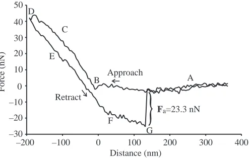
Fig.·4. Original registration of a force–distance curve recorded on a single setule. Points labelled in the diagram are as follows: (A) Probe not in contact with sample. (B) Contact between probe and sample is established. (C) The sample is indented by the probe with a defined force, F. (D) Turning point (F=maximum), retraction begins. (E) During retraction, the force between the probe and the sample decreases. (F) Due to adhesive forces, the probe remains in contact with the sample during retraction. (G) Contact abruptly breaks off (‘pull-off’ event); the registered force value during this sudden cantilever instability represents the adhesive force Fa[here: 23.3·nN;
[image:4.612.44.293.86.153.2]values ranging between 0.17·GPa and 1.5·GPa (Seidl, 2002). By integrating the rubber-like protein resilin in defined locations, further functionally adequate, high elasticities can be obtained in I. ricinus (Dillinger and Kesel, in press). Providing that the cuticular material of the attachment system possesses similar characteristics, it is supplied not only with the structural elements but also with the material properties required for a detailed reproduction of the substrate.
AFM analysis – adhesion and tenacity
As expected, the elaborate attachment system of E. arcuata results in a high adhesive force (2.4×10⁵·N), with absolute values similar to those documented for larger and heavier animals such as the cockroach Periplaneta americana (Pell, cited in Walker, 1993). Thus, the body mass-related safety factor (SF; Fa/Fm) of 160 appears surprisingly high. Insects achieve factors of between 1.5 (P. americana; Pell, cited in Walker, 1993) and 50 (knotgrass leaf beetle Chrysolina polita; Stork, 1980b), although a safety factor of 146 has been reported (cocktail ants Crematogaster spec; Federle et al., 2000). In fact, the safety factor of E. arcuata is only exceeded by that of the beetle Hemisphaerota cyanea, which is temporarily able to adhere with a force 200 times its body mass (Attygale et al., 2000). The adhesive tenacity (τ) produced by insects is reportedly between 2×10³·N·m–2 (great green bush cricket Tettigonia viridissima; Jiao et al., 2000) and 8×10⁴·N·m–2(H. cyanea; estimated from Attygale et al., 2000). Significantly higher values have been obtained when adhesive tenacity is measured parallel to (τp) rather than normal (τn) to the contact surface. Under these circumstances, additional friction forces contribute considerably to adhesion. With an adequate experimental set-up, Walker (1993) registered a τpof 28.6×10⁴·N·m–2for C. vomitoria, which was ten times larger than τn(2.9×10⁴·N·m–2). Thus, the τn(2.24×10⁵·N·m–2) for E. arcuata, gained by adhesion measurements perpendicular to a
contact surface in the present study, lies approximately one order of magnitude above that described for insects. Comparably large adhesive capacities have, until now, only been documented for geckoes. The gecko’s attachment system is remarkably similar to that of E. arcuata. Gecko adhesion is also made possible by a highly structured attachment system of comparable dimensions, and the keratin contact elements, the so-called spatulae, are also reported to be free of adhesive secretions (Ruibal and Ernst, 1965; Hiller, 1968; Stork, 1983). Although adhesive forces are not documented for single spatulae, they are for single setae and, with a τ of 5.76×10⁵·N·m–2, these range within the same order of magnitude as measured here for E. arcuata (Autumn et al., 2002). Analogous to the findings in Calliphora (Walker et al., 1985; Walker, 1993) and Syrphids (Gorb et al., 2001), it was shown that a perpendicular preloading and subsequent pulling of the attachment system parallel to the substrate surface dramatically enhances the adhesive force in geckoes (Autumn et al., 2000). It can thus be concluded that friction forces dominate over all other possible adhesive forces in any attachment system. Nevertheless, dry adhesive mechanisms
seem superior to wet adhesive mechanisms with regard to the adhesive force perpendicular to surfaces.
The physical principle forming the basis for wet adhesion is surface tension of an adhesive secretion between the attachment device and substrate (Bauchhenß, 1979; Walker et al., 1985; Dixon et al., 1990; Walker, 1993). By contrast, van der Waals forces have recently been discussed for the dry adhesive system in geckoes (Autumn et al., 2000, 2002). These short-ranged forces are relatively independent of the materials in contact but demand close proximity (only a few nanometres) of the contacting areas. The ultrastructural design of the spider scopula shown here could allow such a close approach. As previously mentioned, pointspectroscopy, as carried out in this study, is a valid method for determining van der Waals forces (Hartmann, 1991). Thus, the measured 38.12·nN were interpreted as the mean van der Waals force of a single, isolated setule contact area. Admittedly, evidence still has to be provided as to whether adhesion to a substrate results from van der Waals forces in the living system.
In addition, it should be pointed out that the extremely high SF of 160 can only be attained if all 624·000 setules are in full contact with the substrate. This represents the upper limit. The same situation was observed in geckoes, for which the high total adhesive force was calculated from single seta measurements (Autumn et al., 2000, 2002). Experiments on live animals provided significantly reduced figures, with a tenacity of only 8.7×10⁴·N·m–2 (Irschick et al., 1996). Analogous reductions can be expected for E. arcuata. Furthermore, the hunting lifestyle, especially the associated dynamics, substrate contamination, wear of the cuticular attachment devices and numerous other factors should result in a drastic decrease in adhesion. Nonetheless, even a significantly reduced SF should be sufficient to guarantee a secure grip on smooth plant surfaces as well as successful prey capture. As behavioural studies of salticids have shown, prey capture is even possible when hanging in an upside-down position with some of the feet holding on to the substrate while the other feet firmly cling to the prey.
The remarkable adhesive capacities presented for E. arcuata raise a final and important question: how does the spider detach its feet from a substrate? Although it is known that the animals do detach their front legs before jumping, a detailed study of the actual detachment process at the level of the scopula has yet to be performed. Further experiments are planned to address this issue.
List of symbols A contact area
Fa adhesive force (N) Fm weight (N)
l distance of AFM cantilever to sample (nm)
N number of measurements SF safety factor
τn tenacity normal to surface (N·m–2)
τp tenacity parallel to surface (N·m–2)
We would like to thank Prof. Dr Uwe Hartmann (Institute of Experimental Physics, Saarland, University, Saarbrücken, Germany) for access to the atomic force microscope and also two anonymous referees for their critical and important comments on the manuscript. This study was supported by the Federal Ministry of Education, Science and Technology (BMB+F), Germany to A.B.K. (Bionik-Kompetenz-Netz).
References
Attygalle, A. B., Aneshansley, D. J., Meinwald, J. and Eisner, T. (2000). Defense by foot adhesion in a chrysomelid beetle (Hemisphaerota cyanea): characterisation of the adhesive oil. Zoology 103, 1-6.
Autumn, K., Liang, Y. A., Hsieh, S. T., Zesch, W., Chan, W. P., Kenny, T. W., Fearing, R. and Full, R. J. (2000). Adhesive force of a single gecko foot-hair. Nature 405, 684-688.
Autumn, K., Sitti, M., Liang, Y. A., Peattie, A. M., Hansen, W. R., Sponberg, S., Kenny, T. W., Fearing, R., Israelachvili, J. N. and Full, R. J. (2002). Evidence for van der Waals adhesion in gecko setae. Proc. Natl. Acad. Sci. USA 99, 12252-12256.
Bauchhenß, E. (1979). Die Pulvilli von Calliphora erythrocephala (Diptera, Brachycera) als Adhäsionsorgan. Zoomorph. 93, 99-123.
Binnig, G., Quate, C. F. and Gerber, C. (1986). Atomic force microscope. Phys. Rev. Lett. 56, 930-933.
Burnham, N. A. and Kulik, A. J. (1999). Surface Forces and Adhesion. In Handbook of Micro/Nano Tribology (ed. B. Bhushan), pp. 247-271. Boca Raton, FL: CRC Press.
Dillinger, S. C. G. and Kesel, A. B. (2002). Changes in the structure of the cuticle of Ixodes ricinus L. 1758 (Acari, Ixodidae) during feeding. Arthropod Struct. Dev. 31, 95-101.
Dixon, A. F. G., Croghan, P. C. and Cowing, R. P. (1990). The mechanism by which aphids adhere to smooth surfaces. J. Exp. Biol. 153, 243-253.
Eisner, T. and Aneshansley, D. J. (2000). Defense by foot adhesion in a beetle (Hemisphaerota cyanea). Proc. Natl. Acad. Sci. USA 97, 6568-6573.
Emerson, S. B. and Diehl, D. (1980). Toe pad morphology and mechanisms of sticking in frogs. Biol. J. Linn. Soc. 13, 199-216.
Federle, W., Rohrseitz, K. and Hölldobler, B. (2000). Attachment forces of ants measured with a centrifuge: better ‘wax-runners’ have a poorer attachment to a smooth surface. J. Exp. Biol. 203, 505-512.
Foelix, R. F. (1992). Biologie der Spinnen. Thieme, Stuttgart.
Gorb, S. N. (1998). The design of the fly adhesive pad: distal tenent setae are adapted to the delivery of an adhesive secretion. Proc. R. Soc. Lond. B 265, 747-752.
Gorb, S. N. (2001). Attachment Devices of Insect Cuticle. Dordrecht: Kluwer Academic Publishers.
Gorb, S., Gorb E. and Kastner V. (2001). Scale effects on the attachment pads and friction forces in syrphid flies (Diptera, Syrphidae). J. Exp. Biol. 204, 1421-1431.
Green, D. M. (1981). Adhesion and the toe-pads of treefrogs. Copeia 1981, 790-796.
Hanna, G. and Barnes, W. J. P. (1991). Adhesion and detachment of the toe pads of tree frogs. J. Exp. Biol. 155, 103-125.
Hartmann, U. (1991). Van der Waals interaction between sharp probes and flat sample surfaces. Phys. Rev. B 43, 2404-2407.
Hasenfuss, I. (1977). Die Adhäsionsflüssigkeit bei Insekten. Zoomorph. 87, 51-64.
Hill, D. E. (1977). The pretarsus of Salticid spiders. Zool. J. Lin. Soc. 60, 319-338.
Hiller, U. (1968). Untersuchungen zum Feinbau und zur Funktion der Haftborsten von Reptilien. Z. Morph. Tiere 62, 307-362.
Hillerton, J. E., Reynolds, S. E. and Vincent, J. F. V. (1982). On the indentation hardness of insect cuticle. J. Exp. Biol. 96, 45-52.
Irschick, D. J., Austin, C. C., Petren, K., Fisher, R. N., Losos, J. B. and Ellers, O. (1996). A comparative analysis of clinging ability among pad-bearing lizards. Biol. J. Lin. Soc. 59, 21-35.
Jensen, M. and Weis-Fogh, T. (1962). Biology and physics of locust flight: V. Strength and elasticity of locust cuticle. Phil. Trans. R. Soc. Lond. B 245, 137-169.
Jiao, Y., Gorb, S. and Scherge, M. (2000). Adhesion measured on the attachment pads of Tettigonia viridissima (Orthoptera, Insecta). J. Exp. Biol. 203, 1887-1895.
Kreuz, P., Kesel, A. B., Kempf, M., Göken, M., Vehoff, H. and Nachtigall, W. (2000). Mechanische eigenschaften biologischer materialien am beispiel insektenflügel. In Biona-Report 14 (ed. A. Wisser, and W. Nachtigall), pp. 201-202. Mainz: Akad. Wiss. Lit.
Nachtigall, W. (1974). Biological Mechanisms of Attachment. Berlin: Springer.
Radmacher, M., Fritz, M., Cleveland, J. P., Walters, D. A. and Hansma, P. K. (1994). Imaging adhesion forces and elasticity of lysozyme adsorbed on mica with the atomic force microscope. Langmuir 10, 3809-3814. Roscoe, D. T. and Walker, G. (1991). The adhesion of spiders to smooth
surfaces. Bull. Br. Arachnol. Soc. 8, 224-226.
Rovner, J. S. (1978). Adhesive hairs in spiders: behavioral functions and hydraulically mediated movement. Symp. Zool. Soc. Lond. 42, 99-108. Ruibal, R. and Ernst, V. (1965). The structure of the digital setae of lizards.
J. Morph. 117, 271-294.
Scherge, M. and Gorb, S. N. (2001). Biological Micro- and Nanotribology – Nature’s Solutions. Berlin: Springer.
Seidl, T. (2002). Neue ansätze zur charakterisierung biologischer werkstoffe am beispiel der arthropodenkutikula. M.Sc. Thesis. Saarland University, Saarbrücken, Germany.
Stork, N. E. (1980a). A scanning electron microscope study of tarsal adhesive setae in the Coleoptera. Zool. J. Linn. Soc. 68, 173-306.
Stork, N. E. (1980b). Experimental analysis of adhesion of Chrysolina polita (Chrysomelidae: Coleoptera) on a variety of surfaces. J. Exp. Biol. 88, 91-107.
Stork, N. E. (1983). A comparison of the adhesive setae on the feet of lizards and arthropods. J. Nat. Hist. 17, 829-835.
Vincent, J. F. V. and Pentrice, J. H. (1973). Rheological properties of the extensible intersegmental membrane of adult female locust. J. Mater. Sci. 8, 634-640.
Walker, G., Yule, A. B. and Ratcliffe, J. (1985). The adhesive organ of the blowfly, Calliphora vomitoria: a functional approach (Diptera: Calliphoridae). J. Zool. Lond. A 205, 297-307.
Walker, G. (1993). Adhesion to smooth surfaces by insects – a review. Int. J. Adhesion Adhesives 13, 3-7.