Sensitivity of Enhanced MR in Multiple
Sclerosis: Effects of Contrast Dose and
Magnetization Transfer Contrast
Stefano Bastianello, Claudio Gasperini, Andrea Paolillo, Elisabetta Giugni, Olga Ciccarelli, Maria P. Sormani, Mark A. Horsfield, Marco Rovaris, Carlo Pozzilli, Massimo Filippi
BACKGROUND AND PURPOSES: New strategies have been developed to improve the sen-sitivity of contrast-enhanced MR imaging in quantifying disease activity in patients with multiple sclerosis (MS). The goal of the present study was to evaluate the sensitivity of T1-weighted images after injection of a triple dose of contrast material and application of a magnetization transfer (MT) pulse in the detection of enhancing lesions as compared with the conventional approach.
METHODS:Monthly MR images were obtained in 13 patients with relapsing-remitting MS for a period of 3 months. The MR studies were performed on two separate occasions with single-and triple-dose contrast material. In each session, T1- single-and T2-weighted spin-echo images with and without the MT pulse were obtained before and after contrast administration. All images were evaluated in a blinded fashion and scored in random order and consensually by two readers. The number of total and new enhancing lesions and active images was counted.
RESULTS:Eighty-six percent more enhancing lesions and 54% more new enhancing lesions were detected with triple-dose as compared with single-dose non-MT sequences, whereas single-dose MT images depicted 33% more enhancing lesions and 18% more new enhancing lesions than the single-dose non-MT images. Twenty-nine percent more lesions were detected on triple-dose non-MT images than on single-dose MT images. The combination of a triple dose of contrast material and MT did not produce any significant change in detection of enhancing lesions as compared with a triple dose of contrast without MT.
CONCLUSION:The use of a triple dose of contrast material is the best approach to maximize the sensitivity of enhanced MR imaging.
MR imaging is a powerful tool to evaluate the natural course of disease and to assess the efficacy of new treatment in patients with multiple sclerosis (MS) (1, 2). In MS, active lesions are considered to be any new lesion seen on serial proton density– or T2-weighted
images (1) and any lesion that enhances after the administration of contrast agent on T1-weighted im-ages (3). Enhancement represents areas with blood-brain barrier (BBB) breakdown associated with in-flammatory changes (4) and it is a very sensitive marker of disease activity in MS (1, 3, 5). It also increases the specificity of MR imaging in the diag-nosis of MS (4–7) and is partially predictive of the subsequent evolution of the disease (1, 8–10).
Currently, several new strategies have been devel-oped to improve the sensitivity of contrast-enhanced MR imaging in patients with MS (1, 11, 12). Among these, the use of a triple dose of contrast material (11–13) and magnetization transfer (MT) contrast (14–23) are among the most sensitive. Several cross-sectional MR studies have reported an increase in the number of enhancing lesions and active images when one or the other of the two approaches is used. More recently, two cross-sectional studies compared the two approaches and found the use of a triple dose to be more sensitive (14, 24) and reproducible (24),
Received February 27, 1998; accepted after revision July 24. This study was supported by a generous grant from the Associa-zione Italiana Sclerosi Multipla; M. P. Sormani is supported by a grant from TEVA Italy.
From the Department of Neurological Sciences, University “La Sapienza” Rome (S.B., C.G., A.P., E.G., O.C., C.P.); the Depart-ment of Neurology, S. Camillo Hospital, Rome (C.G.); the MS Biosignal Analysis Center, Department of Neurology, Scientific Institute Ospedale S. Raffaele, University of Milan (M.P.S., M.R., M.F.); the Unit of Clinical Epidemiology and Trials, National Institute for Cancer Research, Genoa (M.P.S.); and the Depart-ment of Medical Physics University of Leicester, U.K. (M.A.H.).
Address reprint requests to Stefano Bastianello, MD, PhD, Neu-roradiological Section, Department of Neurosciences, University of Rome “La Sapienza,” Viale Dell’Universita` 30, 00185, Rome, Italy.
©American Society of Neuroradiology
while the combined use of triple dose, MT contrast, and delayed scanning maximizes the depiction of en-hancing lesions (14). At present, however, there are no longitudinal studies comparing the sensitivity of these techniques (ie, standard dose, standard dose plus MT contrast, triple dose, and triple dose plus MT contrast). A longitudinal evaluation is clearly relevant if these techniques are to be used in the context of clinical trials in MS.
The aim of the present longitudinal study was to evaluate the effects of contrast dose and MT contrast on the sensitivity of enhanced MR imaging.
Methods Patients
Thirteen patients (three men and 10 women) with clini-cally definite MS (25) were entered into the study. All patients had a relapsing-remitting course. Their mean age was 35 years (standard deviation, 5.5), the median duration of the disease was 6 years (range, 2 to 10 years), and the median Expanded Disability Status Scale (EDSS) score was 2.0 (range, 0.0 to 4.0) (26). None of the patients had taken immunosuppressive or immunomodulating medications for at least 12 months prior to entry in the study. In addition, they neither had relapses nor had received steroid treatment during the preceding 3 months. For patients who had a relapse during the study that required steroid treatment, the schedule was intravenous methylprednisolone 1 g per day for 3 days. MR imaging was always scheduled either before the start of steroid treatment or 10 days after the end. No other immunosuppressive or immunomodulating treatment was allowed during the study. Approval from the local ethical committees and written informed consent from all the pa-tients were obtained before the study began.
MR Imaging
MR imaging was performed using a 1.5-T superconductive system for all patients every 28 (65) days on four occasions. On each scanning occasion, the MR examination was split into two sessions, separated by an interval of 12 to 24 hours. During the first session, the following imaging studies were obtained: one dual-echo, conventional spin-echo (CSE) sequence with pa-rameters of 2000/30,90/1 (TR/TE/excitations); two noncontrast T1-weighted (560/14/2) CSE images with and without the MT pulse; and two contrast-enhanced T1-weighted images (with the same acquisition parameters as before contrast injection) obtained 5 minutes after the injection of contrast material. During the second session, the following studies were obtained with the same parameters as above: two noncontrast T1-weighted images with and without the MT pulse, and two contrast-enhanced T1-weighted images with and without the MT pulse obtained 5 minutes after the contrast injection. The dose of contrast was randomized so that in the first session the patient received either a single dose (0.1 mmol/kg) or a triple dose (0.3 mmol/kg), with the opposite dose given during the second session. Thus, on each monthly occasion, each patient had four contrast-enhanced T1-weighted images: sin-gle-dose images with and without the MT pulse and triple-dose images with and without the MT pulse. The MT pulse used was a 50-millisecond sinc-gaussian pulse, cut off at the third zero crossing, with a duration of 15 milliseconds, amplitude of 620°, power of 15.7mT, bandwidth of 400 Hz, and frequency offset of 2000 Hz. The MT pulse was applied before each excitation pulse (off resonance). To avoid the bias introduced by the time elapsed since injection, the sequences obtained with and with-out MT were acquired randomly.
For all images, 24 contiguous interleaved axial sections were acquired with a 5-mm section thickness, a 2053256 matrix, and a 220-mm field of view. The same acquisition protocol was used throughout the study. For follow-up studies, the image planes were carefully repositioned according to published guidelines (1).
Safety Assessment
At the end of each scanning session, the patients, who were unaware of the contrast dose, were asked about any discomfort or side effects related to contrast administration. This inquiry was repeated by telephone 2 days after the completion of each monthly visit. The questions were posed by the same observers throughout the study. Every 28 (65) days, blood was collected from each patient in a routine manner prior to the MR exam-ination, and the following types of chemical and hematologic analyses were performed: red and white blood cell count, plate-let count, hemoglobin level, hematocrit, mean corpuscular vol-ume, blood urea and creatinine, levels of aminotransferase aspartate, aminotransferase alanine,g-glutamyltransferase, al-kaline phosphatase, and total bilirubin and iron levels. Abnor-mal results were considered to be those falling outside the normal ranges.
Image Review
The enhanced T1-weighted images were assessed to deter-mine the total number of enhancing lesions and the number of new enhancing lesions in a manner described in previously published guidelines (7). The assessment process had three stages.
Stage 1.—Each of the four sets of contrast-enhanced T1-weighted images from each patient was viewed in a consensual fashion by two of us, with the films presented in random order. The observers were unaware of the contrast dose, the patient to whom the images belonged, and the time point of the acquisi-tion. Because MT images are easily recognizable, an element of unblinding was unavoidable.
Stage 2.—Next, the four sets of images obtained at each time point from each patient were compared to evaluate whether lesions seen on some but not all the images could be seen retrospectively on the others. The observers were still unaware of the contrast dose used, the scanning delay, and the patients’ clinical characteristics. Thus, after this stage, each lesion had been reassessed.
Stage 3.—For each patient and for the two contrast doses and pre- and post-MT images, the four monthly images were evaluated side-by-side to count the new enhancing lesions. Throughout the entire image evaluation process, the corre-sponding dual-echo and noncontrast T1-weighted images were used to confirm possible areas of enhancement.
Statistical Analysis
The Wilcoxon signed rank test was used to assess the effect of contrast dose and MT contrast on the measured number of total and new enhancing lesions. The effect of contrast dose and MT contrast on the images with at least one enhancing lesion, and the images with at least one new enhancing lesions were analyzed by using the McNemar test for paired data.
Results
new enhancing lesions as well as the average number of lesions per month per patient seen on the single-dose and triple-single-dose images obtained with and with-out the application of an MT pulse.
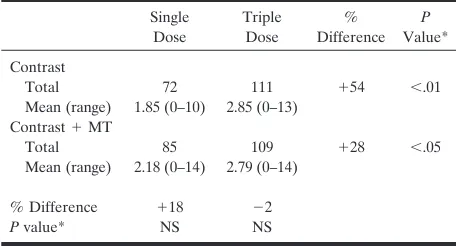
Significant increases of 86% (P,.005) and 54% (P,.01) in the number of total and new enhancing lesions were observed when comparing non-MT single- and triple-dose images, and increases of 37% (P,.01) and 28% (P,.05) were found when comparing MT single-dose images with non-MT triple-dose images. The application of the MT pulse significantly increased the sensitivity of sin-gle-dose images in the detection of the total num-ber of enhancing lesions (P , .01) (Figs 1–3). An 18% increase was also observed for the number of new enhancing lesions, but this increase was not statistically significant. Application of the MT pulse after injection of the triple dose of contrast did not change the sensitivity of the technique (Figs 2 and 3).
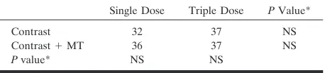
No statistical differences were observed in the number of active images in comparisons of the tech-niques (Table 3). Eighty percent of lesions considered enhancing on the contrast-enhanced MT T1-weighted images were seen as hyperintense lesions on the non-contrast MT T1-weighted images. No patient re-ported discomfort or side effects over the entire du-ration of the study. No permanent or significant changes in the blood test parameters tested were found during the follow-up period.
Discussion
Our study shows that the use of a triple dose of contrast material and the application of an MT pulse to single-dose T1-weighted sequences significantly in-crease the sensitivity of enhanced MR imaging in detecting active lesions in patients with MS. This may have implications in the design of MR-monitored clinical trials, as it may result in a considerable reduc-tion in the number of MR images needed to obtain adequate statistical power. The increased MR imag-ing sensitivity may also improve our understandimag-ing of disease evolution and the strength of the correlation between clinical and MR measures.
There is general agreement as to the value of en-hanced MR imaging for monitoring short-term MS evolution, either natural or modified by treatment (1, 2). In such a context, optimization of image timing, sequences, and contrast dose is essential (1, 3, 6). Several recent cross-sectional studies have docu-mented that the use of a triple dose of contrast ma-terial markedly increases the detection of enhancing lesions in patients with MS (11–14). Our longitudinal study confirms this effect and its magnitude, thus suggesting that the higher sensitivity of triple-dose contrast may be due to the detection of lesions that would never be detected on serial single-dose images. Our study also showed that MT single-dose se-quences are more sensitive than non-MT single-dose sequences. This approach was, however, less sensitive than non-MT triple-dose sequences. A previous study (24) also showed that interrater variability in report-ing enhancreport-ing lesions is much higher when MT im-ages are used. This may be because variable numbers of lesions that are hyperintense on contrast-enhanced MT images are already hyperintense on noncontrast MT images (19, 24), as shown in the present study. A cross-comparison of noncontrast and contrast-en-hanced MT images to count the number of enhancing lesions seems to be mandatory. Clearly, a lower sen-sitivity coupled with a lower reproducibility may re-sult in the need to obtain a greater number of images to show the effects of treatment. Nevertheless, one must also consider the increased costs of MR-moni-tored trials when serial triple-dose images are used.
[image:3.587.54.281.83.207.2]The higher sensitivity of the triple dose versus the MT single-dose technique may be due to the depic-tion of a greater number of lesions with less intense BBB damage on triple-dose images, owing to the higher transmembrane gradient of the contrast mate-rial, which is not reached when the other technique is used. Although the detection of lesions with different degrees of BBB damage may result in a better and more complete understanding of MS evolution, it might be questioned whether the suppression of such lesions due to the effects of treatment may signifi-cantly affect the subsequent evolution of the disease. The use of an MT pulse after the injection of a triple dose of contrast agent does not further increase the sensitivity of enhanced MR imaging. On the con-trary, when we applied the MT pulse, three small periventricular lesions were no longer seen. This TABLE 1: Total and mean number of enhancing lesions per month
per patient detected by the four different MR techniques
Single Dose
Triple Dose
% Difference
P
Value* Contrast
Total 112 208 186 ,.005 Mean (range) 2.15 (0–11) 4.00 (0–18)
Contrast1MT
Total 149 205 137 ,.01 Mean (range) 2.87 (0–16) 3.94 (0–19)
% Difference 133 21
Pvalue* ,.01 NS
* Statistical analysis by Wilcoxon signed rank test.
TABLE 2: Total and mean number of new enhancing lesions per month per patient detected by the four different MR techniques
Single Dose
Triple Dose
% Difference
P
Value* Contrast
Total 72 111 154 ,.01 Mean (range) 1.85 (0–10) 2.85 (0–13)
Contrast1MT
Total 85 109 128 ,.05 Mean (range) 2.18 (0–14) 2.79 (0–14)
% Difference 118 22
Pvalue* NS NS
[image:3.587.54.282.258.381.2]might have been caused by the increased signal inten-sity of the periventricular white matter on the MT images, which may mask small lesions. This observation, on the one hand, suggests that the triple-dose technique depicts the vast majority of lesions with BBB disrup-tion and, on the other hand, confirms the difficulties already encountered with MT-related artifacts.
Our data are discordant with the findings of Silver
et al (14), who reported an 8% (nonsignificant) in-crease in lesion enhancement when triple-dose MT sequences were applied. However, it is notable that the MT sequence of Silver and coworkers was per-formed at three different delay times after contrast injection, ranging from 20 to 60 minutes. In our study, the MT pulse sequence was done within 5 minutes of contrast injection, which might explain the lack of FIG 1. Enhanced axial MR images of the brain obtained without
(left column) and with (right column) an MT pulse, after a single (upper row) and triple (lower row) dose of contrast material. Two enhancing lesions are present on the MT single-dose image (upper right) and one more lesion, located at the level of the left occipital white matter, is seen on triple-dose image. On the triple-dose MT images, enhancing lesions are well depicted with two additional punctate enhancements shown in the right paraventricular white matter.
FIG 2. Enhanced axial MR images of the brain obtained without
(left column) and with (right column) an MT pulse, after a single (upper row) and triple (lower row) dose of contrast material. A paratrigonal enhancing lesion is seen better on the single-dose MT images; two more lesions are present on the triple-dose images, more evident when the MT pulse is applied.
FIG 3. Enhanced axial MR images of the brain obtained without
increase in enhancing lesions. Moreover, the same authors (14) reported that the use of a triple dose caused more adverse events than the single dose (14% versus 4%). However, the results of our study, in which all patients were blinded to the dose of contrast used and were closely controlled for each possible adverse event, indicate the safety of this approach.
Conclusion
Triple-dose contrast is the most sensitive MR tech-nique for detecting enhancing lesions in patients with MS. The application of an MT pulse to T1-weighted sequences after the injection of a single dose of con-trast also results in a higher detection rate of enhanc-ing lesions. Larger and longer longitudinal studies are needed to ascertain what impact the additional le-sions detected have on disease evolution and to es-tablish the relative gains in statistical power that can be achieved by one or the other of the two techniques.
Acknowledgments
We thank Luigi Bozzao and Cesare Fieschi for helpful sug-gestions. We also thank the MR imaging radiographers, Ales-sandro Dell’Anna, Paolo Montanari, and Mario Rossi, who assisted in the study.
References
1. Miller DH, Albert PS, Barkhof F, et al.Guidelines for the use of magnetic resonance technique in monitoring the treatment of mul-tiple sclerosis.Ann Neurol1996;39:6–16
2. Evans AC, Frank JA, Antel J, Miller DH.The role of MR imaging in clinical trials of multiple sclerosis: comparison of image pro-cessing techniques.Ann Neurol1997;41:125–132
3. Miller DH, Barkhof F, Nauta JJP.Gadolinium enhancement in-creased the sensitivity of MR imaging in detecting disease activity in MS.Brain1993;116:1077–1094
4. Katz D, Taubenberger JK, Cannella B, McFarlin DE, Raine CS, McFarland HF.Correlation between magnetic resonance imaging findings and lesion development in chronic, active multiple sclero-sis.Ann Neurol1993;34:661–669
5. Bastianello S, Pozzilli C, Bernardi S, et al.Serial study of gadolin-ium-DTPA MR imaging enhancement in multiple sclerosis. Neu-rology1990;40:591–595
6. Tas MV, Barkhof F, van Walderveen MA, Polman OH, Hommes OR, Valk J.The effect of gadolinium on the sensitivity and speci-ficity of MR imaging in the initial diagnosis of multiple sclerosis.
AJNR Am J Neuroradiol1995;16:259–264
7. Barkhof F, Filippi M, van Waesberghe et al.Improving interob-server variation in reporting gadolinium-enhanced MRI lesions in multiple sclerosis.Neurology1997;49:1682–1688
8. Koudriavtseva T, Thompson AJ, Fiorelli M, et al. Gadolinium enhanced MR imaging predicts clinical and MR imaging disease activity in relapsing-remitting multiple sclerosis.J Neurol Neuro-surg Psychiatry1997;62:285–287
9. Losseff NA, Kingsley DPE, McDonald WI, et al. Clinical and magnetic resonance imaging predictors in primary and secondary progressive multiple sclerosis.Multiple Sclerosis1996;1:218–222 10. Smith ME, Stone LA, Albert PS, et al. Clinical worsening in
multiple sclerosis is associated with increased frequency and area of gadopentate dimeglumine-enhancing magnetic resonance imag-ing lesions.Ann Neurol1993;33:480–489
11. Filippi M, Campi A, Martinelli V, et al.Comparison of triple dose versus standard dose gadolinium-DTPA for detection of MR im-aging enhancing lesions in patients with primary progressive mul-tiple sclerosis.J Neurol Neurosurg Psychiatry1995;59:540–544 12. Filippi M, Yoursy T, Campi A, et al.Comparison of triple dose
versus standard dose gadolinium-DTPA for detection of MR im-aging enhancing lesions in patients with MS.Neurology1996;46: 379–384
13. Haustein J, Laniado M, Niendorf HP, et al. Triple dose versus standard dose gadopentate dimeglutine: a randomized study in 199 patients.Radiology1993;186:855–860
14. Silver NC, Good CD, Barker GJ, et al. Sensitivity of contrast enhanced MR imaging in multiple sclerosis: effects of gadolinium dose, magnetization trasfer contrast and delayed imaging.Brain
1997;120:1149–1161
15. Finelli DA, Hurst GC, Gullapali RP, Bellon EM.Improved con-trast of enhancing brain lesions on postgadolinium T1-weighted spin- echo images with the use of magnetization transfer.Radiology
1994;190:553–559
16. Metha RC, Pike BG, Enzmann DR.Improved detection of enhanc-ing and nonenhancenhanc-ing lesions of multiple sclerosis with magneti-zation transfer.AJNR Am J Neuroradiol1995;16:1771–1778 17. Gass A, Barker GJ, Kidd D, et al.Correlation of magnetization
transfer ratio with clinical disability in multiple sclerosis. Ann Neurol1994;36:62–67
18. Tomiak MM, Rosemblum JD, Prager JM, Metz CE.Magnetization transfer: a potential method to determine the age of multiple sclerosis lesions.AJNR Am J Neuroradiol1994;15:1569–1574 19. Bozzao A, Bastianello S, Ferone E, et al. Enhanced and
unen-hanced MR with magnetization transfer in multiple sclerosis.
AJNR Am J Neuroradiol1996;17:1837–1842
20. Loevner LA, Grossman RI, McGowan JC, Ramer KN, Cohen JA. Characterization of multiple sclerosis plaques with T1-weighted MR and quantitative magnetization transfer.AJNR Am J Neuro-radiol1995;16:1473–1479
21. Petrella JR, Grossman RI, McGowan JC, Campbell G, Cohen JA. Multiple sclerosis lesions: relationship between MR enhancement pattern and magnetization transfer effect.AJNR Am J Neuroradiol
1996;17:1041–1049
22. Elster AD, Mathews VP, King JC, Hamilton CA.Improved detec-tion of gadolinium enhancement using magnetizadetec-tion transfer im-aging.Neuroimaging Clin N Am1994;4:185–192
23. Mathews VP, Caldemeyer KS, Ulmer JL, Nguyen H, Yuh WT. Effects of contrast dose, delayed imaging and magnetization trans-fer saturation on gadolinium-enhanced MR imaging of brain le-sions.J Magn Reson Imaging1997;7:14–22
24. van Waesberghe JHTM, Castelijns JA, Roser W, et al.Single dose gadolinium wth magnetization transfer contrast versus triple dose gadolinium in detecting enhancing multiple sclerosis lesions.AJNR Am J Neuroradiol1997;18:1279–1285
25. Poser CM, Paty DW, Scheimberg L, et al.New diagnostic criteria for multiple sclerosis: guidelines for research protocols.Ann Neu-rol1983;13:227–231
[image:5.587.54.283.85.137.2]26. Kurtzke JF.Rating neurological impairment in multiple sclerosis: an expanded disability status scale (EDSS). Neurology 1983;33: 1444–1452
TABLE 3: Number of active scans detected by the four different MR techniques
Single Dose Triple Dose PValue*
Contrast 32 37 NS
Contrast1MT 36 37 NS
Pvalue* NS NS