EMPEROR PENGUIN OXYGEN CONSUMPTION, HEART RATE
AND PLASMA LACTATE LEVELS DURING GRADED
SWIMMING EXERCISE
G. L. KOOYMAN ANDP. J. PONGANIS
Centre for Marine Biotechnology and Biomedicine, Scripps Institution of Oceanography, University of California, San Diego, La Jolla, CA 92093, USA
Accepted 1 June 1994
Summary
Oxygen consumption (V˙O∑), heart rate and blood chemistry were measured in four
emperor penguins, Aptenodytes forsteri (Gray), during graded swimming exercise. The maximum V˙O∑obtained, 52 ml O2kg21min21, was 7.8 times the measured resting V˙
O∑ of
6.7 ml O2kg21min21 and 9.1 times the predicted resting V˙O∑. As the swimming effort
rose, a linear increase in surface and submerged heart rates (fH) occurred. The highest
average maximum surface and submersion heart rates of any bird were 213 and 210 beats min21, respectively. No increase in plasma lactate concentrations occurred
until V˙O∑was greater than 25 ml O2kg21min21. At the highest V˙
O∑ values measured,
plasma lactate concentration reached 9.4 mmol l21. In comparison with other animals of
approximately the same mass, the aerobic capacity of the emperor penguin is less than those of the emu and dog but about the same as those of the seal, sea lion and domestic goat. For aquatic animals, a low aerobic capacity seems to be consistent with the needs of parsimonious oxygen utilization while breath-holding.
Introduction
Maximum oxygen consumption (V˙O∑max) has not been measured in birds, but V˙O∑is about 10–13 times the resting rate during horizontal flight (LeFebre, 1964; Tucker, 1968; Butler et al. 1977) and is over 15 times the resting rate in hovering hummingbirds (Lasiewski, 1963). In contrast, ducks swimming in a watermill can only increase their V˙O∑ about 3.5–4 times above out-of-water resting rates (Prange and Schmidt-Nielsen, 1970; Woakes and Butler, 1983; Baudinette and Gill, 1985; Butler, 1991). The low V˙O∑ increases for swimming ducks are due to the small amount of leg muscle required for propulsion, compared with the larger flight muscles (Prange and Schmidt-Nielsen, 1970; Butler, 1991). Since penguins ‘fly’ under water, they use a much greater amount of their muscle mass than do swimming ducks and may, therefore, have a metabolic scope closer to that of flying birds. However, it would seem that diving birds, such as the emperor penguin, would be metabolically adapted to O2 conservation and would consequently have a moderate aerobic scope, similar to that measured in seals (Ponganis et al. 1990a)
and sea lions (Ponganis et al. 1991; Williams et al. 1991). Furthermore, the exceptionally low drag of their fusiform body shape (Nachtigall and Bilo, 1980) suggests that penguins may swim at low energetic costs even at high speeds.
We selected emperor penguins, the largest of all aquatic birds, so that we would be able to make comparisons, at high swimming effort, with marine mammals of similar mass that have already been studied within the same type of exercise protocol. This comparison was, in part, prompted by the diving physiology and behaviour of emperor penguins. During dives, heart rate responses of emperor penguins are similar to those observed in phocid seals (Kooyman et al. 1992b). However, for their size, birds appear to be better divers if duration, depth and speed are used as the evaluating criteria. Emperor penguins cruise at a speed about 50 % faster than those of seals and sea lions (Ponganis et al. 1990b; Castellini et al. 1992; Kooyman et al. 1992b). King penguins (Aptenodytes
patagonicus) often dive for longer than 5 min, to depths greater than 200 m, and at speeds
of slightly more than 2 m s21(Kooyman et al. 1992a); emperor penguin diving capacities are about twice those of king penguins (G. L. Kooyman, unpublished observations). In an analysis of several fur seal species, none matched any of these diving standards even though all were at least two times greater in body mass (Gentry and Kooyman, 1986).
We therefore sought to measure the metabolic scope, exercise heart-rate response and alteration in plasma lactate concentration in emperor penguins (Aptenodytes forsteri) during graded exercise. These variables provided us with the information needed to determine whether the birds were near or at V˙O∑max. The results show a close correlation between average heart rate and average V˙O∑. The reluctance or inability of the birds to swim beyond the maximum induced work load and the rapid rise in plasma lactate concentration at this inflection point indicate that an aerobic/anaerobic threshold had been reached. This level of exercise was similar to that at the highest V˙O∑measured previously in seals, sea lions and dolphins (Ponganis et al. 1990a, 1991; Williams et al. 1991, 1993).
Materials and methods
Husbandry
On 12 November 1988, four 4-month-old emperor penguin chicks (CW, M, T, N) weighing between 2.5 and 4.5 kg were collected at Cape Washington (74˚399S, 167˚229E), Antarctica. They were flown by twin-otter aircraft to a sea-ice laboratory near McMurdo Station. After 6 days at the station, they were flown directly to San Diego, California, in a refrigerated US Air Force Starlifter. Brief stops were made at Christchurch, New Zealand, and Honolulu, Hawaii. Within 24 h, the birds were placed in the Sea World polar bird quarantine facility (Mission Bay, San Diego, California). In this environment, they were hand-fed until they were transferred to the Sea World public display facility. The air temperature in the display was 2 ˚C and the water temperature was about 5.5 ˚C.
For each experiment, the birds were transported to Scripps Institution of Oceanography (SIO), University of California, San Diego (UCSD), in upright, padded boxes in a refrigerated van. They remained in the van except when an experiment was conducted. Total time away from Sea World was between 36 and 48 h for each trip.
At the SIO Hydraulics Laboratory, UCSD, the birds were placed individually in the water channel previously described by Davis et al. (1985). All procedures for measuring
V˙O∑, increasing the workload (swim effort) and measuring heart rate were the same as formerly described (Davis et al. 1985; Ponganis et al. 1990a; Williams et al. 1991). Very briefly, water in the water channel (1.1 m square 316 m long) was chilled to 2–4 ˚C the day before the experiment. This water temperature was about the same as that at the Sea World facility. Water flow rate was held constant at 1 m s21while the birds swam in a 2.5 m long test section partitioned by two grates from the rest of the channel.
Water velocity was measured with an electromagnetic flow sensor placed 30 cm above the floor of the flume. A vertical profile of water velocity varied by less than 0.1 m s21 (Davis et al. 1985). The velocity was correlated to the rotation rate of the propellers, which in normal operation was used to set the water speed. Water velocity in the test section was the same as in the rest of the channel.
Metabolic rate
Resting V˙O∑was obtained from three birds held for several hours in a small water bath, temperature regulated at 1.5–6.1 ˚C. During exercise in the water channel, the swim procedure and V˙O∑measurements were as follows. Before releasing a bird into the flume, a narrow epoxy-glue strip painted on the mid-line of the back and about two-thirds of the body length from the head was used as an anchor for attachment of a 5 cm long, 1.5 cm diameter tube to the feathers with nylon ties. The tube had a swivel to prevent an attached load line from becoming twisted when the bird occasionally made turns. The 6 mm diameter, braided nylon line passed through the rear grate in the centre of the channel and made an approximately 45 ˚ turn through a pulley towards the surface. About 1 m above the surface, the line turned vertically through another pulley hanging at the edge of the channel. About 2 m below this second pulley, the desired weight was hung onto the line. A weight of at least 0.25 kg was always applied, to prevent slack in the line. This arrangement produced a horizontal pull on the penguin when it swam in the centre of the channel. Increased swim effort was produced by adding loads ranging from 0.25 to 4 kg.
Once the bird had been released in the test section, the water surface was covered with an acrylic dome mounted on a sheet of 6 mm plywood. The birds breathed under this dome, and air was drawn through the dome at a suitable rate to maintain the O2 concentration above 20 %. Air flow, O2 concentration, calibration of the system and calculation of V˙O∑ were accomplished with an on-line computer system similar to that described by Davis et al. (1985).
began, V˙O∑reached a plateau and remained stable until the end. Average V˙O∑values were determined from the sum of all values from all runs at a specific load. During heart rate and lactate experiments, average V˙O∑values were calculated from the V˙O∑measurements during the specific run in which the heart rate or lactate determination was made.
Heart rate
Prior to fHand lactate measurements, the birds were anaesthetized. The procedure used was similar to that described previously (Kooyman et al. 1992b). In brief, birds were masked with a 1 l plastic bottle in which a neoprene neck ring formed a seal. Induction was at 4 % isoflurane, which was reduced to 1 % during the procedure.
For placing heart-rate electrodes, anaesthesia lasted about 30 min, and after about 5 h of recovery the bird was released into the flume. As in previous studies (Kooyman et al. 1992b), the two electrodes were placed on the dorsal midline; one was placed between the wings and another about two-thirds of the body length behind the head. Heart rates were monitored on a Hewlett-Packard chart recorder. The total heart beat count of each surface and submersion interval was divided by the duration of the interval to obtain fH. The average fHfor a specific V˙O∑was calculated by summing all beats over the entire exercise cycle and dividing by the total time. Heart rate analysis was performed only for periods in which V˙O∑ was stable. Surface and submersion times were manually recorded by an observer and stored on the computer as V˙O∑was measured.
Lactate levels
The placement of catheters required 30–60 min under anaesthesia. An 18 gauge (1.3 mm diameter, 3 cm length) catheter was placed 2.5 cm into the vein of the middle toe. The catheter was held in place with Vetwrap coated with epoxy glue to form a rigid cast that allowed the foot enough mobility for normal positioning while swimming. A 2 m extension was placed on the catheter, the dead space of which was 1.6 ml. This length was necessary to raise the sampling stopcock above the water for sampling during swimming. Sampling duration ranged from 30 s to 10 min and was fastest when the bird was swimming and slowest between runs, when the bird rested in the water and was usually shivering. Samples were analyzed immediately after collection on a YSI model 2300 Stat lactate and glucose analyzer. Heart-rate and blood-sampling experiments were carried out separately to avoid catheter, electrode and load lines becoming tangled when the birds made turns in the flume.
Statistics
All statistics and tests of significance were obtained using the Statistix commercial software package (St Paul, MN). Unless stated otherwise, confidence intervals are expressed as one standard deviation of the mean.
Results
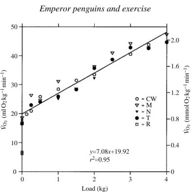
During exercise, V˙O∑ was closely correlated with load, and the coefficients of determination (r2) ranged from 0.93 to 0.99. There was no significant difference among the birds either in the slope or the intercept (analysis of covariance, multiple-comparison test, P<0.05; Zar, 1984). The pooled analysis is shown in Fig. 1.
The maximum mean V˙O∑ when loaded was 47.3 ml O2kg21min21 for M while swimming and pulling a 4 kg load. Maximum values for both CW and T were close to this, at 44.6 ml O2kg21min21and 44.7 ml O2kg21min21respectively, also with a 4 kg load (Fig. 1). The maximum average value for a single run was 52 ml O2kg21min21for M (Figs 2 and 3).
Heart rates
Regression lines for the relationship between fHand V˙O∑during surface swimming and 50
40
30
20
10
0 V˙O
∑
(ml
O2
kg
−
1min
−
1)
V˙O
∑ (
mmol
O2
kg
−
1min
−
1)
0 1 2 3 4
Load (kg)
2.0
CW
y=7.08x+19.92 r2=0.95
M N T R
1.6
1.2
0.8
0.4
0
Fig. 1. Measured V˙O∑of all birds in relation to load. Average resting values fromTable 1 are indicated as upright triangles. Other symbols are for the overall average V˙O∑at each load for each bird. Standard errors of the means fall within the symbols.
Table 1. Resting V˙O∑; samples were taken every 10 s Minimum resting Water
V˙O∑ temperature
Bird (ml O2kg−1min−1) (°C) N
CW 6.2±0.99 1.5 115
M 6.7±1.89 2.4 221
T 6.7±2.09 6.1 185
200 160 120 80 200 160 120 80 23 26 14 15 15 12 12 13 37 36 15 15 17 16 16 16 9 6 22 23 23 23 32 25 23 23 23 30 30 30 31 31 50 51
18 29 43 29 34 28 35 31 16
16 16 16 15 18 17 17 26
119 133 72 15 98 45 43 11
200 160 120 80 12 12 34 21 21 20
19 98 11 23 60 36 26
Heart rate (beats
min
−
1)
15 20 25 30 35 40 45 50 55 CW
M
T
V˙O∑ (ml O2kg−1min−1)
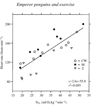
submersion are summarized in Fig. 2 and Table 2. The r2values ranged from 0.07 for M when surfaced to 0.39 for CW and T when submerged. In all three birds, examination of surface versus submerged fHvalues showed that mean fHvalues at the surface and while submerged were significantly different (P<0.001, paired sample t-test, d.f.=23). These mean surface and submerged fHvalues are given in Fig. 2. Submersion times ranged from 5 to 15 s; the lowest values corresponded to the highest V˙O∑values. Surface times ranged from 2 to 6 s. The average heart rate values at specific V˙O∑values for each bird are plotted in Fig. 2 for each bird and in Fig. 3 for pooled results.
Plasma lactate levels
The highest lactate levels were 7 and 9.5 mmol l21in birds CW and M, respectively. Fig. 4 shows the relationship between [lactate] and swimming V˙O∑.
Fig. 2. Submersion and surface heart rates in relation to V˙O∑in three swimming penguins. The dashed regression line was calculated from the mean heart rate and V˙O∑for each run (open circles). Mean surface (filled circles) and submersion (filled triangles) heart rates for all runs were calculated from all surface and submersion rates during each run. The bars show ±1 standard error. V˙O∑ values are the means for each run for which the heart rates were measured. Standard errors fall within the symbols. Values next to each point represent the number of surface and submersion intervals during which heart rate was measured for each run. Numbers above the V˙O∑axis are the number of 10 s samples used to determine mean V˙O∑values.
200
160
120
80 y=2.6x+55.8
r2=0.695
Heart rate (beats
min
−
1)
15 20 25 30 35 40 45 50 55 M CW
T
V˙O∑ (ml O2kg−1min−1)
Discussion
The 10- to 13-fold increase in V˙O∑from resting levels during flight in birds previously cited should not be assumed to represent the entire aerobic scope of birds since V˙O∑max was not measured. Similarly, the highest measured V˙O∑ in this study (52 ml O2kg21min21) may not represent V˙O∑max for the emperor penguin, since no plateau in V˙O∑occurred. However, the observers’ impressions of the swimming effort of the birds when pulling a 4 kg load, as well as the high plasma lactate concentration (Fig. 4) at these loads, suggested that the birds were near their maximum limit. The
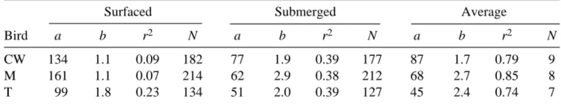
Table 2. Values for the regressions of heart rate (fH; beats min−1) on oxygen
consumption (V˙O∑; ml O2kg−1min−1) during graded swim effort Surfaced Submerged Average
Bird a b r2 N a b r2 N a b r2 N
CW 134 1.1 0.09 182 77 1.9 0.39 177 87 1.7 0.79 9 M 161 1.1 0.07 214 62 2.9 0.38 212 68 2.7 0.85 8 T 99 1.8 0.23 134 51 2.0 0.39 127 45 2.4 0.74 7
The equation is y=a+bx, where y is fH, x is V˙O∑.
N refers to the number of surface and submergence intervals during which heart rate was calculated. In the average fHequation, N is equal to the number of determinations of average V˙O∑.
All r2values are significant at P=0.05.
0 10
8
6
4
2
Plasma lactate concentration (mmol
l
−
1)
20
0 10 30 40 50
M N CW
T
V˙O∑ (ml O2kg−1min−1) 118
18
18
25
50
24
37
42
35 36
102
Fig. 4. The relationship between maximum plasma lactate concentration and the mean V˙O∑
lowest measured resting V˙O∑ of 6.2 ml O2kg21min21 is similar to previous values obtained for emperor penguins resting in air in their thermoneutral zone (6.2–7.3 ml O2kg21min21; Pinshow et al. 1976; Le Maho et al. 1976; Deswasmes et al. 1980). This is greater than the resting rate of 5.7 ml O2kg21min21 for a 20.8 kg bird predicted from the equation given by Aschoff and Pohl (1970). The metabolic scope of the emperor penguin, from our values, is 7.8, and it is 9.1 using the predicted resting V˙O∑. The maximum increase in V˙O∑measured in swimming emperor penguins is less than that measured in birds during flapping flight. However, all flying birds are considerably smaller than emperor penguins (Butler, 1991), so that direct comparisons are difficult. The emu (Dromaius novaehollandiae) is of similar body mass to the emperor penguin, and the highest measured V˙O∑ for this bird (48 ml O2kg21min21) and the resting V˙O∑ (4.2 ml O2kg21min21) gave a metabolic scope of 11.4 (Grubb et al. 1983). As V˙O∑max was not measured, the actual aerobic capacity of the emu is therefore probably much greater than that of the emperor penguin.
The aerobic capacity of the emperor penguin measured in this study is, therefore, lower than that for the running emu or for flying birds. Patak and Baldwin (1993) recently reported that the relative mass and oxidative capacity of the pelvic limb muscle of emus were similar to those of the flight muscles of birds. Although the emperor penguin pectoralis/supracoracoideus muscle mass constitutes a similar percentage of the body mass (P. J. Ponganis, unpublished results) to that of the pelvic limb muscle of the emu (Patak and Baldwin, 1993), muscle oxidative capacity may be less, as previous studies have suggested that emperor penguin muscle is ‘best adapted for anaerobic work’ (Baldwin, 1988). Its lower aerobic capacity in comparison with the emu and with flying birds may, therefore, be related more to differences in mitochondrial density than to relative muscle mass.
The measured exercise capacity of the emperor penguin is also less than the 30- and 12-fold aerobic scopes of dogs and goats, terrestrial mammals of similar body mass to emperor penguins (Taylor et al. 1987). Those values were based on measured V˙O∑maxdata and predicted resting V˙O∑values. The ninefold increase in emperor penguin V˙O∑above predicted resting levels is, however, similar to that measured in three marine mammals. Maximum V˙O∑measured in the bottlenose dolphin (Tursiops truncatus) was 7–11 times the predicted resting rate (Williams et al. 1993). In harbour seals (Phoca vitulina) and sea lions (Zalophus californianus), both of similar mass to the emperor penguin, the maximal measured exercise capacity was 9–10 times both the predicted and measured resting V˙O∑ (Ponganis et al. 1990a, 1991). Although we did not obtain a well-defined V˙O∑max, the measured aerobic capacity of the emperor penguin appears to be most similar to the aerobic capacities measured in diving mammals.
(Williams et al. 1991; Butler et al. 1992). However, the highest V˙O∑levels reported for these mammals was 20–25 ml O2kg21min21. Seals and sea lions show a significant drop in fHbetween surface breathing and when submerged; in the seal, submersion fHis about one-third of the surface rate (Ponganis et al. 1990a). The surface and submersion
fHpattern of the emperor penguin is similar to that of the seal, although absolute fH values were higher.
It was not possible to estimate swimming speeds from the methods used in this study. Flow was turbulent because of the grate, and thrust generation by the wings while swimming in natural conditions may be different from thrust generation while swimming against flowing water in a channel or against flowing water with a load applied. Thus, direct extrapolation of diving in the wild may be limited. Comparisons between video tapes of penguins swimming in the wild and in the channel showed that the amplitude and frequency of the wing stroke appeared to be lower under natural conditions than for the captive birds swimming with the lightest load. Wing-beat frequency of emperor penguins swimming in the wild was about 45 beats min21(G. L. Kooyman unpublished), and an average of 62–70 beats min21 for captive birds swimming with the lightest loads. Similarly, heart rates during natural dives (Kooyman et al. 1992b) are often less than the submerged heart rates observed at the lowest workloads in this study. Therefore, it seems reasonable to assume that V˙O∑of diving birds in the wild would be less than the lowest submerged swimming values obtained in this study, 20 ml O2kg21min21(Fig. 1). If so, then the emperor penguin diving metabolic rate would be less than three times the resting levels we measured, which is less than the swimming V˙O∑ of six times the resting metabolic rate measured in the Adelie penguin (Pygoscelis adeliae) surface swimming in a 20 m water channel at 2.3 m s21(Culik and Wilson, 1991). Such a diving metabolic rate would be consistent with the moderate aerobic capacity measured in this study and may help to explain the emperor penguin’s remarkable diving breath-hold capacity of 15–20 min (Kooyman et al. 1971).
We thank L. Starke, L. Winter, P. Jobsis and S. Eckert for assistance in these studies. This research was supported by grants NSF DPP 87-15863 and USPHS HL 17731.
References
ASCHOFF, S. ANDPOHL, H. (1970). Rhythmic variation in energy metabolism. Fedn. Proc. Fedn Am.
Socs. exp. Biol. 29, 1541–1552.
BALDWIN, J. (1988). Predicting the swimming and diving behavior of penguins from muscle
biochemistry. Hydrobiologia 165, 255–261.
BAUDINETTE, R. V. ANDGILL, P. (1985). The energetics of ‘flying’ and ‘paddling’ in water: locomotion
in penguins and ducks. J. comp. Physiol. 155, 373–380.
BUTLER, P. J. (1991). Exercise in birds. J. exp. Biol. 160, 233–262.
BUTLER, P. J., WEST, N. H. ANDJONES, D. R. (1977). Respiratory and cardiovascular responses of the
pigeon to sustained level flight in a wind-tunnel. J. exp. Biol. 71, 7–26.
BUTLER, P. J., WOAKES, A. J., BOYD, I. L. ANDKANATOUS, S. (1992). Relationship between heart rate and
oxygen consumption during steady-state swimming in California sea lions. J. exp. Biol. 170, 35–42.
CASTELLINI, M. A., KOOYMAN, G. L. ANDPONGANIS, P. J. (1992). Metabolic rates of freely diving
CULIK, B. ANDWILSON, R. P. (1991). Energetics of under-water swimming in Adelie penguins. J. comp. Physiol. B 161, 285–291.
DAVIS, R. W., WILLIAMS, T. M. ANDKOOYMAN, G. L. (1985). Swimming metabolism of yearling and adult harbor seals (Phoca vitulina). Physiol. Zool. 58, 590–596.
DESWASMES, G., LEMAHO, Y., CORNET, A. ANDGROSCOLOS, R. (1980). Resting metabolic rate and cost
of locomotion in long-term fasting emperors. J. appl. Physiol. 49, 888–896.
GENTRY, R. L. ANDKOOYMAN, G. L. (eds) (1986). Fur Seals: Maternal Strategies on Land and at Sea.
New Jersey: Princeton University Press. 291pp.
GRUBB, B., JORGENSEN, D. O. ANDCONNER, M. (1983). Cardiovascular changes in the exercising emu. J. exp. Biol. 109, 193–201.
KOOYMAN, G. L., CHEREL, Y., LE MAHO, Y., CROXALL, J. P., THORSON, P. H., RIDOUX, V. AND
KOOYMAN, C. A. (1992a). Diving behavior and energetics during foraging cycles in king penguins.
Ecol. Monogr. 62, 143–163.
KOOYMAN, G. L., DRABEK, C. M., ELSNER, R. ANDCAMPBELL, W. B. (1971). Diving behavior of the
emperor penguin, Aptenodytes forsteri. Auk 88, 775–795.
KOOYMAN, G. L., PONGANIS, P. J., CASTELLINI, M. A., PONGANIS, E. P., PONGANIS, K. V., THORSON, P. H.,
ECKERT, S. A. ANDLEMAHO, Y. (1992b). Heart rates and swim speeds of emperor penguins diving
under sea ice. J. exp. Biol. 165, 161–180.
LASIEWSKI, R. C. (1963). Oxygen consumption of torpid, resting, active and flying hummingbirds.
Physiol. Zool. 36, 122–140.
LEFEBRE, E. A. (1964). The use of D2O18for measuring metabolism in Columbia livia at rest and in flight. Auk 81, 403–418.
LEMAHO, Y., DECLITTE, R. ANDCHATONNET, J. (1976). Thermoregulation in fasting emperor penguins under natural conditions. Am. J. Physiol. 231, 913–922.
NACHTIGALL, W. AND BILO, D. (1980). Stromungsanpassung des Penguins bein Schwimmen unter
wasser. J. comp. Physiol. 137, 17–26.
PATAK, A. ANDBALDWIN, J. (1993). Structural and metabolic characterization of the muscles used to power running in the emu (Dromaius novachollandiae), a giant flightless bird. J. exp. Biol. 175, 233–249.
PINSHOW, B., FEDAK, M. A., BATTLES, D. R. ANDSCHMIDT-NIELSEN, K. (1976). Energy expenditure for
thermoregulation and locomotion in emperor penguins. Am. J. Physiol. 231, 903–912.
PONGANIS, P. J., KOOYMAN, G. L. ANDZORNOW, M. H. (1991). Cardiac output in swimming california
sea lions, Zalophus californianus. Physiol. Zool. 64, 1296–1306.
PONGANIS, P. J., KOOYMAN, G. L., ZORNOW, M. H., CASTELLINI, M. A. ANDCROLL, D. A. (1990a).
Cardiac output and stroke volume in swimming harbor seals. J. comp. Physiol. 160, 473–482.
PONGANIS, P. J., PONGANIS, E. P., PONGANIS, K. V., KOOYMAN, G. L., GENTRY, R. L. ANDTRILLMICH, F.
(1990b). Swimming velocities in otariids. Can. J. Zool. 68, 2105–2112.
PRANGE, H. D. ANDSCHMIDT-NIELSEN, K. (1970). The metabolic cost of swimming in ducks. J. exp. Biol.
53, 763–777.
TAYLOR, C. R., KARAS, R. H., WEIBEL, E. R. ANDHOPPELER, H. (1987). Adaptive variation in the
mammalian respiratory system in relation to energetic demand. II. Reaching the limits to oxygen flow. Respir. Physiol. 69, 7–26.
TUCKER, V. A. (1968). Respiratory exchange and evaporative water loss in the flying budgerigar. J. exp.
Biol. 48, 67–87.
WILLIAMS, T. M., FRIEDL, W. A. AND HAUN, J. E. (1993). The physiology of bottlenose dolphins
(Tursiops truncatus): heart rate, metabolic rate and plasma lactate concentration during exercise. J. exp. Biol. 179, 31–46.
WILLIAMS, T. M., KOOYMAN, G. L. ANDCROLL, D. A. (1991). The effect of submergence on heart rate
and oxygen consumption of swimming seals and sea lions. J. comp. Physiol. 160, 637–644.
WOAKES, A. J. ANDBUTLER, P. J. (1983). Swimming and diving in tufted ducks, Aythya fuligula, with