Pretransplantation Conditioning Influence on
the Occurrence of Cyclosporine or FK-506
Neurotoxicity in Allogeneic Bone Marrow
Transplantation
Walter S. Bartynski, Zella R. Zeigler, Richard K. Shadduck, and John Lister
BACKGROUND AND PURPOSE:Transplantation conditioning regimens have been shown to affect the brain imaging appearance in patients with cyclosporine or FK-506 neurotoxicity. We assessed whether the occurrence of neurotoxicity was affected by the choice of conditioning regimen used before allogeneic bone marrow transplantation (allo-BMT).
METHODS:An allo-BMT was performed in 290 patients conditioned before transplantation with myeloablative therapy. Neurotoxicity from cyclosporine or FK-506 developed in 21 (7.2%) of these patients, as confirmed with CT or MR imaging. Two hundred seventy-four (94%) of these 290 patients were conditioned with minor variations of one of five fundamental regimens: cyclophosphamide (Cy)/busulfan (n ⴝ 97), Cy/total body irradiation (TBI) (n ⴝ 122), Cy/ thiotepa/TBI (n ⴝ 40), bischloroethylnitrosourea/etoposide/cytarabine/melphalan, or BEAM (n ⴝ 10), and Cy/thiotepa/busulfan (n ⴝ 5). The remaining 16 patients were prepared with variable regimens. The rates of occurrence of cyclosporine or FK-506 neurotoxicity relative to these conditioning regimens were compared.
RESULTS: The lowest rate of cyclosporine or FK-506 neurotoxicity was found in those patients conditioned with Cy (2 days)/busulfan (4 days) (5.1%) or Cy (2 days)/TBI (4 days) (5.9%). Rate of neurotoxicity increased with lengthier conditioning regimens. A high rate of neurotoxicity was present in those patients conditioned with Cy (4 days)/TBI (4 days) (13.7%), and this was statistically significant (P < .05) when compared with Cy (2 days)/busulfan (4 days).
CONCLUSION:The rate of occurrence of cyclosporine or FK-506 neurotoxicity varies with the conditioning regimen used, with lengthier regimens associated with a higher rate of neurotoxicity. As the length of the conditioning regimen equates to the total dose of chemo-therapy administered, it suggests that the intensity of the regimen is correlated to the predis-position to neurotoxicity from cyclosporine or FK-506.
Cyclosporine and FK-506 are immunosuppressive agents used to control transplant rejection and graft-versus-host disease (GVHD). Reports have linked these drugs with central nervous system toxicity, but the eti-ology of this neurotoxicity is not understood (1–42).
Factors under consideration include direct cyclosporine or FK-506 toxicity (2–4,6,8,9,15,16,21,23–25,27,29–32), altered cyclosporine metabolism or drug binding with secondary increase in brain drug levels (18, 24), endo-thelial damage with the release of vasoactive peptides leading to labile blood pressure and vasospasm (28, 32–35), and thrombotic microangiopathy leading to mi-crovascular damage (31, 36). Hypertension with in-creased sympathetic neural activation has been sug-gested (30, 37, 38), and selected reports have considered other potential causes such as high-dose methylpred-nisolone therapy (2, 4), ketoconazole therapy (3), hypo-magnesemia (9), anaphylactic reaction (8), and human leukocyte antigen (HLA) mismatch (33).
Imaging studies describe subcortical and deep white matter changes in the occipital and parietal regions likely representing vasogenic edema (17, 18, Received March 24, 3003; accepted after revision August 8.
From the Departments of Radiology (W.S.B.) and Medicine (Z.R.Z., R.K.S., J.L.), The Western Pennsylvania Hospital, Pitts-burgh.
Presented at the 40th annual meeting of the American Society of Neuroradiology, Vancouver, British Columbia, Canada, May 11– 17, 2002.
Address reprint requests to Walter S. Bartynski, M.D., Depart-ment of Radiology, Division of Neuroradiology, University of Pitts-burgh Medical Center, 200 Lothrop St., D132, PittsPitts-burgh, PA 15213.
©American Society of Neuroradiology
21–23, 26–31). Cortex involvement has been noted and contrast material enhancement occasionally seen (23, 27, 30, 32, 33). This pattern has been referred to as the reversible posterior leukoencephalopathy syn-drome (39–44). Also, nonspecific white matter fea-tures have been noted (45).
A recent report demonstrated that the imaging appearance of cyclosporine or F-506 neurotoxicity varies depending on the conditioning regimen used to eliminate native bone marrow before allogeneic bone marrow transplantation (allo-BMT) (46). White mat-ter lesions were present in patients conditioned with radiation therapy and chemotherapy, whereas cortex lesions predominated in patients conditioned with chemotherapy alone. This suggests that pretransplan-tation conditioning regimens may play a role in the toxicity process.
The purpose of this study was to assess the fre-quency of cyclosporine or FK-506 neurotoxicity rela-tive to the pretransplantation conditioning regimens used, to investigate whether conditioning affects the neurotoxicity process.
Methods
During an 11-year period (January 1991–June 2002), 290 allo-BMT procedures with myeloablative conditioning were performed at our institution. Patients were receiving cyclospor-ine or FK-506 to prevent GVHD. One hundred sixty-eight patients were male and 122 were female. The age distribution was 17–65 years, with an average age of 40 years. The clinical problems requiring allo-BMT are summarized in Table 1. All patients were referred because of initial treatment failure of their primary disease process.
In 21 of these 290 patients, significant neurologic symptoms developed, and imaging studies demonstrated brain changes consistent with previous literature description of cyclosporine or FK-506 neurotoxicity. Twelve patients were female and nine were male with an average age of 34 years (range, 17–49 years). Fifteen of the these patients undergoing allo-BMT received transplants from related donors and six from matched unre-lated donors.
Allo-BMT Procedures
Patients underwent allo-BMT in accordance with treatment protocols approved by the hospital’s institutional review board.
GVHD Prophylaxis.—All patients received either cyclospor-ine or FK-506 combcyclospor-ined with steroid (methylprednisolone or
prednisone) as prophylaxis against GVHD. Cyclosporine 3–5 mg/kg/day or FK-506 0.03 mg/kg/day was administered intra-venously or orally, and the dosages were adjusted to maintain whole blood levels between 350 and 800 ı`g/L (polyclonal fluo-rescence polarization assay) in the case of cyclosporine and 5–20 ı`g/L in the case of FK-506. In patients receiving an unrelated donor transplant, methotrexate was administered at 15 mg/mol/L on day 1 and 10 mg/mol/L on days 3, 6, and 11 after transplantation. Immunosupression was tapered in the posttransplantation period to end at day 360 after transplantion in patients not experiencing GVHD.
Preparative Conditioning Regimens.—A variety of myeloab-lative pretransplantation conditioning regimens were used and are reviewed in Tables 2 and 3. Conditioning regimens included chemotherapy drug combinations, or drug combinations and total body irradiation (TBI) administered in divided doses for several days before allogeneic marrow administration. Bischlo-roethylnitrosourea (BCNU), etoposide, cytarabine (ara-C), and melphalan (BEAM) therapy was used in 10 patients. In four patients, a lower radiation dose was applied, characterized as total lymphocytic irradiation (TLI).
Conditioning dosages administered were as follows: cyclo-phosphamide (Cy) 50–60 mg/kg/day, busulfan 4 mg/kg/day, thiotepa 5 mg/kg/day, TBI 300 cGy/day, TLI 100 cGy/day, BCNU 300 mg/m2/day, etoposide 200 mg/m2/day, ara-C 200
mg/m2, melphalan 140 mg/m2/day, carboplatin 200 mg/m2/day,
and cisplatin 50 mg/m2/day. A limited number of regimens were
used in most patients as follows: Cy/busulfan (n ⫽97), Cy/ thiotepa (n⫽40), Cy/thiotepa/busulfan (n⫽5), Cy/TBI (n⫽
[image:2.603.306.534.70.224.2]122), Cy/thiotepa/TBI (n⫽39), and BEAM therapy (n⫽10).
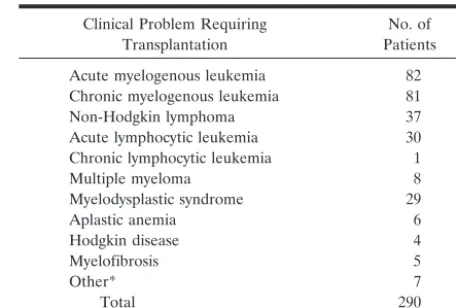
TABLE 1: Reasons for transplantation
Clinical Problem Requiring Transplantation
No. of Patients Acute myelogenous leukemia 82 Chronic myelogenous leukemia 81
Non-Hodgkin lymphoma 37
Acute lymphocytic leukemia 30 Chronic lymphocytic leukemia 1
Multiple myeloma 8
Myelodysplastic syndrome 29
Aplastic anemia 6
Hodgkin disease 4
Myelofibrosis 5
Other* 7
Total 290
* Included biphenotypic expression (features of both acute myelog-enous leukemia and acute lymphocytic leukemia) in two patients, and Fanconi anemia, Waldenstrom macroglobulinemia, hairy cell leukemia, breast carcinoma, and hypereosinophilic syndrome in one patient each.
TABLE 2: Major conditioning regimens
Regimen
No. of Patients
(n⫽274) Neurotoxicity*
Onset of Neurotoxicity*
Average Survival after Neurotoxicity (days) Early Mid Late
Cy 2 days, busulfan 4 days 97 5 (5.1) 3 1 1 58
Cy 2 days, busulfan 3 days, thiotepa 3 days 5 1 (20) 1
Cy 1 day, TBI 4 days 3 0 (0)
Cy 2 days, TBI 4 days 68 4 (5.9) 3 0 1 51
Cy 2 days, TBI 4 days, thiotepa 1 day 39 3 (7.7) 3 25
Cy 4 days, TBI 4 days 51 7 (13.7)† 3 2 2 49
Cy 4 days, thiotepa 4 days, carboplatnium 4 days 1 0 (0)
BEAM therapy 10 1 (10) 1
* Data are number of patients. Numbers in parentheses are percentages.
[image:2.603.55.537.585.717.2]Imaging Procedures
CT scans were obtained with 5-mm contiguous images ob-tained through the posterior fossa and 10-mm images obob-tained to the vertex. Contrast material, when used, consisted of a bolus of 150 mL of iothalamate meglumine (Conray 60; Mallinckrodt, St. Louis, MO) infused through a peripheral venous access.
MR imaging was obtained with a 1.5-T unit and included sagittal and axial T1-weighted images (600/25/1 [TR/TE/exci-tations]) with 5-mm section thickness, and axial proton-den-sity– and T2-weighted images (2500/25 and 80/2 [TR/TE]) with 5-mm section thickness. Contrast-enhanced T1-weighted im-ages were obtained with 0.1 mmol/L/kg gadopentatate dime-glumine (Magnevist; Berlex Laboratories, Wayne, NJ) by using typical T1-weighted parameters as mentioned above. Fluid-attenuated inversion-recovery (FLAIR) images were obtained in two patients (10,000/149/2200 [TR/TE/TI]).
Supratentorial lesions were most commonly identified in four primary locations: frontoparietal junction, parietal region, occipital poles, and inferior temporal-occipital junction. The lesions are demonstrated in Fig 1. These typically conformed to the watershed distribution. Additional lesions were identified less frequently in the cerebellar hemispheres, splenium of the corpus callosum, corona radiata, and frontal lobes.
Small nonspecific focal white matter change was occasion-ally identified. These lesions appeared random in location and no attempt was made to itemize these areas.
Clinical Review
Inpatient and outpatient records of these 21 patients were retrospectively reviewed. Factors implicated in cyclosporine or FK-506 toxicity were identified and consisted of the following: elevated blood pressure; elevated levels of cyclosporine or FK-506, magnesium, or cholesterol; HLA matching; GVHD; veno-oclusive disease; and bone marrow transplant thrombotic microangiopathy (BMT-TM). The presence or absence of sei-zure activity was noted, as was the timing of neurotoxicity relative to brain imaging. Baseline blood pressure and blood pressure at the time of toxicity were recorded. The presence of BMT-TM, GVHD, or veno-oclusive disease was noted and graded by using techniques previously described (47–50).
BMT-TM and Endothelial Injury.—Blood vessel endothelial injury is suggested clinically when evidence of BMT-TM is identified in patients undergoing allo-BMT (47). The methods of defining and categorizing BMT-TM are reviewed.
Clinical BMT-TM (grades 2–4) was diagnosed and graded if the lactate dehydrogenase level was increased in association with 1.3–4.8% schistocytes for grade 2, 4.9–9.6% schistocytes for grade 3, and 9.7% or greater schistocytes for grade 4 BMT-TM, as previously described (46). The presence of BMT-TM is an indicator of endothelial injury.
To determine the percentage of fragmented erythrocytes, a single observer counted 500 red blood cells on blinded smears.
The percentage fragmented red cells were then calculated. A fragmented erythrocyte was defined as a schistocyte (crescen-tic, helmet shaped, or triangular).
Staging System for GVHD.—Acute GVHD was diagnosed and staged from I to IV according to the Seattle criteria (48, 49). This was confirmed histologically by skin, gut, or liver biopsy.
Veno-occlusive Disease of the Liver.—Veno-occlusive disease of the liver was diagnosed if the bilirubin value was 2 mg/dL or greater with two of three of the following conditions: hepato-megaly, ascites, or weight gain of 5% or greater as proposed by Jones et al (50). Veno-occlusive disease was graded as mild (resolved without therapy), moderate (resolved with treat-ment), or severe (did not resolve or the patient died before day 100 after allo-BMT).
Statistical Analysis
Statistical assessment was accomplished with the statistical analysis functions accompanying the Excel (Microsoft, Red-mond WA) software package. Chi-square assessment of the incidence of neurotoxicity between groups receiving different conditioning regimens was compared. A difference ofP⬍.05 was considered significant.
Results
Pretransplantation conditioning regimens and rel-ative frequencies of neurotoxicity are presented in Tables 2 and 3. Neurotoxicity documented by abnor-mal CT or MR imaging findings occurred in 21 (7.2%) of 290 patients. Five conditioning regimens were used for most allo-BMT procedures. A number of the conditioning regimens did not demonstrate neurotoxicity, but their use was infrequent (Table 3). The frequency of cyclosporine or FK-506 neurotox-icity appeared to increase with greater complexity of the conditioning regimen. The lowest toxicity level was present in patients preconditioned with Cy (2 days)/busulfan (4 days) (5.1%) or Cy (2 days)/TBI (4 days) (5.9%). A high rate of neurotoxicity was en-countered with Cy (4 days)/TBI (4 days) (13.7%), and this was statistically significant (P⬍ .05) when com-pared with Cy (2 days)/busulfan (4 days).
The frequency of neurotoxicity noted with Cy/TBI increased progressively when additional chemother-apy was added. With Cy (2 days)/TBI (4 days), the toxicity rate was 5.9%. With Cy (2 days)/TBI (4 days)/ thiotepa (1 day), the rate increased to 7.7%, and with Cy (4 days)/TBI (4 days) the rate increased to 13.7%. The frequency of toxicity with Cy (1 day)/TBI (4 days) was 0%, but only three patients were given this regimen.
A similar tendency was apparent with the chemo-therapy regimens. With Cy (2 days)/busulfan (4 days), the rate of toxicity was low (5.1%) but increased dramatically with additional dosages of chemotherapy such as Cy (2 days)/bulsulfan (3 days)/ thiotepa (3 days) (20%) and BEAM (10%).
Neurotoxicity occurred at three distinct time points after transplantation: early, intermediate, and late onset. The overall onset of neurotoxicity occurred between 5 and 480 days (average, 68 days) after allo-BMT, as noted in Table 4. Early-onset toxicity oc-curred in 14 patients (67%) between 5 and 27 days (average, 18.9 days) after transplantation.
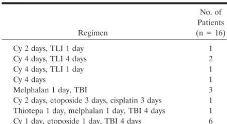
Intermedi-TABLE 3: Other conditioning regimens
Regimen
No. of Patients (n⫽16)
Cy 2 days, TLI 1 day 1
Cy 4 days, TLI 4 days 2
Cy 4 days, TLI 1 day 1
Cy 4 days 1
Melphalan 1 day, TBI 3
Cy 2 days, etoposide 3 days, cisplatin 3 days 1 Thiotepa 1 day, melphalan 1 day, TBI 4 days 1 Cy 1 day, etoposide 1 day, TBI 4 days 6
[image:3.603.54.280.70.193.2]ate-onset toxicity occurred in three patients (14%) between 55 and 78 days (average, 69 days) after trans-plantation. Late-onset toxicity occurred in four pa-tients (19%) between 151 and 480 days (average, 269 days) after transplantation. Onset of neurotoxicity was early or intermediate for most treatment groups (Table 5). In the Cy (4 days)/TBI (4 days) group, more patients were noted to develop toxicity at the intermediate onset (two of seven) or late onset (two of seven) time point.
BMT-TM was present at the time of demonstrated neurotoxicity in 20 patients who were tested. More severe grade of BMT-TM was associated with a worse long-term outcome. In one patient (patient 15), BMT-TM parameters were not obtained at the time of toxicity, but lactate dehydrogenase was low sug-gesting absent or subclinical (grade 0–1) BMT-TM. Four patients became long-term survivors; one with indeterminate BMT-TM grade (patient 15), two with grade 2, and one with grade 3 BMT-TM. In the remaining 17 patients, BMT-TM was grade 3 or 4, suggesting moderate to severe systemic endothelial injury.
Twelve of 18 patients evaluated developed acute GVHD (stages III–IV), and another patient devel-oped chronic GVHD. Four patients develdevel-oped veno-occlusive disease.
Patient survival after cyclosporine or FK-506 neu-rotoxicity is presented in Tables 4 and 5. Four pa-tients are long-term survivors of transplantation, and all developed neurotoxicity early after allo-BMT (day 5–27). In the remaining 17 patients, the average sur-vival after onset of neurotoxicity was 95 days. Patients who presented with early toxicity are either still sur-vivors (four patients) or had a longer average survival (120 days) after toxicity (14 patients). Survival dura-tion in this early-onset toxicity group was also vari-able, with five patients demonstrating short survival (4–33 days, average 16 days), one patient demonstrat-ing intermediate survival (64 days), and four patients demonstrating long survival (115–392 days, average 263 days). Patients who developed neurotoxicity in the intermediate or late period demonstrated only short or intermediate survival.
Discussion
The imaging findings in cyclosporine or FK-506 neurotoxicity include areas of attenuation or signal intensity abnormality in the parietal region, occipital poles, and to a lesser extent in the frontal lobes, inferior temporal-occipital junction, and cerebellar hemispheres (Fig 1). White matter abnormality is seen more than cortex involvement, and the lesions FIG 1. Images in a 40-year-old woman
with non-Hodgkin lymphoma who pre-sented 27 days after allo-BMT with a seizure. Conditioning regimen was BEAM therapy. Blood pressure was 114/64 mm Hg and FK-506 level at the time of toxicity was 12.7 ı`g/L (normal range, 5–20 ı`g/l).
A–D,FLAIR images demonstrate ab-normal signal intensity in the cortex and subcortical white matter of the left infe-rior temporal-occipital junction (arrow-head inA), occipital poles (large arrows inA), parietal region (short arrows inB
andC), and frontal lobes (long arrows in
may appear edematous and become confluent. When areas of abnormality are separate, a watershed pat-tern is apparent and vasospasm has been noted at MR angiography (28, 31, 46). Contrast enhancement is occasionally demonstrated, with a stipple-like pattern noted in adjacent cortex (30, 46).
The term posterior reversible encephalopathy syn-drome (PRES) has been used to describe this imaging appearance, because of predominance of parietal and occipital lobe abnormalities and frequent reversibility of the imaging findings (39–44). This pattern is noted in patients with preeclampsia or eclampsia, as well as systemic disease such as lupus. MR diffusion-weighted imaging demonstrates vasogenic edema in the areas of signal intensity abnormality that only rarely develops restricted diffusion indicating infarc-tion (42).
The cause of the PRES pattern is not clear. The imaging literature has focused on increased sympathetic neurovascular reactivity in the posterior circulation or uncontrolled systemic hypertension. Blood pressure in-stability is a common problem in the allo-BMT recipient but hypertension is not present in all patients who de-velop cyclosporine or FK-506 neurotoxicity or PRES (43, 46). The most likely causes of cyclosporine or FK-506 toxicity appear to be direct toxicity from the im-mune suppressive drugs or endothelial damage intrinsic to the transplantation process (34–36).
[image:5.603.57.534.71.393.2]The rate of cyclosporine or FK-506 neurotoxicity in our patients was dependent up the conditioning reg-imen. Cy (4 days)/TBI (4 days) was associated with a high rate of toxicity (13.7%), whereas other regimens such as Cy (2 days)/busulfan (4 days) and Cy (2 days)/ TBI (4 days) were associated with significantly lower rates of neurotoxicity. This difference appears dosage dependent with an increase in toxicity associated with additional dosages of chemotherapy. The effect sug-gests that the conditioning regimen (chemotherapy and/or TBI) plays a role in the etiology of the toxicity process. The average rate of toxicity of 7.2% in our patients was consistent with that of previous reports by Reece et al (27) 4.2%, Zimmer et al (33) 10%, Wijdicks (35) 10%, and Fung et al (51) 8.4%.
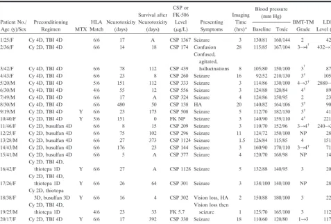
TABLE 4: Clinical summary of patients undergoing allo-BMT
Patient No./ Age (y)/Sex
Preconditioning
Regimen MTX
HLA Match
Neurotoxicity (days)
Survival after Neurotoxicity
(days)
CSP or FK-506 Level (g/L)
Presenting Symptoms
Imaging Time (hrs)*
Blood pressure (mm Hg)
BMT-TM Grade
LDH Level (u/L) Baseline Toxic
1/25/F Cy 4D, TBI 4D 6/6 17 A CSP 1367 Seizure 3 130/81 160/144 2 426
2/36/F Cy 2D, TBI 4D 6/6 14 18 CSP 174 Confusion 28 115/85 167/104 334† 43231980
3/42/F Cy 4D, TBI 4D 6/6 78 112 CSP 439
Confused, agitated,
hallucinations 8 105/80 150/100 3† 875
4/43/F Cy 4D, TBI 4D 6/6 23 8 CSP 260 Seizure 16 92/52 210/130 3† 1055
5/20/M Cy 4D, TBI 4D 5/6 151 112 CSP 333 Seizure 3 114/86 130/100 433† 28803546
6/30/M Cy 4D, TBI 4D 4/6 55 12 CSP 556 Seizure 3 124/88 120/84 4† 890
7/49/M Cy 4D, TBI 4D 6/6 17 A CSP 324 Seizure 4 124/86 150/95 2 237
8/30/M Cy 4D, TBI 4D 6/6 480 50 CSP 138 HA 20 140/82 164/106 3† 900
9/19/M Cy 2D, TBI 4D Y 6/6 23 173 CSP 508 Seizure 5 112/70 182/130 3† 415
10/40/F Cy 2D, TBI 4D Y 5/6 151 0 FK NP Seizure 3 140/90 159/110 4† 2214
11/46/F Cy 2D, busulfan 4D 6/6 8 15 CSP 209 Seizure 3 110/70 152/96 334† 24032680
12/25/F Cy 2D, busulfan 4D 6/6 75 102 CSP 296 Seizure 11 124/72 150/100 NP 285
13/28/M Cy 2D, busulfan 4D 6/6 27 373 CSP 1124 Seizure 1.5 126/84 115/85 4 1515
14/43/M Cy 2D, busulfan 4D 6/6 176 23 CSP 144 Seizure 3 160/90 170/110 334† 718
15/41/M Cy 2D, busulfan 4D 6/6 5 A CSP 377 Seizure 4 120/70 168/98 NP 143
16/42/F
Cy 2D, TBI 4D,
thiotepa 1D Y 6/6 27 A CSP 1128 Seizure 5 132/88 140/95 3 204
17/26/F
Cy 2D, TBI 4D,
thiotepa 1D Y 6/6 26 64 CSP 301 Seizure 3 138/100 140/100 NP 283
18/38/F
Cy 2D, thiotopa
3D, busulfan 3D Y 6/6 16 4 CSP 302 Vision loss, HA 2 150/88 180/100 3 222
19/25/M
Cy 2D, TBI 4D,
thiotepa 1D 4/6 23 33 FK 5.7
Vision loss then
seizure 1 125/70 165/100 3 1125
20/17/F Cy 2D, TBI 4D Y 6/6 17 392 CSP 330 Seizure 18 110/60 120/80 133 1179
21/40/F BEAM Y 6/6 27 115 FK 2.7 Seizure 6 108/60 114/64 2 2495
Note.—MTX indicates methotrexate; CSP, cyclosporine; LDH, lactate dehydrogenase; D, number of days of regimen; HA, ?; Y, yes; NP, not performed; A, alive survivor.
* Time after patient presentation with symptoms that imaging study was performed.
†Patient undergoing apheresis for BMT-TM.
TABLE 5: Onset of neurotoxicity
Onset
No. (%) of Patients
(n⫽21)
Average Time from Transplantation to Neurotoxicity (days)
Average Survival after Neurotoxicity (days)
Early 14 (67) 19 120
Mid 3 (14) 69 75
[image:5.603.54.282.461.534.2]An additional unexpected finding was that cyclo-sporine or FK-506 neurotoxicity appears to occur at three distinct time points after transplantation (Table 5). The reason for the three time points is not clear. Early-onset toxicity was seen in 14 patients (67%), occurring within the first month after transplantation, whereas intermediate-onset toxicity was seen in three patients (2–3 months after transplantation) and late-onset toxicity occurred in four patients (5–13 months after transplantation). This suggests that different factors contribute to neurotoxicity at different time points or that the toxic agents manifest their effects at many times after transplantation.
In standard cancer management, chemotherapy and radiation therapy are dosed to avoid myelosup-pression while preserving the antitumor effect. In contrast, the conditioning regimens used in prepara-tion for allo-BMT, during the time period of this study, were designed to be toxic to both tumor cells and native bone marrow. The conditioning regimen (TBI and chemotherapy) performed two essential tasks: kill tumor cells and eliminate the native bone marrow in preparation for allo-BMT. TBI was deliv-ered in high dose fractions (300 cGy/day⫻ 4 days), and the individual chemotherapeutic drugs were de-livered in high doses over several days for a powerful and acute effect. These are not ordinary applications of either chemotherapy or radiation. During the time of this study, nonmyeloablative conditioning regimens were not routinely used. Currently, a mixture of my-eloablative and nonmymy-eloablative (reduced intensity) conditioning regimens are used at our institution.
The chemotherapy drugs interfere with cell division and induce cell arrest or apoptosis (52–56). The al-kylating agents used in allo-BMT (cyclophosphamide, busulfan, thiotepa, BCNU, and melphalan) contrib-ute alkyl groups to DNA bases, corrupting DNA rep-lication and interfering with DNA synthesis and cell division (52). Cyclophosphamide’s active forms (phosphoramide mustard and 4-HC-aldophospha-mide), busulfan (an alkyl sulfonate), melphalan (an amino acid derivative of nitrogen mustard), and BCNU (a nitrosourea) all have actions similar to the nitrogen mustard agents leading to DNA cross-link-ing. Thiotepa, a compound similar to nitrogen mus-tard, is active in vitro causing DNA crosslinkage and is also modified in the liver to triethylenephosphor-amide (TEPA) and causes single-strand binding of DNA. Cisplatin and carboplatin form single-strand and interstrand DNA connections (53). The antime-tabolites ara-C and methotrexate affect normal pu-rine or pyrimidine metabolic pathways, interfering with DNA chain elongation (54). Etoposide (VP-16) is a topoisomerase, an enzyme active in the regulation of DNA synthesis and organization, cleavage, and strand passage (55). Although different chemically, these drugs operate by a similar mechanism, and it is likely that their effects on cells would be additive.
Radiation therapy is well known to induce tissue injury. Radiation affects tissue through the deposition of high-energy photons with resultant electron ejec-tion (57). This leads to direct molecular injury to
DNA or secondary chemical by-products, such as free radicals, that cause molecular alteration of DNA. Intracellular effects of radiation include direct cellu-lar injury with cell death and apoptosis, aberrant cell division and failed cell division, accelerated cell se-nescence, and terminal differentiation. Cell death is related to radiation sensitivity and demonstrates a logarithmic dose-response curve, similar to that with chemotherapeutic drugs (57). The median lethal dose in 60 days (LD50/60) for whole-body radiation is 350– 1050 cGy, and exposure at these levels leads to a series of acute complications including marrow fail-ure, gastrointestinal epithelial injury, and reproduc-tive failure (57). The TBI whole-body dose (300 cGy/ day x 4 days) used in the conditioning regimen delivers a daily radiation dose approaching the
LD50/60lower threshold.
The cellular effects of radiation and chemotherapy are similar. In both treatments, the primary action is either injury to DNA or corruption of DNA synthesis, and their dose responses are similar with a fractional rate of tumor cells killed and a logarithmic dose response (56, 57). Although the exact mechanism of cellular or DNA injury may be different, their effects are likely additive. Therefore, high-dose chemother-apy and/or TBI, as is used in the conditioning regi-mens, likely contribute in an additive fashion to any resultant toxicity. This could explain the multidrug multiple dose increase in cyclosporine or FK-506 neu-rotoxicity identified in our patients.
A separate finding in our patients who developed neurotoxicity was that survival appears to be related to the severity of endothelial injury that occurred. BMT-TM is a complication of marrow transplanta-tion characterized by red cell fragmentatransplanta-tion (schisto-cytes, which indicates endothelial injury) along with elevation of lactate dehydrogenase levels (indicating hemolysis) (36, 46, 47). Other markers of endothelial cell damage are known to be present and elevated in patients with BMT-TM including thrombomodulin, an endothelial cell transmembrane receptor for thrombin (46, 58).
Four (19%) of 21 patients who developed cyclo-sporine or FK-506 neurotoxicity in our patient popu-lation remain alive as long-term survivors after allo-geneic transplantation. These four patients developed subclinical BMT-TM that was controlled during the course of transplantation management. Seventeen (81%) of 21 patients who developed cyclosporine or FK-506 neurotoxicity did not survive. All of these patients developed severe (grade 3–4) BMT-TM in conjunction with neurotoxicity with marked shisto-cytosis and significant elevation of lactate dehydroge-nase levels. These features strongly suggest that cyclosporine or FK-506 neurotoxicity and an endo-thelial injury process (BMT-TM) may be related.
moder-ate, with in vivo and in vitro studies demonstrating an average turnover rate of 0.1–0.3% per day (59–66). There is, however, a variation in individual vessels with areas of high cell turnover adjacent to other areas of lower turnover (61, 62). High-turnover zones demonstrate turnover rates as great as 1.5% per day (61, 62). The endothelial cell turnover rate is higher in children; reaches a lower steady state with matu-rity; and increases with endothelial injury, shear stress or turbulence, and hypertension (60, 62, 67).
Radiation therapy induces endothelial injury in vitro and in vivo (68–73). In vitro, endothelial cells demonstrate a typical dose-related kill effect with a 37% survival fraction dose (D0) 120–150 cGy (69– 71). In vivo, early and late endothelial cell damage is demonstrated. Radiation therapy has long been asso-ciated with vascular complications in patients (74– 76). The brain response to radiation is time depen-dent and divided into acute reactions, early delayed reaction, and late delayed reaction (74–76). Acute reactions are usually related to cellular edema in the first day of therapy and this typically resolves. Early delayed reaction typically demonstrates areas of vas-cular inflammation with leukocyte and plasma cell infiltration and may lead to blood-brain barrier com-promise and tissue edema. Late delayed reactions include areas of vascular damage, necrosis, and edema and may include an element of mass effect (74–76).
Since the effects of radiation therapy and chemo-therapy are similar, with targeted DNA injury and corruption of cell turnover, it is likely that chemother-apy also affects the endothelium. Although the doses applied in routine chemotherapy are low in compar-ison to the myeloablative doses used for allo-BMT, neurotoxicity that resembles cyclosporine or FK-506 neurotoxicity has been seen in association with che-motherapy alone, such as cisplatin, tiazofurin, ara-C, and mixed chemotheraputic regimens (35, 77–79).
In addition, the drugs cyclosporine and FK-506 are believed to induce endothelial injury (34–36, 80–82). With an intact blood-brain barrier, cyclosporine and FK-506 are known to accumulate in the endothelial cell. Neurotoxicity with cyclosporine or FK-506 ap-pears to require breakdown of the blood-brain barrier to allow the drugs to enter the brain (35). Cyclospor-ine and FK-506 lead to neurotoxicity in solid-organ transplantation, in particular liver transplantation, thereby suggesting that endothelial cell injury and tox-icity clearly occur in the absence of applied radiation or chemotherapy (35, 51). Neurotoxicity occurred in sev-eral of our patients receiving nonmyeloablative condi-tioning, suggesting the immune suppressive regimen continues to be an etiologic factor.
A diffuse endothelial toxic response would explain most of the features of cyclosporine or FK-506 neu-rotoxicity. Endothelial function includes regulation of vascular tone, platelet adhesion and coagulation, the immune response, synthesis of subendothelial stromal macromolecules, and angiogenesis (59, 83–85). Once systemic endothelial injury occurs, a series of endo-thelial and vascular reactions would likely take place.
Alteration of vascular tone with release of vasoactive peptides such as endothelin could lead to systemic vascular spasm and fluctuating blood pressure. Endo-thelial injury would likely lead to systemic fluid leak-age and organ edema, including the brain. Surface injury would lead to release of endothelial cell surface molecules into the circulation, such as thrombomodu-lin, as well as subendothelial macromolecules, such as fibronectin. Endothelial injury would lead to vessel surface alterations that would allow platelet aggrega-tion and consumpaggrega-tion.
Vasospasm with brain hypoperfusion would lead to hypoxia with vulnerability in the watershed zones (oc-cipital poles, parietal region, frontal lobes, inferior temporal-occipital junction, and cerebellum), the typ-ical locations of the brain lesions in cyclosporine or FK-506 neurotoxicity (28, 31, 42). Regional hypoxia could induce vascular endothelial growth factor (pre-viously known as vascular endothelial permeability factor), leading to endothelial cell permeability alter-ation with capillary leakage of macromolecules and fluid (86–91). Vasospasm and watershed vulnerabil-ity, if sufficiently severe, could lead to more perma-nent ischemic changes and brain infarction. Hyper-tension frequently accompanying the toxicity process may be related to systemic vascular injury and vaso-spasm that could worsen the brain endothelial injury. The conditioning regimen, the immunosuppressive regimen used after transplantation, and the immune cells of the graft are all potentially toxic to the endo-thelium. High-dose chemotherapy and radiation ther-apy could lead to injury of actively turning over en-dothelial cells. Cyclosporine and FK-506 appear to have a direct endothelial toxic effect. Direct or by-stander immune reaction of the graft against the en-dothelial cell could lead to damage and death. The degree of HLA match has been shown to be a factor in the toxicity process (33). Hypertension, a reflection of the systemic vascular injury and vasospasm, could further aggravate the brain vessel endothelial surface, accelerating the injury process. Once established, a complex systemic response would likely occur, which may be the process of BMT-TM. Cyclosporine or FK-506 neurotoxicity is likely the brain manifestation of a systemic toxicity process. Early toxicity may be related to the acute phase of chemical and radiation toxicity and could be associated with initial immune response of the engrafted cells. Intermediate and late toxicity could be related to other causes such as late GVHD.
Conclusion
probably induced by high-dose chemotherapy, is likely the cause of the systemic response and would in part explain the complex superimposed findings, including watershed location of the brain lesions, vasogenic edema, occasional watershed infarction, and systemic hypertension. Toxicity could be further aggravated by immune factors related to the graft such as HLA mis-match and the development of GVHD.
References
1. Joss DV, Barrett AJ, Kendra JR, Lucas CF, Desai S.Hypertension and convulsions in children receiving cyclosporin A[letter].Lancet 1982.i:906
2. Boogaerts MA, Zachee P, Verwilghen RL.Cyclosporin, methyl-prednisolone, and convulsions[letter].Lancet1982.ii;1216–1217 3. Dieperink H, Moller J.Cyclosporin A, methylprednisolone, and
convulsions (letter).Lancet1982.ii;1217
4. Durrant S, Chipping PM, Palmer S, Gordon-Smith EC (letter). Lancet1982;829–830
5. Noll RB, Kulkarni R.Complex visual hallucinations and cyclospor-ine.Arch Neurol1984;41:329–330
6. Shah D, Rylance PB, Rogerson ME, Bewick M, Parsons V. Gen-eralized epileptic fits in renal transplant recipients given cyclo-sporin A.BMJ1984;289:1347–1348
7. Atkinson K, Biggs J, Phil D, et al.Cyclosporine-associated central-nervous-system toxicity after allogeneic bone-marrow transplanta-tion.N Engl J Med1984;38:527
8. Kahan BD, Wideman CA, Flechner S, Van Buren CT. Anaphylac-tic reaction to intravenous cyclosporin(letter).Lancet1984.i;52 9. Thompson CB, Sullivan KM, June CH, Thomas ED.Association
between cyclosporin neurotoxicity and hypomagnesemia. Lancet 1984.ii:1116–1120
10. Powell-Jackson PR, Carmichael FJL, Calne RY, Williams R.Adult respiratory distress syndrome and convulsions associated with administration of cyclosporine in liver transplant recipients.
Transplantation1984;38:341–343
11. Wilczek H, Ringden O, Tyden G.Cyclosporine-associated central nervous system toxicity after renal transplantation.Transplantation 1984;39:110
12. Atkinson K, Biggs J, Darveniza P, Boland J, Concannon A, Dodds A.Cyclosporine-associated central nervous system toxicity after allogeneic bone marrow transplantation.Transplantation1984;38: 34–37
13. Sloane JP, Lwin KY, Gore ME, Powles RL, Smith JF.Disturbance of blood-brain barrier after bone-marrow transplantation(letter). Lancet1985.ii;280–281
14. Velu T, Debusscher L, Stryckmans PA. Cyclosporin-associated fatal convulsions(letter).Lancet1985.i:219
15. Beaman M, Parvin S, Veitch PS, Walls J.Convulsions associated with cyclosporin A in renal transplant recipient.BMJ1985;290:139–140 16. Berden JHM, Hoitsma AJ, Merx JL, Keyser A.Severe
central-nervous-system toxicity associated with cyclosporin (letter).Lancet 1985.i:219–220
17. Rubin AM, Kang H.Cerebral blindness and encephalopathy with cyclosporin A toxicity.Neurology1987;37:1072–1076
18. de Groen PC, Aksamit AJ, Rakela J, Forbes GS, Krom RAF.
Central nervous system toxicity after liver transplantation: the role of cyclosporin and cholesterol.N Engl J Med1987;317:861–866 19. Bhatt BD, Meriano FV, Buchwald D.Cyclosporine-associated
cen-tral nervous system toxicity.N Engl J Med1988;318:788–789 20. Hoefnagels WAJ, Gerritsen EJA, Brouwer OF, Souverijn JHM.
Cyclosporin encephalopathy associated with fat embolism induced by the drug’s solvent.Lancet1988:901
21. Wilson SE, de Groen PC, Aksamit AJ, Wiesner RH, Garrity JA, Krom RAF.Cyclosporin A-induced reversible cortical blindness.
J Clin Neuroophthalmol1988;8:215–220
22. Lane RJM, Roche SW, Leung AAW, Greco A, Lange LS. Cyclo-sporin neurotoxicity in cardiac transplant recipients.J Neurol Neu-rosurg Psychiatry1988;51:1434–1437
23. Scheinman SJ, Reinitz ER, Petro G, Schwartz RA, Szmalc FS.
Cyclosporine central neurotoxicity following renal transplantation: report of a case using magnetic resonance images.Transplantation 1990;49:215–216
24. Lucey MR, Kolars JC, Merion RM, Campbell DA, Aldrich M, Watkins PB.Cyclosporin toxicity at therapeutic blood levels and cytochrome P-450 IIIA.Lancet1990;335:11–15
25. Ghalie R, Fitzsimmons WE, Bennett D, Kaizer H.Cortical blind-ness: a rare complication of cyclosporine therapy.Bone Marrow Transplant1990;6:147–149
26. Lloveras JJ, Larrue V, Suc E, Fourtanier G, Durand D. Leukoen-cephalopathy after cyclosporine in a liver transplant.Clin Trans-plant1990;4:58–62
27. Reece DE, Frei-Lahr DA, Shepherd JD, et al.Neurologic compli-cations in allogeneic bone marrow transplant patients receiving cyclosporin.Bone Marrow Transplant1991;8:393–401
28. Truwit CL, Denaro CP, Lake JR, DeMarco T.MR imaging of reversible cyclosporin A-induced neurotoxicity.AJNR Am J Neuro-radiol1991;12:651–659
29. Nussbaum ES, Maxwell RE, Bitterman PB, Hertz MI, Bula W, Latchaw RE.Cyclosporine A toxicity presenting with acute cere-bellar edema and brainstem compression.J Neurosurg1995;82: 1068–1070
30. Schwartz RB, Bravo SM, Klufas RA, et al.Cyclosporine neurotox-icity and its relationship to hypertensive encephalopathy: CT and MR findings in 16 cases.AJR Am J Roentgenol1995;165:627–631 31. Bartynski WS, Grabb BC, Zeigler Z, Lin L, Andrews DF.
Water-shed imaging features of clinical vascular injury in cyclosporin A neurotoxicity.J Comput Assist Tomogr1997;21:872–880
32. Jansen O, Krieger D, Krieger S, Sartor K.Cortical hyperintensity on proton density–weighted images: an MR sign of cyclosporine-related encephalopathy.AJNR Am J Neuroradiol1996;17:337–344 33. Zimmer WE, Hourihane JM, Wang HZ, Schriber JR.The effect of
human leukocyte antigen disparity on cyclosporin neurotoxicity after allogeneic bone marrow transplantation.AJNR Am J Neuro-radiol1998;19:601–608
34. Zoja C, Furci L, Ghilardi F, et al.Cyclosporin-induced endothelial cell injury.Lab Invest1986;55:455–462
35. Wijdicks EFM.Neurologic manifestations of immunosuppressive agents.In: Wijdicks EFM, ed.Neurologic Complications in Organ Transplant Recipients.Boston, MA: Butterworth-Heinemann, 1999; 127–140
36. Holler E, Kolb HJ, Hiller E, et al.Microangiopathy in patients on cyclosporine prophylaxis who developed acute graft-versus-host disease after HLA-identical bone marrow transplantation.Blood 1989;73:2018–2024
37. Scherrer U, Vissing SF, Morgan BJ, et al.Cyclosporine-induced sympathetic activation and hypertension after heart transplanta-tion.N Engl J Med1990;323:693–699
38. Mark AL.Editorial: Cyclosporine, sympathetic activity, and hyper-tension.N Engl J Med1990;323:748–750
39. Hinchey J, Chaves C, Appignani B, et al.A reversible posterior leukoencephalopathy syndrome.N Engl J Med1996;334:494–500 40. Provenzale JM, Petrella JR, Cruz LC, et al.Quantitative
assess-ment of diffusion abnormalities in posterior reversible encepha-lopathy syndrome.AJNR Am J Neuroradiol2001;22:1455–1461 41. Mukherjee P, McKinstry RC.Reversible posterior
leukoencepha-lopathy syndrome: evaluation with diffusion-tensor MR imaging.
Radiology2001;219:756–765
42. Covarrubias DJ, Leutmer PH, Campeau NG.Posterior reversible encephalopathy syndrome: prognostic utility of quantitative diffusion-weighted MR images.AJNR Am J Neuroradiol2002;23:1038–1048 43. Ay H, Buonanno FS, Schaefer PW, et al.Posterior
leukoencepha-lopathy without severe hypertension: utility of diffusion-weighted MRI.Neurology1998;51:1369–1376
44. Ito Y, Arahata Y, Goto Y, et al.Cisplatin neurotoxicity presenting as reversible posterior leukoencephalopathy syndrome.AJNR Am J Neuroradiol1998;19:415–417
45. Osborn AG.Infection, white matter abnormalities, and degenera-tive diseases (part 4).In: Osborne AG, ed.Diagnostic Neuroradi-ology. St. Louis, MO: Mosby-Year Book, Inc., 1994:764–766 46. Bartynski WS, Zeigler Z, Spearman MP, Lin L, Shadduck RK,
Lister J.Etiology of cortical and white matter lesions in cyclospor-in-A and FK-506 neurotoxicity.AJNR Am J Neuroradiol2001;22: 1901–1914
47. Zeigler ZR, Shadduck RK, Nemunaitis J, Andrews DF, Rosenfeld CS.Bone marrow transplant-associated thrombotic microangiopa-thy: a case series.Bone Marrow Transplant1995;15:247–253 48. Silva VA, Frei-Lahr D, Brown RA, Herzig GP.Plasma exchange
and vincristine in the treatment of hemolytic uremic syndrome/ thrombotic thrombocytopenic purpura associated with bone mar-row transplantation.J Clin Apheresis1991;6:16–20
49. Carlson K, Smedmeyr B, Hagberg H, Oberg G, Simonsson B.
50. Jones RJ, Lee KSK, Beschorner WE, et al.Venocclusive disease of the liver following bone marrow transplantation.Transplantation 1987;44:778
51. Fung JJ, Todo S, Tzakis A, et al.Current status of FK 506 in liver transplantation.Transplant Proc1991;23:1902–1908
52. Colvin OM.Antitumor alkylating agents.In: DeVita VT, Hellman S, Rosenberg SA, eds.Cancer Principles & Practice of Oncology, 6th ed. Philadelphia, PA: Lippincott Williams& Wilkins, 2001:363–376 53. Johnson SW, Stevenson JP, O’Dwyer PJ.Cisplatin and its ana-logues.In: DeVita VT, Hellman S, Rosenberg SA, eds. Cancer Principles & Practice of Oncology6th ed. Philadelphia, PA: Lippin-cott Williams& Wilkins, 2001:376–388
54. Chu E, Mota AC, Fogarasi MC.Antimetabolites. In: DeVita VT, Hellman S, Rosenberg SA, eds.Cancer Principles & Practice of Oncology6th ed. Philadelphia, PA: Lippincott Williams& Wilkins, 2001:388–415
55. Stewart CF, Ratain MJ.Topoisomerase interactive agents.In: De-Vita VT, Hellman S, Rosenberg SA, eds. Cancer Principles & Practice of Oncology 6th ed. Philadelphia, PA: Lippincott Wil-liams& Wilkins, 2001:415–452
56. Ratain M.Pharmacokinetics and pharmacodynamics.In: DeVita VT, Hellman S, Rosenberg SA, eds.Cancer Principles & Practice of Oncology6th ed. Philadelphia, PA: Lippincott Williams& Wilkins, 2001:335–345
57. Hellman S.Principles of cancer management: radiation therapy.
In: DeVita VT, Hellman S, Rosenberg SA, eds.Cancer Principles & Practice of Oncology 6th ed. Philadelphia, PA: Lippincott Wil-liams& Wilkins, 2001:265–289
58. Zeigler ZR, Rosenfeld CS, Andrews III DF, et al. Plasma von Willebrand factor antigen (vWF:AG) and thrombomodulin (TM) levels in adult thrombotic thrombocytopenic purpura/hemolytic uremic syndromes (TTP/HUS) and bone marrow transplant-asso-ciated thrombotic microangiopathy (BMT-TM). Am J Hematol 1996;53:213–220
59. Fajardo LF.The complexity of endothelial cells.Am J Clin Pathol 1989;92:241–250
60. Wright HP.Mitosis patterns in aortic endothelium.Atherosclerosis 1972;15:93–100
61. Schwartz SM, Benditt EP.Cell replication in the aortic endothelium: a new method for study of the problem.Lab Invest1973;28:699–707 62. Schwartz SM, Benditt EP.Aortic endothelial cell replication:
ef-fects of age and hypertension in the rat.Circ Res1977;41:248–255 63. Spaet TH, Lejnieks I.Mitotic activity of rabbit blood vessels.Proc
Soc Exp Mol Biol Med1967;125:1197–1201
64. Tannock IF, Hayashi S.The proliferation of capillary endothelial cells.Cancer Res1972;32:77–82
65. D’Amore PA, Braunhut SJ.Stimulatory and inhibitory factors in vascular growth control.In: Ryan US, ed.Endothelial CellsBoca Raton, Fl: CRC Press; 1988:13–36
66. Heimark RL, Schwartz SM.Endothelial morphogenesis. In: Simio-nescu N, SimioSimio-nescu M, eds.Endothelial Cell Biology in Health DiseaseNew York, NY: Plenumm Press; 1988:123–143
67. Davies PF, Remuzzi A, Gordon EJ, Dewey CR Jr.Turbulent fluid shear stress induces vascular endothelial cell turnover in vitro.
Proc Natl Acad Sci U S A1986;83:2114–2117
68. Reinhold HS, Buisman GH.Radiosensitivity of capillary endothe-lium.Br J Radiol1973;46:54–57
69. Martin DF, Fischer JJ. Radiation sensitivity of cultured rabbit aortic endothelial cells.Int J Radiat Oncol Biol Phys1984;10:1903– 1906
70. Penhaligon M, Laverick M.Radiation response of endothelial cells in vitro.Br J Radiol1985;58:913–914
71. Rhee JG, Lee I, Song CW. The clonogenic response of bovine aortic endothelial cells in culture to radiation.Radiat Res 1986; 106:182–189
72. Fajardo LF, Stewart JR. Experimental radiation-induced heart disease.Am J Pathol1970;59:299–314
73. Phillips TL.An ultrastructural study of the development of radia-tion injury in the lung.Radiology1966;87:49–54
74. Sheline GE, Wara WM, Smith V.Therapeutic irradiation and brain injury.Int J Radiat Oncol Biol Phys1980;6:1215–1228 75. Fajardo LF.Morphology of radiation effects on normal tissues.In:
Principles and Practice of Radiation Oncology,3rd ed. Philadelphia, Pa: Lippencott-Raven; 1977:143–154
76. Sauer R.Radiation therapy of brain tumors.In:Therapy of Malig-nant Brain Tumors.New York, NY: Springer-Verlag; 1987:250–273 77. Rippe DJ, Edwards MK, Schrodt Bognanno JR, D’Amour PG, Boyko OB.Reversible cerebral lesions associated with Tiazofurin usage: MR demonstration.J Comput Assist Tomogr1988;12:1078– 1081
78. Heran F, Defer G, Brugieres P, et al.Cortical blindness during chemotherapy: clinical, CT and MR correlations.J Comput Assist Tomogr1990;14:262–266
79. Vaughn DJ, Jarvik JG, Hackney D, Peters S, Stadtmauer EA.High dose cytarabine neurotoxicity: MR findings duriung acute phase.
AJNR Am J Neuroradiol1993;14:1014–1016
80. Mihatsch MJ, Thiel G, Ryffel B.Histopathology of cyclosporine nephrotoxicity.Transplant Proc1988;20:759–771
81. Remuzzi G, Bertani T.Renal vascular and thrombotic effects of Cyclosporine.Am J Kidney Dis1989;13:261–272
82. Kon V, Sugiura M, Inagami T, Harvie BR, Ichkawa I, Hoover RL.
Role of endothelin in cyclospoine-induced glomerular dysfunction.
Kidney Int1990;37:1487–1491
83. Shireman PK, Pearce WH.Endothelial cell function: biological and physiologic functions in health and disease.AJR Am J Roentgenol 1996;166:7–13
84. Jaffe EA.Cell biology of endothelial cells.Hum Pathol1987;18: 234–239
85. Davies MG, Hagen PO.The vascular endothelium.Ann Surg1993; 218:593–609
86. Shweiki D, Ahuva I, Soffer D, et al.Vascular endothelial growth factor induced by hypoxia may mediate hypoxia-initiated angiogen-sis.Nature1992;359:843–845
87. Levy AP, Levy NS, Wegner S, et al.Transcriptional regulation of the rat vascular endothelial growth factor gene by hypoxia.J Biol Chem1995;270:13333–13340
88. Kevil CG, Payne DH, Mire E, et al.Vascular permeability factor/ vascular endothelial cell growth factor-mediated permeability oc-curs through disorganization of endothelial tight junctional pro-teins.J Biol Chem1998;273:15099–15103
89. Schoch HJ, Silvia F, Marti HH.Hypoxia-induced vascular endo-thelial growth factor expression causes vascular leakage in the brain.Brain2002;125:2549–2557
90. Chavez JC, Agani F, Pichiule et al. Expression of hypoxia-inducible factor 1␣in the brain of rats during chronic hypoxia.J Appl Physiol 2000;89:1937–1942