With 6 figures Urinted in Great Britain
LOCUSTS USE THE SAME BASIC MOTOR PATTERN IN
SWIMMING AS IN JUMPING AND KICKING
BY HANS-JOACHIM PFLUGER* AND MALCOLM BURROWS
Department of Zoology, University of Cambridge, Downing Street, Cambridge CBz 2EJ, England
{Received 21 October 1977)
SUMMARY
1. When on the surface of water, locusts {Schistocerca or Locustd) adopt a characteristic posture with the front legs pointing forwards and the middle and hind legs backwards.
2. Swimming is accomplished by the rapid extension of both hind tibiae. The legs move at the same time and not alternately as they do in walking. The swimming strokes are repeated at 2-3 Hz and give the locust a forward velocity of some 13 cm s- 1.
3. The motor pattern that brings about the extension of the hind tibiae is described from extracellular recording in the tibial muscles during swimming. The tibia is first flexed and held flexed by spikes of the flexor motoneurones. Then the extensor motoneurones begin to spike so that there is a period when flexor and extensor motoneurones are both spiking. This leads to co-contraction of the muscles. The flexor motoneurones then stop, but the extensors continue, with the result that the tibia extends rapidly.
4. The same basic motor pattern for the movement of the tibia is used by the locust in jumping, kicking and swimming.
INTRODUCTION
If a locust {Schistocerca or Locusta) is thrown into water it is able to swim well, by rapid and powerful extensions of its hind legs. Our interest in this behaviour was aroused by the apparent similarity of the movements of the hind tibiae during swimming and those used in kicking and jumping. It is the purpose of this paper to show by behavioural and electrophysiological analysis that the movements of the hind tibiae, used in the three diverse behaviours, are derived from the same basic motor pattern. It is shown in swimming, as in jumping and kicking (Heitler & Burrows, 1977 a, b) that there is a co-contraction of extensor and flexor tibiae muscles, followed by a relaxation of the flexor, which allows the stored energy to produce the rapid movements of the tibiae.
At first sight, inducing swimming in what is a desert animal, might seem to be one of the more perverse delights of an experimentalist. In their normal habitat, however, locusts (usually Locusta) are sometimes forced to swim; marching bands of hoppers have been seen swimming across the river Danube in Rumania (Knechtel, 1938),
across lakes in Russia (NikoPskii, 1925) and across water channels in Ethiopia (Jannone, 1940), Persia and Kenya (Kennedy, 1945), and East Africa (Johnston ^ Buxton, 1949). There are also grasshoppers from South America (Marellia remipes and Paulinia acuminata) which live on broad floating leaves of aquatic plants and thus can be expected to occasionally fall into the water. The tibiae and tarsi of their hind legs are oar-like, enabling them to swim on the surface of the water, walk on the ground under water or climb upon submerged water plants (Carbonell, 1959). The East African Gryllid Tridactylus madecassus, when disturbed, jumps on to the water surface (Wickler, 1966) where it can swim actively (Miller, 1972). Mantids, Sphodromantis lineola, will also swim in the laboratory (Miller, 1972). The grasshopper Melanoplus differentiate swims well and the pattern of leg movements has been described (Franklin, Jander & Ele, 1977).
The ability of locusts to swim must indicate an evolutionary advantage and field observations give some indication of what this might be. In the Danube delta Locusta migratoria breeds on sandy islands, so that when all the grass is eaten, they are forced to find new feeding grounds. As the hoppers cannot fly the only way is to swim, but this involves a considerable journey in which heavy losses are likely. Knechtel (1938) saw one hopper band being carried 12 km downstream before reaching the other river bank. In Russia there are often fires in their natural habitat and escape can be effected only by entering lakes, where sth-instar hoppers have survived for 13-28 h (Nikol'skii, 1925). Only one observation casts doubt on the ability of the locust to swim in what must have been their ultimate test (Exodus, x. 19): 'And the Lord turned a mighty strong west wind which took away the locusts and cast them into the
Red Sea: there remained not one locust in all the coasts of Egypt'.
MATERIALS AND METHODS
Behavioural observations were made on ist~5th instar and adult Schistocercagregaria renamed Schistocerca americana gregaria by Dirsh (1974) of either sex, and on adult, male and female Locusta migratoria. High speed motion pictures, using a Hycam camera operating at 500 frames/s, were made of sustained swimming sequences of Schistocerca in a water container measuring 420 x 720 mm and 70 mm deep. Video-tape recordings were also made.
which gave a d.c. voltage proportional to the angular movement (Sandeman, 1968). All recordings were stored on FM magnetic tape for later analysis. All experiments were performed at an air and water temperature of 20-22 °C.
RESULTS
Behavioural description of swimming
Ten larval (1st instar~5th instar) and 30 adult Schistocerca were observed. Locusta was also observed but no differences from Schistocerca were found. When thrown into the water the initial behaviour of the locust takes four possible forms; first, all the legs make struggling or searching movements, each unsuccessfully attempting to grasp something; second, the legs are motionless and the insect merely floats; third, both pairs of wings are opened in a vain attempt to fly from the surface of the water; fourth, active swimming movements begin at once. After variable periods of time all the locusts begin to swim in a way that appears directed towards the nearest edge of the water container.
First and second instar locusts, which are no more than a few mm long, make co-ordinated swimming movements but the rapid extensions of their hind legs engender no forward progress against the surface tension of the water. When in their third instar and about 15 mm long, forward progress becomes possible. In one adult, the forward velocity of swimming was 10-7 cm s- 1 140 ms after the first stroke and rose to 13-8 cms"1 after the second. The attitude that the body adopts in the water is variable; the usual posture is for approximately half the height of thorax to be below the surface of the water. Successful swimming can nevertheless be accomplished even when the body is completely submerged, or rolled partially over on its long axis. In the usual posture, the abdominal spiracles are below the surface of the water and only those of the meso- and meta-thorax could permit gaseous exchange.
Posture of the legs
When the locust is floating upon the water, its legs assume a characteristic posture (Fig. 1). Front legs: the anterior angle between the femur and the long axis of the body, as viewed from above, is small (50°), and the femoral tibial joint is partially flexed. The tibia is thus held protracted, almost parallel to the long axis of the body and with the tibia and the tarsus pointing forward. Middle legs: the angle between the femur and the body is large (ioo°) so that the whole leg is retracted. The femoral tibial joint is fully extended (170°). Hind legs: the angle between the femur and the body is large (ioo°) so that the femur is almost parallel with the long axis of the body. The femoral tibial joint can be either flexed or extended; it is movements of this joint that provide the major forward thrust in swimming. The posture is very similar to the landing posture adopted by locusts at the end of flight (Cooter, 1973).
The swimming stroke
532 1008 1144 1188 1260 ins
Fig. i. Swimming in Schistocerca viewed from above. The tracings were made from film taken at 500 frames/s and show one complete swimming cycle lasting 1260 ms. The numbers indicate the time in ms between frames. The swimming was performed on the surface of the water, its
direction being indicated by the arrow.
duration of flexion, as measured from 18 cycles on video-tape, is 305-6169-1 ms (range 180-500 ms), that of extension, as measured from 31 cycles on high speed film, is 50-5 + 15-4 ms (range 39-90 ms). If the five long lasting extensions that started from incomplete flexions are excluded, the mean value of extension falls to 44-9 ± 7-9 ms (range 30-56 ms). If the femoral-tibial joint is not fully flexed, reaching an angle of only 2O°, then the extension that follows is not as rapid. During one sustained swimming sequence the durations of extensions may vary, but not in a predictable way: for example the first stroke may last 42 ms, the sixth 78 ms; in another, the duration may be quite constant with the first stroke lasting 45 ms, the twelfth 48 ms, while in another, the duration may decrease with the first stroke lasting 45 ms, the eighth 30 ms.
Co-ordination of the legs during swimming
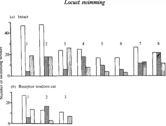
Both hind legs normally participate in swimming: if only one hind leg is used repeatedly, the locust moves in a curve, there being no compensatory movements by the other legs. The two hind legs may move together, they may alternate or one leg may sometimes be used repeatedly for a few cycles (Figs. 1, 3). In 482 swimming strokes of eight locusts, 261 involved the use of one leg (114 the left, 107 the right) (Fig. 2a). In all strong swimming sequences the two hind legs were used together.
Only a few locusts consistently move their middle legs in swimming. When they do, the femoral tibial joint remains extended at about 1700 with, perhaps, a slight flexion during protraction. The whole limb is thus protracted and retracted by a slight rotation of the coxa about the thorax, so that it acts like an oar (Fig. 3). Both middle legs usually move together but can provide little in the way of thrust. Their role is presumably to provide lateral stability to the body during the powerful thrusting of the hind legs.
(a) Intact
40-
20-1
I
Q.
1
I
(/;) Receptor tendons cut I 2 3
20-Fig. 2. Modes of co-ordination of the hind legs of Schistocerca during swimming. The open bars indicate that the two hind legs move together, the hatched bars that the left hind leg moves alone, and the stippled bars the right hind leg. (a) Eight intact locusts, in six of which there is a preference for the use of both hind legs together. (6) The receptor tendons of the chordotonal organs in both hind legs were cut but there is no effect upon the co-ordination.
A locust that is swimming by the use of all its legs moves them in a sequence that begins with the protraction of the front legs and ends with protraction of the hind legs. The onset of protraction in the two hind legs may vary but there is no consistent tendency for one leg to lead the other. The sequence of leg movements is quite different from that in walking where legs of one segment alternate, thereby setting up the typical alternating tripod gait. In swimming the legs of a segment usually move together.
[image:5.451.66.397.73.325.2]492
Fig. 3. Swimming in Schistocerca viewed from the side. This locust swam completely below the surface of the water which lessened the diffraction caused by the movements and thus allowed easier viewing of the individual legs. The tracings were selected from film taken at 500 frames/s and show two cycles of swimming lasting a total of 1180 ms. The frames are arranged in two vertical columns and the numbers indicate the time in ms. between each. The arrow indicates the direction of swimming.
The motor pattern of the swimming stroke
Flexor
4U(^^^
Flex. Extend
[image:7.451.93.372.59.339.2]> Extensor Extensor
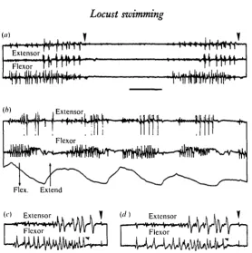
Fig. 4. Electrical activity in the extensor and flexor tibiae muscles of a hind leg during swim-ming, (a) Free swimming: flexor activity starts first (lower trace) to be followed by the slow extensor (upper trace) and then the fast extensor (middle trace). (6) Tethered swimming, in which the movements of the tibia are also monitored, (c, d) Expanded portions of two swim-ming cycles to show the cessation of flexor activity (small arrows) before the end of extensor activity and the commencement of the movement (vertical arrows). Time scale (a, 6) 200 ms;
(c, d) 100 ms.
begin to spike, so that there is a period when the flexor and extensor motoneurones are spiking at the same time. The flexor motoneurones then stop spiking but the extensor motoneurones continue, and rapid extension of the tibia follows.
Flexor muscle
Extensor muscle
There are only two excitatory motoneurones innervating the extensor muscle, but there is such an enormous discrepancy in the size of their synaptic potentials as recorded extracellularly that the large (from the fast motoneurone) usually swamps the small (from the slow). The SETi starts to spike first but the number of its spikes during one swimming cycle is difficult to determine. The better recordings reveal at least six spikes before they are masked by spikes of the FETi (Fig. 4a). In tethered swimming the FETi motoneurone usually spikes four times (s.E. = + 1-5, range 1-8, n = 219) per stroke (Fig. 4b). In some free swimming sequences the number of FETi spikes is often higher (Fig. 4a). The mean duration of the burst of FETi spikes is 106-7 i 5^4 ms (n = 292). The first FETi spike occurs about 90 ms (average 92-1 + 51-9 ms, n = 95) after the first large flexor spike. The average frequency attained during the burst is 33 Hz and the highest 52-6 Hz. The average interval is 30-1 ±9-7 ms (n = 57). Occasionally there may be an acceleration in the frequency throughout the burst but usually comparison of the first with the last interval reveals no significant difference.
In most swimming strokes, the last FETi spike occurs before the movement has been initiated, but in some it may occur after the movement (Fig. 46). In recordings where the movement transducer was not used, the extension is marked by a movement artifact on the recordings from the muscles. The average time difference between the last FETi spike and the movement is 24-1 + 34-8 ms (n = 129).
Motor activity of both hind legs
In each leg the number of FETi spikes is different even when both legs are moved at about the same time (Fig. 5 a). The bursts of spikes may occur at the same time or there may be a considerable time difference, so that the asynchrony of the resulting tibial movements is visible to the eye (Fig. 5 c). Occasionally one leg does not move and there is no electrical activity in the muscles (Fig. 56, c). We have not recorded abortive activity in the muscles during swimming like that known to occur during jumping (Godden, 1975; Heitler & Burrows, 1977a). At other times a single FETi spike is recorded in one leg accompanying a full burst in the other (Fig. $b). The leg is then slowly and weakly extended from an already partially extended position.
In the majority of swimming strokes the speed of flexion is the same on the two sides. Sometimes, and usually at the start of a swimming sequence, or during sporadic strokes, there may be a very slow flexion of one tibia and a more rapid flexion of the other which is followed by extension of both tibiae at about the same time (Fig. 5 d).
DISCUSSION
L. FETi R. FETi
L. FETi
R. FETi
L. ext.
T
-mv^-t
LUiJ-L. flex. R. flex. R. cxi.
L, ext. L. flex. R. flex.
[image:9.451.100.368.52.330.2]R, ext. 200 ms
Fig. s. The electrical activity in the femora of both hind legs during swimming, (a) The bursts of FETi spikes in the left and right legs are similar and the legs move at about the same time.
(b) The legs first move together and then the left leg moves alone. This is followed by a weak
extension of the right leg and a rapid movement of the left leg. (c, d) Flexor and extensor muscles of both legs, (c) There is first a movement of both legs together, then the left leg alone moves, (d) A slow flexion of the left leg is accompanied by a rapid flexion of the right, whereupon both legs extend at about the same time. Arrows in (c, d) indicate tibial extension.
are few. Mantids can swim well (Miller, 1972). The grasshopper Melanoplus has a swimming pattern very similar to that of Schistocerca (Franklin et al. 1977). We have tested several species of cockroaches and two species of crickets (Pfliiger & Burrows, unpublished). Periplaneta americana, for example showed either a distinct swimming pattern in which the two legs of one segment moved together, or struggling movements that result in little forward progress. Crickets, Gryllus campestris or Gryllus domesticus by contrast, are excellent swimmers using all three pairs of legs in a stereotyped pattern.
of load upon them was a sufficient stimulus. We will show in the following paper (Pfluger & Burrows, 1978) that, after swimming, the locust grooms itself to rid its body of water.
Second, how is the locust able to breathe during swimming? Normally the pro-and meso-thoracic spiracles are above water but whether gaseous exchange through these alone is sufficient to explain the long survival in water observed by Nikol'skii (1925) seems doubtful. When all spiracles are completely submerged any explanation must involve either a role for the tracheae as a physical lung, or the synchronization of ventilation and leg movements. Since the thrust of the hind legs pushes the head and anterior part of the thorax above water at each stroke, this might allow sufficient time for the anterior spiracles to open.
Comparison of swimming, kicking and jumping
The most striking feature of swimming is the similarity in the movements of the tibiae of the hind legs with those used in kicking and jumping. From behavioural observations of leg movements, Franklin et al. (1977) concluded that there must be common features in the motor patterns underlying swimming and jumping. The available information on the three behaviours is summarized in Table 1. A revealing demonstration of the similarity in the underlying motor patterns is to record from one locust performing all three movements (Fig. 6). We describe only the typical per-formance of each behaviour. The jumps were elicited by movements of a hand in the visual field of the locust (Fig. 6a, b); the kick, from a standing position, was elicited by pricking the abdomen with a sharp pin (Fig. 6c); the swimming was tethered (Fig. 6d)-The common features are the following. First, the motoneurones used are the same: the FETi, not used in walking, is active in each; many flexor motoneurones are active, but one, identified by the amplitude of its synaptic potential in the muscle, is active in all behaviours. Second, the sequence of action of the motoneurones is the same. The flexor motoneurones are always active first ensuring that the tibia is tightly flexed about the femur. The flexion is an essential prerequisite for a jump because there could not otherwise be a co-contraction of sufficient duration to achieve the energy required. If the leg is not fully flexed, weak swimming strokes or half-hearted kicks can still occur. Preventing full flexion of the leg with a mechanical stop does not prevent swimming (Franklin et al. 1977). Next the extensor motoneurones start to spike so that there is a period of co-contraction of the flexor and extensor muscles. The flexor muscles then relax while the extensor motoneurones continue to spike overcoming the lock at the femoral tibial joint (Heitler, 1974). The energy stored during the period of co-contraction is rapidly converted into the extension of the tibia. Third, in all three behaviours the hind legs may move together but the synchronization of the two is not absolute.
Table I. Comparison of available data on jumping, kicking and swimming The data are taken from this paper unless suffixed by a letter in parentheses indicating that they are from the following papers: (a) Heitler & Burrows (1977~); (b) Godden (1975); (c) Brown (1967); (d) Bennet-Clark (1975). Swimming
..
Kicking A Jumping I A \ Flexor Extensor Flexor Extensor , > FETi SETi eof 1.5 > 6 (range: 1-8) Flexor Extensor FETi SETi 19.1 f 3.9 (a)-
70 (a) (range: 13-25) - 20 FETi SETi Number of spikesSeveral unidentified units
active Fast:
-
30 (a) Slow:-
IOO (a) Several 10-20 (b) - unidentified 17.2 f 5.9 udts active (range : I 1-26) Fast: N 60 (a) Slow:-
200 (a) 60-100 (a) 130 (0) First five spikes : 18.9 (53 k9.7) Last five splkes: 142.9 (7 f 2.9) Spike frequency (Hz). Inter-spike intervals (ms) and standard error in brackets - First five spikes: - 25.9 (38f 22.2) Last five spikes : b I I 5.4 (8.7 f 2.6)a
409.9 f 81 -5-
300 (b)5
(range : 385.2 f I 16.2 250-5 10) (range : -2
210-690)s-
3
3- h¥ Tibial
(a) Jump Extensor , , , , . , . , . . . „ ' Extension
100 ms
(h) Jump Extensor
(c) Kick Extensor
Swimming T Extensor Flexor
Fig. 6. Jumping, kicking and swimming. All recordings were made from the same locust within I h. (a, b) Jumps in response to visual stimuli, (c) A kick from the standing position in response to an abdominal prick, (d) Two cycles of tethered swimming. All records are aligned vertically at the moment of rapid tibial extension (vertical arrows). See text for detailed description.
co-contraction phase in a kick or in a jump is 500 ms, but in a swim is only 80 ms. In a kick or a jump there may be 10-20 FETi spikes which can sometimes reach a final instantaneous frequency of 200 Hz just before the movement. This is an extremely high frequency for a motoneurone that can produce a rapid and powerful muscle twitch even with a single spike. The high frequency of spikes leads to a diminution in the height of the muscle potentials, due either to antifacilitation, or movement of the wire electrodes. These high frequencies are never seen in a swim-ming locust. Typically in swimswim-ming there are 1-8 spikes which occur at a maximum frequency of 53 Hz. There is little difference in the delay between the end of flexor activity and the start of the tibial extension in any of the three behaviours. In jumping and kicking (Fig. 6 a, b, c) a motor unit is recorded in the flexor muscle at the end of the spiking of the excitatory flexor motoneurones and before the tibial movement. This unit could be an inhibitory motoneurone, for it occurs at the time that these neurones are known from intracellular recordings to be active (Heitler & Burrows, 1977a). The same electrode placement during swimming fails to give convincing evidence that this unit is active.
It should be emphasized that there is not an absolute distinction between the movements of the hind tibiae in swimming, kicking and jumping. Rather the move-ments represent a spectrum in which a powerful jump represents one extreme and a weak kick or swimming stroke the other. For example, there may be little difference in the motor pattern underlying a weak kick and a powerful swimming stroke. The results of this paper suggest that each piece of behaviour may not require a separate motor pattern, but instead may be built from pre-existing patterns used for other movements. Our contention is that swimming, jumping and kicking all result from the same basic motor pattern. The complex movements of, for example, grasshoppers during courtship (Eisner, 1973), or the eclosion movements of crickets (Carlson & Bentley, 1977) are also built from the juxtaposition of pre-existing patterns used in other movements. What we need now to determine are the mechanisms that select and modify the basic patterns for the different behaviours.
This work was supported by S.R.C. and Nuffield Foundation grants to M.B. H.-J.P. was a European Science Exchange Programme Fellow supported by a grant from the D.F.G. (Pf 128/1).
REFERENCES
BXSSLER, U. (1968). Zur Steuerung des Springens bei der Wanderheuschrecke, Schistocerca gregaria.
Kybernetik 4, 112.
BENNET-CLARK, H. C. (1975). The energetics of the jump of the locust Schistocerca gregaria. J. exp. Biol. 63> S8-83.
BROWN, R. H. J. (1967). The mechanism of locust jumping. Nature, Land. 214, 939.
CARBONELL, C. S. (1959). The external anatomy of the South American semiaquatic grasshopper
Marellia remipes Uvarov (Acridoidea, Pauliniidae). Smithson. misc. Collns 137, 61-97.
CARLSON, J. R. & BENTLEY, D. (1977). Ecdysis: neural orchestration of a complex behavioural per-formance. Science, N.Y. 195, 1006-1008.
COOTER, R. J. (1973). Flight and landing posture in hoppers of Schistocerca gregaria (Forsk). Acrida 2, 307-317.
DIRSH, V. M. (1974). Genus Schistocerca (Acridomorpha, Insecta). The Hague: Dr W. Junk, B.V. ELSNER, N. (1973). The central nervous control of courtship behaviour in the grasshopper
Gomphocerip-pus rufus L. (Orthophera: Acrididae). Neurobiology of Invertebrates, Tihany 1971, 261-287.
FRANKLIN, R., JANDER, R. & ELE, K. (1977). The coordination, mechanics and evolution of swimming by a grasshopper, Malanoplus differentialis (Orthoptera: Acrididae). jf. Kansas Ent. Soc. 50, 189-199. GODDEN, D. H. (1975). The neural basis for locust jumping. Comp. Utochem. fhysiol. 51A, 351-360. HEITLER, W. J. (1974). The locust jump: specializations of the femoral-tibial joint. J. comp. Physiol. 89,
93-104.
HEITLER, W. J. & BURROWS, M. (1977a). The locust jump. I. The motor programme. J. exp. Biol. 66,
203-219.
HEITLER, W. J. & BURROWS, M. (19776)- The locust jump. II. Neural circuits of the motor programme.
J. exp. Biol. 66, 221-241.
JANNONE, G. (1940). Relazioni fra corsi d'acqua e spostamenti delle larve e ninfe di cavallette in A.O.I.
Boll. Idrobiol. Cacc. Pesca. Afr. Orient. Ital. I, 70-76.
JOHNSTON, H. B. & BUXTON, D. R. (1949). Field observations on locusts in Eastern Africa. Anti Locust
Bull, 5, 73 pp.
KENNEDY, J. S. (1945). Observations on the mass migration of desert locust hoppers. Trans. R. ent. Soc.
Lond. 95, 247-262.
KNECHTEL, W. K. (1938). Ueber die Wanderheuschrecke in RumSnien. Bull. ent. Res. 29, 175—183. MILLER, P. L. (1972). Swimming in mantids. J. entomology. 46, 91-97.
NIKOL'SKII, V. V. (1925). The asiatic locust Locusta migratoria L. A monograph. Trudy Otd. prikl. Ent. 12 (2), 1-332. (In Russian.)
PFLUGER, H.-J. & BURROWS, M. (1978). How the locust dries itself. .7. exp. Biol. 75, 95-100.
SANDEMAN, D. C. (1968). A sensitive position measuring device for biological systems. Comp. Biochevi.
Physiol. 24, 635-638.
WICKLER, W. (1966). Freilandbeobachtungen an der Uferschrecke Tridactylus madecassus in Ostafrika.
Z. f. Tierpsychol. 23, 845-852.