J. exp. Bwl. 169, 251-254 (1992) 2 5 1 Printed in Great Britain © The Company of Biologists Limited 1992
SHORT COMMUNICATION
SARCOMERE LENGTH CHANGES DURING FISH SWIMMING
BY RICHARD L. LIEBER
1, RAJNIK RAAB
1, SERGEI KASHIN
2AND V. REGGIE EDGERTON3
1
Department of Orthopaedics, Biomedical Sciences Graduate Group and 2Scripps Institute of Oceanography, Veterans Administration Medical Center and
University of California, San Diego, CA 92161, USA and ^Department of Kinesiology, University of California, Los Angeles, CA 90024, USA
Accepted 1 April 1992
The first noninvasive measurements of muscle sarcomere length were made by laser diffraction during swimming in the glass catfish {Kryptopterus bicirri). Sarcomere length changes were recorded at a relatively constant swimming speed (approximately 3.2 body lengths s ~ \ tailbeat frequency 5-7 beats s"1) just dorsal to the vertebral column. 18 s of continuous diffraction data were obtained and yielded cyclic sarcomere length changes of only 65±10nm. Based on published fish morphology, architecture and behaviour, we suggest that these values represent white muscle cyclic sarcomere shortening and elongation during swimming powered by red muscle.
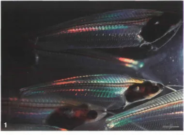
The interaction between actin and myosin represents the fundamental force-producing mechanism in all skeletal muscles. Interaction between analogous proteins also occurs within cells, resulting in movement of subcellular organelles. Although considerable detail is known about the muscle sarcomere length-tension relationship and how isometric force potential is a function of myofilament overlap (Gordon et al. 1966), there are no direct measurements of myofilament movement during locomotion. We have taken advantage of the transparent morphology of the glass catfish {Kryptopterus bicirri, Fig. 1) to investigate noninvasively sarcomere length changes which occur during free swimming.
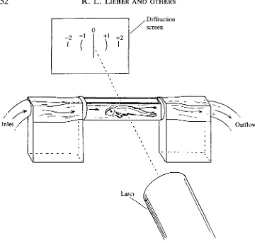
An acrylic 'fish mill' was designed allowing fish to swim against a regulated water flow (average speed 3.2 body lengths s"1) while remaining stationary relative to a laser beam (Fig. 2). (At higher flow rates, fish maintained their position by abrupt changes in swimming speed, suggesting the recruitment of white muscle. Thus, for the purposes of this investigation, we assume that the locomotion was powered by the red muscle.) The sides of the chamber were constructed of flat glass that permitted transillumination of the fish during swimming by a helium-neon laser beam. The resultant diffraction pattern was imaged onto a translucent screen and video taped at 60FIz. Off-line, the position of the first-order diffraction line was measured and- sarcomere length calculated- according -to the-grating equation:
252 R . L . LlEBER AND OTHERS
Diffraction screen
Outflow
[image:2.451.45.405.55.398.2]Laser
Fig. 2. Experimental apparatus used in this study. The fish is placed in the acrylic chamber and transilluminated by a HeNe laser. The resultant diffraction pattern is composed of different orders (0, ±1, ±2, ...). The angular position of a diffracted order is recorded and sarcomere length is calculated according to the grating equation: nX=dsind, as defined in the text.
n\=dsin9, where n is the diffraction order number (1 in this experiment), A is the
1.95
1.85
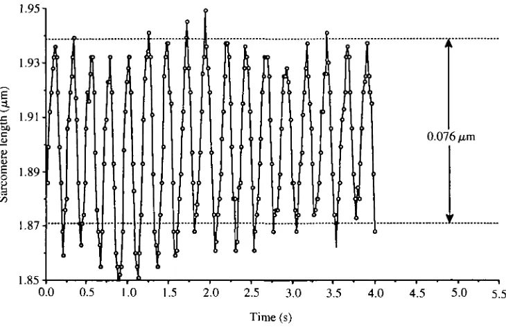
[image:5.451.39.404.78.315.2]Sarcomere changes during swimming
253Fig. 3. Sarcomere length during 4s of uninterrupted measurement. Each data point represents a single diffraction pattern analysis. Sarcomere length change frequency was 4.19±0.04Hz, while tailbeat frequency was 4.20±0.03Hz. Average sarcomere excursion was 76±4nm.
Sarcomere length excursion during swimming was always less than 0.1 (Fig. 3). Assuming that the actin and myosin filament lengths were identical to those found by Sosnicki et al. (1991) in carp (0.99/zm and 1.54 ^m respectively) and thus that the maximum sarcomere length was 3.52(xm while minimum sarcomere length was 1.54[im, sarcomeres shortened by only 3.3% of their potential range. Average sarcomere excursion was 65±10nm (mean±s.D., N=9). Average minimum sarcomere length (1.829±0.021/im; mean±s.E., N=9) was significantly less (P<0.001) than average maximum sarcomere length (1.894±0.023/an). In three fish, sarcomere length change was measured along with tailbeat frequency. These tailbeat frequencies were not significantly different from sarcomere length change frequencies (range 4-10 Hz, mean difference= 0.03±0.06Hz, P>0.8).
The relatively small sarcomere length change reported here is consistent with that provided by Rome and Sosnicki (1991) if one assumes that measurements were obtained from the white muscle during swimming powered by red muscle. Since sarcomere length changes for red muscle are four times those of white muscle for a given spinal curvature, much larger sarcomere length changes would have been expected if records had been obtained from active red muscle (Alexander, 1969; Rome et al. 1988).
254 R . L. LlEBER AND OTHERS
muscles from which sarcomere length was measured have not been definitely identified. The argument presented here is based on a synthesis of available information from the literature. It is possible that active white muscle was being recorded. Future experiments will be required to elucidate this phenomenon further. Second, there is some uncertainty as to the nature of laser light diffraction from the fish itself. The laser traverses two halves of the fish. It is, therefore, not clear precisely why the diffraction pattern obtained appears to represent only one side of the fish. Our working hypothesis is that the diffraction pattern created by the muscle closest to the laser beam is then diffracted again by the second half of the fish at relatively high angles. Empirical support for this hypothesis was obtained by diffracting the laser beam using two blocks of fish muscle embedded in plastic, one of which was fixed at a long sarcomere length (2.1 /mi) and one at a short sarcomere length (1.9/im). When the position of these blocks was inter-changed, the sarcomere length measured was that of the muscle block closest to the screen, including its fine structure. Average sarcomere length measured with the 'shorter' block closest to the screen was 1.93±0.04/an, which was significantly less than that measured with the 'longer' block closest to the screen (2.15±0.05 jum, P<0.01). A second argument against multiple diffraction is that the relative ratios of the zero: first: second order were found to be 1.0:0.05:0.01, which is similar to what would be expected from a thin grating. If significant multiple diffraction effects occurred, we would expect to see intensity 'dumping' to the higher orders, as has been demonstrated experimentally by Magnusson and Gaylord (1977).
References
ALEXANDER, R. MCN. (1969). The orientation of muscle fibers in the myomeres of fishes. J. mar. biol. Ass. 49, 263-289.
CLEWORTH, D. R. AND EDMAN, K. A. P. (1972). Changes in sarcomere length during isometric tension development in frog skeletal muscle. /. Physiol., Lond. 221, 1-17.
GORDON, A. M., HUXLEY, A. F. AND JULIAN, F. J. (1966). The variation in isometric tension
with sarcomere length in vertebrate muscle fibers. /. Physiol., Lond. 184, 170-192.
KAWAI, M. AND KUNZ, I. D. (1973). Optical diffraction studies of muscle fibers. Biophys. J. 13, 857-876.
MAGNUSSON, R. AND GAYLORD, T. K. (1977). Analysis of multiwave diffraction of thick gratings J. opt. Soc. Am. 67, 1165-1170.
ROME, L. C , FUNKE, R. P., ALEXANDER, R. MCN., LUTZ, G., ALDRIDGE, H., SCOTT, F. AND FREADMAN, M. (1988). Why animals have different muscle fiber types. Nature 335, 824-827. ROME, L. C. AND SOSNICKI, A. A. (1991). Myofilament overlap in swimming carp. II. Sarcomere
length changes during swimming. Am. J. Physiol. 260, C289-C296.
SOSNICKI, A. A., LOESSER, K. E. AND ROME, L. C. (1991). Myofilament overlap in swimming carp. I. Myofilament lengths of red and white muscle. Am. J. Physiol. 260, C283-C288. YEH, Y., BASKIN, R. J., LIEBER, R. L. AND ROOS, K. P. (1980). Theory of light diffraction by