With 9 text-figures Printed in Great Britain
ORGANIZATION OF THE GIANT AXONS OF THE
COCKROACH PERIPLANETA AMERICANA
BY M. E. SPIRA, I. PARNAS AND F. BERGMANN
Department of Zoology, the Hebrew University and Department of Pharmacology, the Hebrew University—Hadassah Medical School, Jerusalem
{Received 13 August 1968)
INTRODUCTION
Several authors have reported the presence of ascending giant fibres (AGF) in the nerve cord of the American cockroach, Periplaneta americana (Pumphrey & Rawdon-Smith, 1937; Roeder, 1948; Hess, 1958). Roeder (1948) presented a scheme showing a synaptic link (in the 6th abdominal ganglion) between the cereal sensory neurones and the AGF, the latter then ascending as continuous fibres to the meta-thoracic ganglion (T3). Part of the ascending pathways continue further and terminate at the mesothoracic ganglion (T2), while only a small fraction of the fibres reach the prothoracic (Tj) and the suboesophageal ganglion (So). Thus, in Roeder's scheme the AGF synapse at each thoracic ganglion.
Since stimulation of a single cereal nerve evoked only 6-8 spikes in the ipsilateral and 2-3 in the contralateral connectives in the abdomen, Roeder (1948) claimed that the number of spikes elicited by a cereal stimulus is smaller than the number of giant axons (usually 8—10) in the abdominal cord. On this basis he proposed that some of these axons may not be activated by cereal stimulation and might conduct descending information.
Hess (1958) found that sectioning of the nerve cord between the 5th and 6th abdominal ganglia resulted in degeneration of all the dorsal and most of the ventral giant axons in the abdomen, anteriorly to the point of severance, but two GFs of the ventral group remained intact after the operation. He therefore suggested that these two axons might serve as a descending or even as a common descending-ascending route. This hypothesis has not been supported so far by experimental evidence. Hess found no sign of GF degeneration in the thorax and thus confirmed the scheme of Roeder (1948). Pipa, Cook & Richards (1959), however, traced the ventral giant group throughout the thorax, but did not exclude the possibility of a polysnaptic chain (Bullock & Horridge, 1965).
In the present study an effort was made to demonstrate the existence of giant descending pathways. On the basis of the observations to be reported, the possibility of 'bi-directional conduction' will be considered.
MATERIALS AND METHODS
616 M. E. SPIRA, I. PARNAS AND F. BERGMANN
attached to the ventral body wall. By careful dissection, the nerve cord was freed from cerci to head and mounted on wax in a small moist Lucite chamber. Pairs of fine copper-hook electrodes, insulated except at the tips, were inserted between the ganglia as illustrated in Fig. i. By means of a switching device (SB), each electrode pair could be used either for stimulation or for recording. The nerve was stimulated with short square pulses (0-05 msec.) and the responses were recorded—directly or via a low-level Tektronix 122 a pre-amplifier—on a Tektronix 502 A cathode-ray oscilloscope (CRO).
The preparation was moistened with the bathing solution described by Yamasaki & Narahashi (i960), which contained Na+ 159-6 mM; K+ 3-1 HIM; Ca+ i-8mM;
[image:2.474.105.378.231.376.2]Cl- 160-1 mM; HjjPOi" 0 2 mM and HPOj" i-8 mM. Nicotine sulphate (Hopkins and Williams Ltd.) was dissolved in this solution when applied to the whole preparation.
Fig. 1. Experimental arrangement, showing placement of electrodes on the connectives. Suboesophageal ganglion So, thoracic ganglia Tb T,, T,, abdominal ganglia At-A*. CN, Cereal
nerves. Connectives are designated by the symbols of the two adjacent ganglia. Electrode pairs, inserted at different levels of the cord, served either for stimulation or recording by use of a switch (SB). CRO, Cathode-ray oscilloscope.
RESULTS
Conduction of descending impulses
The two connectives SQ-TJ^ were stimulated together but the evoked potentials were recorded from only a single connective at Ag-Ag. Three groups of responses could be distinguished. The group of lowest threshold was characterized by small spikes with a conduction velocity of 2-4 m./sec. (group I in Fig. 2A). With gradual increase of the stimulus strength a second group of spikes appeared with a sharply defined threshold. This group was composed of no more than two spikes and showed the highest amplitude and conduction velocity (6-7 m./sec.) (group I I in Fig. 2B, C). A further increase of the stimulus strength resulted in the gradual appearance of a third group, composed of several spikes propagating at 4-5 m./sec. (group III in Fig. 2C-F). Because of their large amplitude and high conduction velocity, groups II and I I I are assumed to arise from giant axons.
at Ag-Afl. On plotting conduction time versus distance a linear relation was obtained for abdominal conduction (5-7 m./sec. for groups II and III). These results are in accordance with the earlier observations of Roeder (1948) for ascending conduction in abdominal GFs. By contrast, delays were noted in the thoracic ganglia. The average delay for descending impulses was 0-67 msec, per thoracic ganglion, a value much lower than that reported for the delay of ascending impulses at Ag ( I - I - 6 msec.: Roeder, 1948; Yamasaki & Narahashi, i960).
1
f "ff rtfflw
[image:3.471.138.333.165.329.2]'I I
Fig. 2. Descending responses evoked by graded bilatenal stimulation at So-T^ Recording electrodes unilaterally at Aj-A,. A, Lowest threshold group I composed of slow-conducting fibres. B-E, With a gradual increase in stimulus strength, groups II and III appear in succession; the number of spikes increases to a maximum in E. Scale: 10 msec., 0-25 mV.
Are the descending fibres of the two sides inter-connected?
In order to establish whether connexions exist between the GFs of the two sides, either the left or right connective SQ-TJ was stimulated and the responses were re-corded separately from each of the abdominal connectives AB-Aa. The descending giant (DG) impulses (group II and III) propagated only ipsilaterally (Fig. 3). In addition, small potentials appeared also at the contralateral electrodes. Since these small potentials were simultaneous with or even preceded the earliest ipsilateral responses, they probably represent electrical pick-up by the contralateral electrodes. The use of a wet preparation increases the likelihood of electrotonic spread, facilitating pick-up.
When a left and right stimulus was applied simultaneously again no interference with the contralateral potentials could be recognized (Fig. 3 C). The same result was also obtained with various intervals between the two stimuli. These observations exclude the possibility of either inhibitory or excitatory collaterals between the GFs of the two sides.
Ascending impulses
618 M. E. SPIRA, I. PARNAS AND F. BERGMANN
of the nerve cord the conduction velocity was again constant, while the average delay at each thoracic ganglion for ascending GFs was o-6 msec, a value similar to the figure reported above for the descending giant potentials.
Fig. 3. Ipsilateral and contralateral responses. Either the left (A) or the right (B) connective So-Tt was stimulated and the responses were recorded simultaneously at the left (L) and right (R) connective Aj-A,. Note that a giant response is observed only in the ipsilateral connective. (C) Stimulation of both connectives did not cause a change in the recorded impulses. Scale: 10 msec, 0-5 mV.
CN
S.-T,
T , - T ,
[image:4.470.104.371.116.305.2] [image:4.470.95.374.374.523.2]
«-*•-+J *|
»fr T,-A,
Fig. 4. Effect of nicotine on gangliomc conduction. Recording from A6-A». Stimulation at
Effect of nicotine
It has been reported that the response to stimulation of the cereal nerve recorded at Ag-Afl was blocked by 1 mg./ml. nicotine, while higher concentrations were required to block the AG potentials above the metathoracic ganglion (Roeder, 1948). In our experiments we were able to block the AG potentials at Ag with much lower concentra-tions (5/4g./ml.). The same concentration blocked also the descending impulses, evoked at SQ-TJ^, Tj-Ta and T2—T3 and recorded at As-Ag. The blockade, however, did not appear at all ganglia at the same time. Thus Ag was blocked first and the thoracic ganglia followed in the order cited (Fig. 4). The abdominal axons of group II and III were not blocked at all by 5 /ig./ml. and only slowly by a much higher concentration of the alkaloid (1 mg./ml.). The ganglionic delays and the block of ascending conduc-tion by nicotine were accepted by Roeder (1948) as evidence for the presence of synapses in the thoracic ganglia. If this assumption is correct, then one is led to assume that the descending impulses are propagated in separate pathways, which also form a synapse at each thoracic ganglion. However, as shown in Fig. 2, a large number of descending responses can be recorded at the abdominal connectives. On the other hand, Roeder (1948) and Hess (1958) concluded that only few (two) giant axons may subserve descending conduction. These contradictory observations suggest that some of the pathways may be shared by ascending and descending impulses. Such a possibility is incompatible with Roeder's conclusion on thoracic synapses, since synapses are generally assumed to conduct unidirectionally. This problem is dis-cussed below.
Ascending-descending interaction
The possibility that some or all of the GFs conduct bi-directionally was tested by studying the interaction of ascending and descending impulses. If the two pathways are separated, ascending and descending potentials cannot influence each other what-ever the time-interval between the opposing stimuli. Collateral connexions would be expected to produce interactions only at sharply defined regions of the axons. However, the presence of collaterals has been excluded by the experiments of Fig. 3. On the other hand, sharing of a pathway may be detected by mutual extinction at all points, if ascending and descending stimuli are properly spaced.
The experimental arrangement is shown diagrammatically at the bottom of Fig. 5 A. Stimulating electrodes were placed at the cereal nerves (Ex) and at the connective Sp-Ti (Ee). Parameters of stimulation were adjusted so as to produce only a single large spike at E2 for a descending impulse (see record 2 in Fig. 5B), i.e. a single abdominal giant fibre was activated by the descending stimulus used. Recordings were made at various positions along the nerve cord (from Ea to E6 in Fig. 5 A).
6 2 0 M. E. SPIRA, I. PARNAS AND F. BERGMANN
2t"
*r*
\
b
+-VW*
8
-*w*?—
Asc
Des.
Asc
Since in the scheme of Fig. 5 A the total conduction time from Ex to E8 is about 9 msec., interaction—if any—can be expected only for stimulation intervals equal to or shorter than this period. (The absolute refractory period was ignored as it is quite brief compared to the delays used.) This is shown by drawing broken lines parallel to F-Fl f either with a ' positive' delay less than 9 msec. (B-D on the right ordinate, where the descending stimulus follows the ascending one), or with a 'negative' delay (G-K, where stimulation at SQ-TJ precedes the stimulus at the cereal nerves). Collision of opposing stimuli will take place at Bj, Cj and so forth.
In the actual experiment (Fig. 5 B) only recordings by E2 (upper tracing) and E4 (lower tracing) are shown in order to simplify interpretation of the results. Control records were obtained by a single ascending stimulus at E1 (Fig. 5B, 1) or by a single descending shock applied to E6 (Fig. 5B, 2). It should be noted that the descending response at E4 consists of a whole complex of potentials originating from the multitude of thoracic giant axons (see the following paper). Records (a)-(k) were obtained by varying the delay between excitation of Ej and E6. In record (a) the ascending shock preceded the descending one by 12 msec. Here, both electrodes E2 and E4 registered the ascending as well as the descending impulse, as marked by point and arrow. When the delay was reduced to 9 msec., Fig. 5 A predicts that the ascending impulse will arrive at E6 just in time to suppress initiation of the descending giant potential. Accordingly in Fig. 5 B (b) the descending spike has disappeared at E2. In E4, however, only one potential, marked by a double arrow, has been reduced in size. It is assumed that this spike represents the sum of several component potentials, at least one of which has been suppressed by the ascending impulse. When the delay is reduced to 5-5 msec, line C-Cj^ in Fig. 5 A indicates that the descending pulse is extinguished rostrally to electrode E4 and consequently should not be registered by either E2 or E4. Therefore record (c) is similar to record (b).
However, the results change when the interval between the two stimuli is in the range of + 2 to — 1 msec. Intersection of curves D—D1( F—Fx and G— Gx with o—B takes place at positions between the electrodes E2 and E4 (Fig. 5A). Consequently, E2 can record only the ascending pulse and E4 only the descending one. This is borne out by Fig. 5 B (d,f, g). The reappearance of the descending potential at E4 is indicated by the return of the spike, marked by a double arrow, to its original amplitude. Similarly, the single ascending spike (marked by a dot) which has been recorded by E4 previously, is now missing (see open circle in record/andg).
For a delay of — 3 msec, the descending impulse arrives at E2 just in time to suppress the ascending signal (see curve H-Hx in Fig. 5 A). Accordingly in Fig. 5B (h) E4 registered only the descending potential and the latter now also reappeared in the upper beam (see arrow there).
Fig. 5. Ascending-descending interaction. A, Graphic representation of impulse propagation and collision points At—Kt; for further details see text. B, Ascending responses evoked at CN,
and descending evoked at So—Ti with various delays between them; the responses recorded
by E, (upper beam) and E4 (lower beam), (i) Control, ascending responses evoked at Et (CN).
(2) Control, descending response evoked by E, (at So-TJ. Delays between the ascending and descending stimuli in msec, were (a) +10, (6) + 9 , (c) +5-6, (d) +2, (/) o, (g) —1, (h) —3, (0 — s, (k) —6-5. For further details see text. • , Ascending spike recorded by E4. O, Position
of the 'missing' ascending spike. Single arrow indicates a descending spike recorded by Et.
Double arrow indicates a descending spike recorded by E4. Dotted double arrow shows position
of the 'missing' descending spike. Scale: 10 msec., 0-25 mV.
622 M. E. SPIRA, I. PAKNAS AND F. BERGMANN
Analogous results were obtained with an interval of — 5 msec, (record t). According to Fig. 5 A, intersection of line I-Ij with curve 0-B takes place caudal to electrode E2. It should be borne in mind that since only one descending spike reached the abdomen, only a change, and not a complete extinction, is expected in the ascending complex. When the delay was further increased to —6-5 msec, the descending impulse reached
Fig. 6. Number of descending spikes as a function of the delay after a preceding ascending impulse. The descending impulses were initiated at Sff-T, in column A (electrode E,), at T1- T , (Ei) in column B and at T , - T , (E4) in column C. Ascending impulses were evoked at
the cereal nerves (see inset at bottom). Recording of impulses at Aj-A, (electrode E,). Range of delays for partial to complete block were: for So—T1( 17—12 msec.; for Tt—T,, 12—8 msec.;
E2 a sufficient time before the ascending signal so that no block occurred (record k) (cf. curve K-Kx in Fig. 5 A).
These observations demonstrate clearly that interaction of ascending and descending potentials can take place at any point along the nerve cord, the exact localization de-pending on the time interval between the opposing stimuli.
However, since conduction velocity varies from one fibre to the other, the point of collision for a given stimulus interval differs for each fibre. Therefore, in the
10 8 6 2 -0 10 8 -o
| 4
I 2
0 10 8 6 4 2 0 -• t1 I.I 1 1 1
1 1
1 1
1
<
• 1
1 1 1 1
»
>• •
1 1 1 1
1 1 1 1
1 1
1 1
So-T,
1 1 1 1 1 1 1
T.-T,
i i l i i i i
T.-T,
10 12 14 16 18
msec.
[image:9.472.105.366.169.542.2]20 22 24 26
Fig. 7. Graphic representation of results of Fig. 6. Ordinate: number of large descending spikes; abscissa; time-interval between ascending and descending impulses.
experiment of Fig. 5, we are satisfied to have demonstrated the interaction of at least one ascending and descending potential. The results of Fig. 5 give clear evidence that a single AG fibre can subserve bi-directional propagation from A, to a point rostral to ganglion Tx.
It will now be shown that all abdominal giant axons ascend to SQ-TJ and conduct
624 M. E. SPIRA, I. PARNAS AND F. BERGMANN
in both directions, in contrast to the results predicted from the scheme of Roeder (1948). In the experiment of Fig. 6 the following electrodes were used (see scheme at bottom of Fig. 6): Ex for ascending stimulation at the cereal nerves, followed by descending stimulation at E6, Es and E4. A recording electrode (E2) was placed at Aj-Ag. The time interval after the ascending stimulus was shortened progressively. The tracings in Fig. 6 demonstrate that shortening the delay extinguished more and more components of the descending complex. In column A of Fig. 6, electrode E6 served for descending stimulation. Here partial block was already apparent with a delay of about 17 msec, (second tracing), while complete block of all descending giants was achieved after reduction of the delay to 12-00 msec, (bottom tracing). In column B (stimulation at E6), no marked change in the recorded potentials was discernible in the first three tracings. Beginning with a delay of 11 msec. (4th tracing), spike number and amplitude decreased, and with an interval of 8 msec, complete block was reached (bottom record). In column C (stimulation at E4), a pronounced decrease in descending giant potentials became manifest in the 6th tracing, and complete extinction was attained at a delay of 7 msec, (see bottom line of column C). In column C, the number of the descending spikes is smaller than in A or B; their amplitude, however, is higher. It should be borne in mind that the distance between electrodes E4 and E2 is shorter than, for example, between E2 and E6; therefore spreading of the descending spikes is more restricted in column C than in A or B.
In Fig. 6 only a few representative records have been selected for each sequence. In Fig. 7 all results obtained in this series have been summarized by plotting the number of spikes, recorded at E2, as a function of the interval between ascending and descending stimuli. Three features are noteworthy: (a) the maximal delay, which can still produce complete block, decreases with diminishing electrode distance; (b) in all three sets of points in Fig. 7 the effect progresses from partial to complete block, when the stimulus interval decreases; (c) the range within which this occurs is 17-12-5 msec, for E6 and 11-5-8 msec, for Es; for E4 a complete block occurred at 7 msec., i.e. the range was shortened with decreasing electrode distance.
The experiments of Figs. 6 and 7 thus demonstrate that the two spike complexes can block each other all along the nerve cord and that the pathway is shared by all ascending and descending giant potentials from Ag to S^T^
DISCUSSION
The potentials evoked by stimulation of the connective SQ-TJ^ are recorded by abdominal electrodes. The response to unilateral ascending or descending stimulation is composed of a group of spikes (up to 8) with an average conduction velocity of 5-7 msec, for fibres of groups II and III, a value typical for insect giant axons (Roeder, 1948; Boistel & Coraboeuf, 1954; Boistel, i960; Narahashi, 1963; Pumphrey & Rawdon-Smith, 1937; Roeder, 1963; Roeder, Kennedy & Samson, 1947).
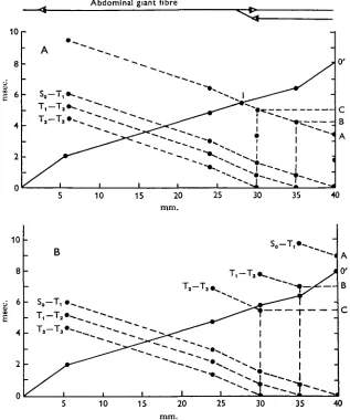
The significance of this conclusion will become clear if we analyse various hypo-thetical situations. For instance, in Fig. 8 (inset at top) we have assumed that only the abdominal part of the pathway is shared by ascending and descending impulses. In such a case, interaction would be possible only in the abdominal cord; once the ascending spikes have reached the synapse at T3, they cannot influence the descending
Abdominal giant fibre
[image:11.470.78.395.145.525.2]10 15 20 25 30 35 40
Fig. 8. Analysis of interaction of descending and ascending responses in a model shown diagrammatically above the graph. A, In this model only the abdominal section of the ascending and descending pathways is common. From T, and rostrally the two pathways are separated and conduct either ascending or descending responses alone. The theoretical analysis of such a case is demonstrated in Fig. 9 A. It is shown that above a delay of 5-5 msec, (point C) no inter-action between ascending and descending impulses should occur. Furthermore, the maximal delay (less than 5-5 msec.) for block of descending responses initiated at So-T,, T j - T , and Tt
626 M. E. SPIRA, I. PARNAS AND F. BERGMANN
impulses any longer. Figure 8 analyses such a model. Line o-o' shows the propagation of the ascending impulse, as established experimentally. The propagation of the descending impulse evoked at So-T1; Tj-T2 or T2-T3 is demonstrated by three broken lines, and these again were established experimentally. According to this model, if a stimulus is applied to S^T]^ with a delay of 3-4 msec., the potential will descend along line A-I to meet the ascending impulse just below T3. A longer delay will prevent any interaction between the two signals because the descending signal will arrive at T8 after the ascending one has passed. In the same way, lines Tx-T2 and T2-T3 have to be shifted into position A-I to determine the maximal delay per-mitting complete block of an ascending impulse. The theoretical values found are 4-2 msec, for stimulation at T ^ T j and 5 msec, for T2-T8 (Fig. 8 A, points A, B and
Fig. 9. Schematic representation of three models, suggested by the experimental findings. A, The ascending pathway is continuous from A, to the suboesophageal ganglion, tapering towards the head, with ' necks' at each ganglion. B, Ascending/descending pathway with double synaptic connections at each of the thoracic ganglia. C, The continuous pathway is interrupted by septa. For further details see text.
C, respectively), i.e. the delay decreases with increasing electrode distance. However, the experimental data presented in Fig. 8B show just the opposite, i.e. the maximal delay capable of producing complete block of the descending response increases with increasing electrode distance. The actual figures for the maximal delay were as follows: S0-Tx, 9 msec; T ^ T j , 7 msec; T2-TB, 5-5 msec. Essentially the same results were obtained in the experiments described in Figs. 6 and 7. These observations show clearly that collision can take place anywhere along the thoracic connectives, thus implying a common route all along the nerve cord.
Our experimental findings can be explained by various hypothetical schemes (Fig. 9A-C).
(A) Continuous fibres ascend from A^ to the suboesophageal ganglion, but taper towards the head (Fig. 9A).
(B) The common pathway is interrupted at each thoracic ganglion by axo-axonal synapses enabling bi-directional conduction (Fig. 9 B).
(C) The common pathway is interrupted by septa or other elements of low safety factor arranged in series (Fig. 9C).
into the thorax. The model in Fig. 9B, on the other hand, would be in agreement with the findings of Hess. Regarding the model in Fig. 9C, septate giant axons are well known in arthropods. According to this scheme a pathway may be physiologically continuous, but cutting at any point can cause degeneration only as far as the nearest septum.
The effect of small doses of nicotine may be due either to synaptic blockade (model B in Fig. 9) or to an action on thin fibres (all models of Fig. 9). Giant fibres of large diameter can be blocked only by high concentrations of the alkaloid, but the narrow parts may be more sensitive. At this point one should recall the sequence of the block by nicotine. First, the synapses at Ag were obstructed; subsequently the thoracic responses followed in the order SQ-T^ Tx-T2, Ta-T3. Evidently this observation is hard to reconcile with synaptic schemes, but may find a satisfactory explanation if indeed nicotine blocks thoracic conduction at the narrow parts of the giant axons. We shall return to this problem in the following paper.
SUMMARY
1. Stimulation of the connectives between the suboesophageal and prothoracic ganglia of the American cockroach induced ipsilateral descending spikes in the abdominal giant axons with an average delay of o-6 msec, per thoracic ganglion.
2. Nicotine at 5 /ig./ml. had no effect on conduction in the abdomen but blocked ascending responses sequentially at the 6th abdominal ganglion then at the levels of T1; T2, and T8.
3. Simultaneous descending and ascending impulses resulted in mutual extinction along the nerve cord with the point of collision depending on the interval between stimuli.
4. It is suggested that a common pathway subserves ascending and descending giant impulses and models for bi-directional conduction are discussed.
REFERENCES
BOISTEL, J. (i960). CaracUristupus fonctionneUes de fibres nerveuses et des recepteurs tactile* et olfactifs
des insectes. Pans: Libraine Arnette. 147 pp.
BOISTEL, J. & CORABOEUF, E. (1954). Potentiel de membrane et potentiels d'action de nerf d'insecte recueillis a l'aide de microelectrode intracellulaires. C. r. hebd. Sianc. Acad. Sci., Paris 338, 2116-18. BULLOCK, T. H. & HORRIDGE, G. A. (1965). Structure and Function in the Nervous Systems of
Inverte-brates. W. H. Freeman and Co., San Francisco and London: p. 1200 (Giant Fibers), p. 139 (Safety Factor).
HESS, A. (1958). Experimental anatomical studies of pathways in the severed central nerve cord of the cockroach. J. Morph. 103, 479-502.
PIPA, R. L., COOK, E. F. & RICHARDS, A. G. (1959). Studies on the hexapod nervous system. II. The
histology of the thoracic ganglia of the adult cockroach, Penplaneta amencana (L). J. comp. Neurol. 113,
4°i-23-PUMPHREY, R. J. & RAWDON-SMITH, A. F. (1937). Synaptic transmission of nerve impulses through the last abdominal ganglion of the cockroach. Proc. R. Soc. B iaa, 106-18.
ROEDER, K. D. (1948). Organization of the ascending giant fiber system in the cockroach Penplaneta
americana. J. exp. Biol. 108, 243—62.
ROEDER, K. D. (1963). Nerve Cells and Insect Behaviour. Cambridge, Mass. Harvard University Press.
ROEDER, K. D., KENNEDY, N. K. & SAMSON, E. A. (1947). Synaptic conduction to giant fibers of the
cockroach and the action of antichohnesterases. J. Neurophysiol. 10, 1—10.
NARAHASHI, T. (1963). The properties of insect axons. In Advances in Insect Physiology, pp. 175-256. Ed. J. W. L. Beament, J. E. Treherne and V. B. Wigglesworth. London and New York: Academic Press.