1015
MS
CNS Involvement in Neuro-Behc¸et Syndrome:
An MR Study
Naci Koc¸er, Civan Islak, Aksel Siva, Sabahattin Saip, Canan Akman, Orhun Kantarci, and Vedat Hamuryudan
BACKGROUND AND PURPOSE: Behc¸et disease (BD) is a multisystem vasculitis of unknown origin in which neurologic involvement has been reported in the range of 5% to 10% in large series. Reports on clinical and radiologic aspects of neuro-Behc¸et syndrome (NBS) are in gen-eral limited in number. Our purpose was to determine the MR patterns in patients with NBS who had neural parenchymal involvement and to correlate our findings with possible vascular pathophysiology.
METHODS: The MR images of 65 patients with NBS and neural parenchymal involvement were reviewed. In a subgroup of patients who had serial MR studies, we evaluated the ana-tomic-radiologic location and distribution of the lesions and whether they corresponded to any vascular territory, and studied their extension, enhancement patterns, and temporal course.
RESULTS: The most common imaging finding in NBS patients who had neural parenchymal involvement was a mesodiencephalic junction lesion with edema extending along certain long tracts in the brain stem and diencephalon in 46% of the patients. The next most common location of involvement was the pontobulbar region, seen in 40% of the cases. Three primary cervical spinal cord lesions and one case of isolated optic nerve involvement were observed.
CONCLUSION: The parenchymal distribution of lesions in NBS appears to support the hypothesis of small-vessel vasculitis; mainly, venular involvement. The anatomic distribution of intraaxial veins of the CNS explains the predominant involvement of the brain stem struc-tures observed in our patients. This pattern of lesion distribution might help to differentiate NBS from other vasculitides as well as from the inflammatory-demyelinating diseases of the CNS, such as multiple sclerosis.
Behc¸et’s disease (BD) is a multisystem vasculitis of unknown origin. The classical triad of oral and genital ulcerations with uveitis was originally de-scribed by a Turkish dermatologist Hulusi Behc¸et in 1937 (1). Other structures reported to be in-volved through the course of the disease are the cardiovascular, CNS, pulmonary, and gastrointes-tinal systems (2–4).
Early neuroimaging reports on neuro-Behc¸et syndrome (NBS) were based on studies done with either CT or, rarely, cerebral angiography. To our knowledge, the largest series in which neuroimag-ing findneuroimag-ings were based on CT studies were those reported by Inaba (5), in which 63 patients were
Received August 27, 1998; accepted after revision February 8, 1999.
From the Departments of Radiology (N.K., C.I., C.A.), Neu-rology (A.S., S.S., O.K.), and Internal Medicine (V.H.), Cer-rahpasa School of Medicine, Istanbul, Turkey.
Address reprint requests to Naci Koc¸er, MD, Department of Radiology, Cerrahpasa School of Medicine, 34300 Cerrahpasa Istanbul, Turkey.
qAmerican Society of Neuroradiology
examined, and by Siva et al (6), who described the findings in 42 patients with NBS examined at this institution. With the introduction of MR imaging in clinical practice, articles reporting the MR findings in NBS have appeared; however, most of these are limited to either single cases or to relatively small series (7–11). The most common CNS findings re-ported in these studies, including ours, were a pref-erence for brain stem–diencephalic involvement and a tendency to resolve over time. Cerebral ve-nous thrombosis is another common neuroimaging finding reported in NBS (12).
In this study, we included only NBS patients who had CNS parenchymal involvement on MR imaging studies in order to define the MR patterns of this form of the disease and to correlate those findings with the results of vascular anatomic-pathologic investigation.
Methods
AJNR: 20, June/July 1999 1016 KOC¸ ER
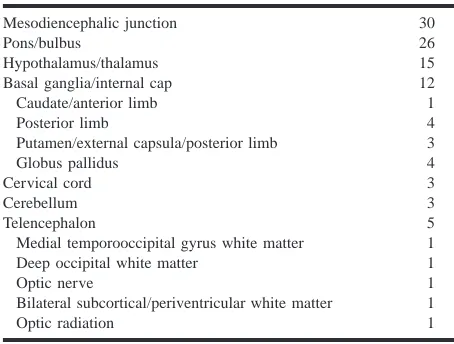
Table 1: Lesion distribution in 65 patients with neuro-Behc¸et syn-drome
Mesodiencephalic junction Pons/bulbus
Hypothalamus/thalamus Basal ganglia/internal cap
30 26 15 12 Caudate/anterior limb Posterior limb
Putamen/external capsula/posterior limb Globus pallidus 1 4 3 4 Cervical cord Cerebellum Telencephalon 3 3 5 Medial temporooccipital gyrus white matter
Deep occipital white matter Optic nerve
Bilateral subcortical/periventricular white matter Optic radiation 1 1 1 1 1 from the Behc¸et’s Disease Research Center of our university
hospital. Criteria established by the International Study Group for Behc¸et Disease (13) were used for diagnosis. Neurologic involvement was evaluated and defined by the neurology con-sultants of the Research Center from the department of neu-rology, who referred the patients for MR examination. Sixty-three patients had fulfilled the criteria for BD before the onset of neurologic involvement and two at the time of neurologic involvement. The study period ranged from January 1, 1990, to December 31, 1997.
The patients included 18 women and 47 men, ages 19 to 51 years (mean age, 36 years). Patients with normal MR findings (21 cases) and with isolated dural sinus thrombosis (13 cases) were excluded from the study, because our primary goal was to evaluate parenchymal involvement. In 48 cases (74%), a 1.0-T MR imager was used, whereas the remaining patients were studied with different MR systems, ranging from 0.5 T to 1.5 T, as some patients had been examined prior to the time of consultation. The imaging was assumed to have been done in the acute stage if the patient was studied within a week of the onset of the neurologic episode or subacute if within a month. MR studies consisted of sagittal and axial T1-weighted spin-echo (SE) sequences and coronal and axial proton den-sity– or T2-weighted fast SE sequences with 3- to 6-mm con-tiguous sections over the whole brain. Additional cervical spi-nal cord examinations with sagittal T1- and T2-weighted SE and axial T2-weighted fast SE sequences were performed in three patients, because their clinical picture suggested cervical spinal involvement. A section thickness of 3 mm was used for spinal cord examinations. Contrast material was used in 11 cases. Twenty-three patients underwent follow-up MR exami-nations (more than one follow-up in eight cases).
The distribution of the lesions and whether they correspond-ed to any vascular territory, their extension and enhancement patterns, as well as the temporal course in the subgroup of 23 patients with serial MR examinations were studied and classified.
Results
The mean age of onset of neurologic symptoms was 26.867.6 (SD) in patients with BD and 30.2 68.5 for those with NBS. The male to female ratio was 2.6 to 1.0, showing a clear male preponder-ance. Fifty-two (80%) of the patients were exam-ined in the acute/subacute phase and 13 (20%) in the chronic phase. Six patients (9%) had a primary-progressive course and nine (14%) had converted to a secondary-progressive course at the time of their MR study. The remaining patients, who had either a single attack or a relapsing-remitting form of the disease (77%), were studied during such an episode. Symptoms at onset in the order of fre-quency were headache, 85%; weakness, 57%; brain stem and/or cerebellar involvement, 50%; cognitive and behavioral disorders, 16%; and disorders of consciousness, 7%.
Distribution of Lesions
A total of 94 lesions were identified in the 65 patients. The most commonly affected region was the mesodiencephalic junction (MDJ), seen in 30 patients (46%), followed by the pontobulbar region in 26 (40%), the hypothalamic-thalamic region in 15 (23%), the basal ganglia in 12, the telencephalon in five, the cerebellum in three, and the cervical
cord in three (Table 1). There was no difference in the distribution of lesions between the acute/sub-acute or chronic phases.
Of the 30 MDJ lesions, 11 showed a marked upward extension involving the diencephalic struc-tures and 18 had a prominent downward extension involving the pontobulbar region. In six, the exten-sion was both upward and downward (Fig 1). In a single case, studied in the chronic stage, the MDJ lesion was isolated, suggestive of a sequela.
Of the 26 lesions with pontine and bulbar in-volvement, three had no association with other le-sions (Fig 2). Twelve were associated with teg-mental and superior cerebellar peduncular extension, and six showed corticospinal tract in-volvement in continuity with an MDJ lesion. In one patient with additional telencephalic lesions, the right side of the pons was hyperintense on long TR/ TE images that did not cross the midline, sugges-tive of an arterial lesion (Fig 3). In two patients with pontine tegmental lesions, there was an asso-ciated middle cerebellar peduncle and deep cere-bellar white matter involvement (Fig 4). Two others had associated cervical lesions.
Cervical cord lesions were seen in three patients. They were predominantly located in the postero-lateral part of the cord, continuing over at least two vertebral segments, with a tendency to reach the inferior cerebellar peduncles superiorly (Fig 5). One of them also had concomitant MDJ and pon-tobulbar region lesions.
FIG1. A, Coronal T2-weighted image (4000/90/2 [TR/TE/excitations]) at the level of the crus cerebri nicely shows heterogeneous left
MDJ lesion with extensive edema, sparing the red nucleus.
B, Coronal noncontrast T1-weighted image (300/15/3) at the same level as A reveals a hemorrhagic focus in the lesion.
C, Coronal T2-weighted image (4000/90/2) posterior to A shows extension of perilesional edema caudally to the superior cerebellar peduncle and pontine tegmentum, and upward to the white matter of the temporal lobe, external capsule, and thalamus.
D, Caudal extension of the edema toward the pontine tegmentum is seen on axial T2-weighted image (4000/90/1).
E, Two years later, after another relapse of the disease, coronal T2-weighted image (4000/90/1) reveals a contralateral MDJ lesion. The left-sided lesion now has shrunk to a small hypointense area.
F, Contrast-enhanced T1-weighted image (660/17/1) shows enhancement of the new right MDJ lesion.
G and H, Similar extension of edema as observed in C and D is seen in the right mesencephalopontine region of the brain stem on T2-weighted images (4000/90/1).
Telencephalic lesions were seen in only five pa-tients (8%) and had a wide asymmetric distribution within the brain, involving the optic nerves and the subcortical and deep periventricular white matter (Fig 6). In the patient with optic neuropathy, the only finding was right optic nerve enhancement. In the other four patients, the hemispheric lesions were located in the subcortical and deep
AJNR: 20, June/July 1999 1018 KOC¸ ER
FIG 2. Axial T2-weighted image (2400/
110/2) shows inhomogeneous hyperin-tense lesion located in the mid pons.
FIG 3. Axial T2-weighted image (2400/
110/2) at the pontine level shows a right-sided pontine lesion that does not cross the midline.
FIG4. A and B, Axial (A) and coronal (B) T2-weighted images (4000/90/2) reveal hyperintense lesions bilaterally in the mid-dle cerebellar peduncles and deep cere-bellar white matter.
FIG5. A–C, Midsagittal cervical lateral T2-weighted image (2200/80/1) (A) and axial T2-weighted image (4000/90/2) through the cervical medullary junction (B) show posteriorly located paracentral hyperintense lesion with mild cervicomedullary enlargement. The pattern of extension up to the inferior cerebellar peduncle suggests involvement of the dorsal columns. Midsagittal cervical lateral T1-weighted image (300/15/3) (C) shows slight enlargement of cervical cord.
Characteristics of the Lesions
The lesions observed during an acute/subacute episode were surrounded by a more hyperintense rostral and caudal extension on long-TR sequences, which was directly related to the location of the lesion. When located posteriorly at the MDJ,
FIG6. A, Axial T2-weighted image (3500/
90/2) shows a well-defined deep right oc-cipital white matter lesion (asterisk) and a subcortical linear hyperintensity (arrow).
B, Coronal T2-weighted image (3500/90/ 1) in a different patient shows multiple sub-cortical white matter and right MDJ and pontine lesions (asterisk and arrowheads).
FIG7. A, T2-weighted image (4000/90/2) shows chronic left MDJ lesion (double ar-rowhead) and ipsilateral lenticulostriatal le-sions (arrow).
[image:5.612.60.288.450.543.2]B, Wallerian degeneration of the optic radiation is evident on paraatrial section of the same sequence (double arrowheads).
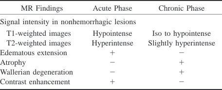
Table 2: MR findings in acute and chronic phase
MR Findings Acute Phase Chronic Phase
Signal intensity in nonhemorrhagic lesions T1-weighted images
T2-weighted images Edematous extension Atrophy
Wallerian degeneration Contrast enhancement
Hypointense Hyperintense
1 2 2 1
Iso to hypointense Slightly hyperintense
[image:5.612.311.542.451.486.2]2 1 1 2
Table 3: Distribution of hemorragic lesions
Mesodiencephalic junction Tectum
Posterior perforate substance
3 1 1
seen with the acute lesions was characterized by involvement of the posterior limb of the internal capsulae, accompanied by involvement of the glo-bus pallidus in four and the putamen and external capsule in three. In lesions of the cervical spinal cord, the bulbar involvement was through exten-sion via sensory long fiber tracts (fasciculus gracilis and cuneatus). The cranial and caudal extensions were found to disappear or decrease in size, leaving a residual lesion on follow-up examinations, as dis-cussed below. These residual lesions were iso- to hypointense on T1-weighted images and slightly hyperintense on proton density– and T2-weighted images, with or without atrophy (Table 2).
Serial MR examinations were performed in 23 cases. Of those, 18 were for follow-up purposes,
whereas in five the subsequent studies were done because of a relapse or worsening in neurologic status. These MR examinations revealed resolution or decrease in the size of the lesions, 17 of which had shrunk to small areas with nearly total disap-pearance of the perilesional extension.
Sixty patients had nonhemorrhagic lesions while five had hemorrhage (Table 3). Nonhemorrhagic le-sions showed prolonged T1 and T2 relaxation times. They were mostly heterogeneous and asym-metric. Of the five patients with hemorrhagic le-sions, three were in the subacute phase, as they appeared hyperintense on T1-, proton density–, and T2-weighted sequences, whereas the other two, which were hypointense on all sequences owing to the existence of hemorrhagic degradation products, as previously described, were in the chronic stage (14).
AJNR: 20, June/July 1999 1020 KOC¸ ER
FIG8. A, Contrast-enhanced coronal
T1-weighted image (600/15/1) shows bilateral optic nerve atrophy and marked enhance-ment of the prechiasmatic segenhance-ment of the right optic nerve (arrow).
B, Contrast enhancement of the nerve disappeared after treatment (asterisk).
enhancing lesions were located at the MDJ, corre-sponding to subacute hemorrhagic foci in three. In one case, the enhancement was confined to the right optic nerve, extending to the prechiasmal bor-der without any other parenchymal involvement (Fig 8).
Discussion
Currently, the most widely used diagnostic cri-teria of BD is the International Study Group’s clas-sification, which requires recurrent oral ulcerations plus two of the following in order to establish a definite diagnosis: recurrent genital ulcerations, skin or eye lesions, or a positive pathergy test (13). The epidemiology of disease shows geographic variation, encountered more commonly along the Silk Road, which extends from the Mediterranean region to Japan (15). This is coupled with a similar variation in HLA B51 (human leukocyte antigen), which has been reported to be strongly associated with the disease in the high prevalence areas (16– 19). Despite broadened clinical understanding of this disease, the etiologic factors remain obscured and speculative: viral agents, immunologic factors, genetic causes, bacterial factors, and fibrinolytic defects have all been implicated (3, 20–25). Vessel wall and perivascular mononuclear cell infiltration, which is consistent with vasculitis involving both arterial and venous systems, has been shown in his-topathologic studies (20, 21). It has been postulated that genetic susceptibility together with a possible trigger by an extrinsic factor, such as an infectious agent, is responsible for the observed vasculitis (24, 26).
Neurologic involvement in BD, which is not in-cluded in the current International Study Group’s classification, was first reported in 1941 by Knapp (27), and the term neuro-Behc¸et syndrome was in-troduced by Cavara and D’Ermo in 1954 (28). The reported rate of development of neurologic in-volvement among BD patients ranges from 4% to 49% (9, 29). This rate was found to be 6.7% in our large nonselective series of patients referred from the Behc¸et’s Disease Research Center (30).
NBS may present as acute focal or multifocal CNS dysfunction, and the clinical picture of NBS may resemble multiple sclerosis (MS) (7, 31–35).
It has been observed that a substantial number of patients with NBS will have a relapsing-remitting course while others may develop a secondary-pro-gressive course; some NBS patients have an insid-ious onset, with primary-progressive CNS dysfunc-tion, and others may display symptoms attributable to intracranial hypertension associated with dural venous sinus thrombosis (12, 36, 37). Although nonneurologic involvement generally precedes neu-rologic findings, the nonneuneu-rologic involvement may go unrecognized in some cases or it may ap-pear late in the patient’s course, thus posing diag-nostic difficulties (38–40). Peripheral nerve in-volvement, although reported in NBS, is relatively uncommon (41).
In our study, the most common imaging lesion seen in the acute or subacute stage of NBS was an asymmetric MDJ lesion, similar to previously pub-lished reports (7–9, 33). These lesions extended along long fiber tracts and spared the red nucleus, suggesting that this downward extension was due to edema. The reversibility of the extension, leav-ing small residual lesions at the center, as observed on follow-up MR studies, further supports their edematous nature (Fig 1). This feature was also noted in earlier publications (9, 11, 42). Accord-ingly, with the exception of the three cases of iso-lated lesions, pontobulbar involvement was an ex-tension of lesions located at other sites, particularly the MDJ. The distribution and intensity changes of the residual lesions closely corresponded to patho-logic descriptions of secondary demyelination (20, 21, 43, 44). In the chronic cases, the intensity changes extended to the cervical cord, along the corticospinal tract, and might be explained by wal-lerian degeneration (Fig 9). This was noted in some pathologic studies as well (20, 43, 44).
FIG9. A, T2-weighted image (2400/110/
2) shows round hyperintense lesion in the posterior and middle third of the right co-rona radiata (asterisk) associated with rather poorly defined periatrial T2 hyper-intensities (arrowheads).
B, Coronal T2-weighted image (4000/90/ 2) shows signal intensity changes along the right corticospinal tract, representative of secondary wallerian degeneration (dou-ble arrowheads), along with basal ganglia and pontine lesions (asterisks).
In four cases (6%), an asymmetric subcortical and deep periventricular white matter distribution without cortical involvement was observed (Fig 6). These patients also had concomitant MDJ lesions, and in one case, wallerian degeneration of the optic radiation associated with primary MDJ involve-ment was detected (Fig 7). In the majority of re-ported cases with hemispheric involvement, the le-sions were located subcortically, particularly within temporal and occipital regions (7–11, 34, 39, 45, 46). The frequency of telencephalic involvement in this study is lower than the rate reported in the literature (7, 8, 10, 44). We can find no explanation for this discrepancy, although it might be attribut-able to the regional differences of the disease’s pre-sentation, as seen with other systemic findings of BD. On the other hand, radiologic and pathologic studies might have different sensitivities, which could also account for this difference (7–10, 21, 34). Another explanation could be related to the fact that the cases reported in the pathologic series were chronic, whereas most of the patients in the present study were imaged during an acute or sub-acute stage. In most of these pathologic studies, the disease observed included primary as well as sec-ondary changes, such as wallerian degeneration, gliosis, and demyelination (4, 20, 21, 43, 44, 47, 48). There have been only a few autopsy studies performed after an acute episode, and in these the prominent lesions reported were venous thrombosis and secondary parenchymal necrosis (21, 49).
Optic neuropathy is a rare finding in NBS (50), and we had a single patient in our series in whom the only MR finding was right optic nerve enhance-ment (Fig 8).
A lesion thought to be of arterial origin was ob-served in a single patient in whom the lesion within the pons did not cross the midline, consistent with involvement of the penetrating arteries (Fig 3). Al-though such lesions, resulting from small or me-dium-sized intracranial arteries, have been reported either microscopically or radiologically in NBS, they are not as common as the arterial lesions ob-served with the involvement of other systems (20, 21, 40, 43, 47, 48, 50–52). There are also a few publications concerning the involvement of large
intracranial arteries (40, 51–53); however, no such involvement was observed in our study. The hem-orrhagic lesions seen in our patients most likely resulted from ‘‘diapedesis of red cells around veins,’’ as already reported, and were not of arterial origin (21).
Vasculitis is regarded as the key feature in BD (3, 26), as biopsy specimens from mucous and cu-taneous lesions show those changes (54, 55). Ar-terial and venous large vessel involvement, such as narrowing, occlusion, and aneurysmal formation, has been reported in up to 27% to 35% of cases, with 12% arterial and 88% venous (52). An even greater proportion of patients with BD may have small vessel vasculitis, and recently this has been validated as the pathologic basis of various histo-logic changes observed in different organ systems (26).
Autopsy studies and biopsy specimens of the CNS lesions are consistent with vasculitis as well, and they show a clear venous predominance (20, 21, 49) (Fig 10). Radiologic studies support this finding, in that lesions seen in NBS are not com-patible with arterial territories. Furthermore, signif-icant perilesional edema with a tendency to disap-pear or to leave disproportionally small residua on follow-up studies has been reported. This feature is consistent with venous infarction, since not all sig-nal intensity changes seen in venous occlusive dis-ease necessarily represent infarction, but rather an accumulation of water within interstitial spaces (56, 57). This information, together with our observa-tions, supports the probable inflammatory-venous pathogenesis for the CNS lesions seen in BD.
ad-AJNR: 20, June/July 1999 1022 KOC¸ ER
FIG10. Histologic section of NBS shows a totally thrombosed medium-sized venous vessel. Focal fibrinoid necrosis (arrowheads) and moderate amount of lymphocyte and plasma cell infiltration is visible in the vessel wall. At the right side of the vessel, necrotic brain tissue with some newly thrombosed small vessel and mononuclear inflammatory infiltration (mainly lymphocytes and histiocytes) is seen (arrows). On the left, there is severe astrogliosis with some gemistocytic differentiation (H and E, original magnification3100).
FIG11. Schematic representation of the intraaxial venous system of normal parenchyma. Supratentorially, a medullary vein (1) permits
bidirectional flow. In the brain stem, especially at the mesencephalic level, intraaxial veno-venous anastomosis (2) is sparse and venous flow is centrifugal, toward the pial veins (pv). ev indicates ependymal vein.
dressed according to regional hemodynamic prop-erties. It is well known that telencephalic structures are drained by superficial and deep venous systems, both of which anastomose via medullary veins (Fig 11). They interconnect superficial pial veins to the internal cerebral vein and the basal vein of Rosen-thal, the former being more common than the latter (59), whereas in the brain stem, intraparenchymal radial and longitudinal anastomotic channels are nearly absent (58) (Fig 11). In the spinal cord, in-traaxial anastomoses are claimed to be prominent at the thoracic level (60). The particular arrange-ment of veins in the telencephalon permits them to flow in both directions via medullary veins, as seen with certain disorders, such as deep arteriovenous malformations, Sturge-Weber disease, and the de-velopmental venous anomalies (60), possibly ex-plaining the small diameter and unimportance of parenchymal lesions when such a vein is throm-bosed. At the mesencephalic, diencephalic, and pontine levels, thrombosis of small veins might be accompanied by a very large, sometimes hemor-rhagic lesion, since there is nearly no collateral ve-nous pathway. The same anatomic arrangement might also explain the vulnerability of the cervical spinal cord (7, 44). It appears, therefore, that the variability of venous anatomic arrangements at dif-ferent levels of the CNS might explain the predi-lection of lesions for different regions.
Vasculitis, like inflammation in other tissues, is caused by many different agents and pathogenic
mechanisms; however, these different causes pro-duce only a limited number of histologic expres-sions of injury. The major type of injury to nervous tissue in vasculitis is ischemia. Therefore, the same clinical manifestations can result from etiologically and pathogenetically different vasculitic diseases. In vasculitic processes, location, extension, and dis-tribution of vascular involvement might point to a specific diagnosis, such as Takayasu or temporal arteritis (26, 61–65). In NBS, lesions therefore ap-pear secondary to the small vessel vasculitis, and the anatomy of those intraaxial venous structures explains the dominant involvement of the upper brain stem and diencephalic structures.
Pathologically proved small vessel arteritis, ei-ther alone or with venous inflammation, has also been reported in conjunction with NBS, and it is probable that some of the telencephalic and mid-brain lesions might result from small vessel arteritis (62, 63). In our study, there appeared to be at least one lesion that occurred within a probable pontine arterial territory, and the hemispheric lesions seen in NBS were not significantly different from the CNS lesions of other vasculopathies of accepted arteritic origin (Fig 3).
un-likely in systemic lupus erythematosus (SLE) and non-Behc¸et vasculitides. CNS involvement due to SLE and other systemic vasculitis tends to involve arterial territories, and as a result, cortical involve-ment is frequently observed (34, 66, 67). We have not observed cortical involvement in NBS, despite pathologic studies in which such involvement has been reported (21). These changes, however, are minor in NBS, which may explain the radiologic-pathologic discrepancy. Periventricular and ovoid lesions suggestive of MS are not expected to be seen in NBS. Extensive confluent periventricular changes that are seen in MS and occasionally in sarcoidosis were not observed in our patients with NBS. Posterior fossa lesions, particularly those lo-cated around the fourth ventricle with or without the associated supratentorial lesions seen in MS, are not similar to the NBS lesions described above. Brain stem lesions in MS are usually small, even in the acute stage, and prominent brain stem and/ or cerebellar atrophy without cerebral volume loss, which is observed in the chronic phase of NBS, is unusual in MS (7, 34). When one considers cer-vical involvement, this rarely extends more than a few vertebral segments in MS (68), unlike the more extensive lesions we observed in NBS. Leptomen-ingeal contrast enhancement is a typical finding of sarcoidosis (69). We did not encounter this finding in our series; however, contrast-enhanced studies were performed in only 11 patients. Devlin et al (38) reported abnormal leptomeningeal enhance-ment in two of their patients with NBS. Abnormal meningeal enhancement secondary to dural venous occlusion or to lumber puncture should be excluded before attributing it to the disease itself (70).
Inflammatory demyelinating diseases, such as MS, and inflammatory vascular disorders (vascu-litides), such as NBS and SLE, can affect the CNS primarily or secondarily, and onset tends to occur in young adulthood. Although the clinical presen-tation of these diseases may be similar, the radio-logic findings of NBS are quite distinct, which may help differentiate it from other disorders, even in the absence of overt systemic involvement.
Conclusion
The parenchymal distribution of lesions in NBS seems to support the hypothesis of small vessel vasculitis, mainly venular involvement. The known anatomic arrangement of CNS intraaxial veins ex-plains the predominant involvement of the brain stem structures observed in our patients. This pat-tern of lesion distribution might help to differenti-ate NBS from other vasculitides as well as from the inflammatory-demyelinating diseases of the CNS, such as MS. Our experience with NBS has caused us to consider NBS in the differential di-agnosis of patients who have brain stem and/or diencephalic lesions that extend along the long tracts and have a tendency to resolve on subsequent
imaging studies, whether or not they are associated with periventricular and subcortical lesions.
Acknowledgments
We thank Sait Albayram, Gu¨lgu¨n Atilla, Alp Dinc¸er, and Bu¨ge O¨ z for help with the manuscript.
References
1. Behc¸et H. Uber residivierende, aphto¨se, durch ein virus verur-sachte Geschwu¨re am Mund, am Auge und an den Genitalien.
Derm Woschenscr 1937;105:1152–1157
2. Yazici H, Yurdakul S, Hamuryudan V. Behc¸et’s syndrome. In: Klippel J, Dieppe P, eds. Rheumatology. London: Gower Medical; 1997;7.26:1–6
3. O’Duffy JD. Vasculitis in Behc¸et’s disease. Rheum Dis Clin North Am 1990;16:423–431
4. Lakhanpal S, Tani K, Lie JT, Katoh K, Ishigatsuba Y, Ohokubo T. Pathological features of Behc¸et’s syndrome: a review of Jap-anese autopsy registry data. Hum Pathol 1985;16:790–795 5. Inaba G. Clinical features of neuro-Behc¸et syndrome. In:
Leh-ner T, Barnes CG, eds. Recent Advances in Behc¸et’s Disease. In-ternational Congress and Symposium Series Number 103. Lon-don: Royal Society of Medicine Services; 1986:235–246 6. Siva A, Necdet V, Yurdakul S, Yardim M, Denktas¸ F, Yazici H.
Neuro-radiologic findings in neuro-Behc¸et syndrome. In: O’DuffyI D, Kokmen E, eds. Behc¸et’s Disease: Basic and Clinical Aspects. New York: Dekker; 1991;323–329
7. Morrisey SP, Miller DH, Hermaszewski R, et al. Magnetic reso-nance imaging of the central nervous system in Behc¸et’s dis-ease. Eur Neurol 1993;33:287–293
8. Wechsler B, Dell’sola B, Vidailhet M, et al. MRI in 31 patients with Behc¸et’s disease and neurological involvement: prospec-tive study with clinical correlation. J Neurol Neurosurg
Psychi-atry 1993;56:783–789
9. Al Kawi MZ, Bohlega S, Banna M. MRI findings in neuro-Behc¸et’s disease. Neurology 1991;41:405–408
10. Banna M, El-Ramahi K. Neurologic involvement in Behc¸et dis-ease: imaging findings in 16 patients. AJNR Am J Neuroradiol 1991;12:791–796
11. Fukuyama H, Kameyama M, Nebatame H, et al. Magnetic res-onance images of neuro-Behc¸et syndrome show precise brain stem lesions: report of a case. Acta Neurol Scand 1987;57:70–73 12. Wechsler B, Vidailhet N, Piette JC, et al. Cerebral venous throm-bosis in Behc¸et’s disease: clinical study and long-term follow-up of 25 cases. Neurology 1992;42:614–618
13. The International Study Group for Behc¸et’s Disease. Evaluation of diagnostic (‘‘classification’’) criteria in Behc¸et’s disease: to-wards internationally agreed criteria. Br J Rheumatol 1992;31: 299–308
14. Osborne AG. Diagnostic Neuroradiology. St Louis: Mosby; 1994: 154–197
15. Ohno S. Behc¸et’s disease in the world. In: Lehner T, Barnes CG, eds. Recent Advances in Behc¸et’s Disease. London: Royal Society of Medicine Service; 1986:181–186
16. Yazici H, Akhan G, Yalc¸in B, Mu¨ftu¨ogˇlu A. The high preva-lence of HLA-B5 in Behc¸et’s disease. Clin Exp Immunol 1977; 30:259–261
17. Yurdakul S, Gu¨naydin I, Tu¨zu¨n H, et al. The prevalence of Beh-c¸et’s syndrome in a rural area in northern Turkey. J
Rheu-matol 1988;15:820–822
18. Ohno S, Ohguchi M, Hirose S, Matsuda H, Wakisaka A, Aizava M. Close association of HLA-BW51 with Behc¸et’s disease. Arch Opthalmol 1982;100:1455–1458
19. Mizuki N, Inoko H, Ohno S. Molecular genetics (HLA) of Beh-c¸et’s disease. Yonsei Med J 1997;38:423–427
20. McMenemey WH, Lawrence BJ. Encephalomyelopathy in Beh-c¸et’s disease: report of necropsy findings in two cases. Lancet 1957;24:353–358
AJNR: 20, June/July 1999 1024 KOC¸ ER
23. Hirohata S, Tekeuchi A, Miyamoto T. Association of cerebro-spinal fluid IgM index with central nervous system involve-ment in Behc¸et’s disease. Arthritis Rheum 1986;29:793–796 24. Kansu E. Endothelial cell dysfunction in Behc¸et’s disease. In:
Ansell BM, Bacon PA, Lie JT, Yazici H, eds. Vasculitides. Lon-don: Chapman & Hall; 1996:207–221
25. Yazici H. The place of Behc¸et’s syndrome among the autoim-mune diseases. Int Rev Immunol 1997;14:1–10
26. Lie JT. Primary (granulomatous) angiitis of the central nervous system: a clinicopathologic analysis of 15 new cases and a re-view of the literature. Hum Pathol 1992;23:164–171
27. Knapp P. Beitrag zur Symptomatologie und Therapie der re-zidiverenden Hypopyoniritis und der begleitenden aptho¨sen Schleimhauterkrankungen. Shweiz Med Wochenschr 1941;71: 1288–1290
28. Cavara V, D’Ermo E. A case of neuro-Behc¸et’s syndrome. Acta XVII Concili Ophtalmologici 1954;3:–1489
29. Serdaroglu P. Behc¸et’s disease and the nervous system. J Neurol 1998;245:197–205
30. Siva A, Saip S, Kantarci O, et al. Neuro-Behc¸et syndrome (NBS): clinical and imaging correlates. J Neurol 1997; 244(Suppl 3):545
31. O’Duffy JD, Goldstein NP. Neurologic involvement in seven pa-tients with Behc¸et’s disease. Am J Med 1976;61:170–178 32. Motomura S, Tabira T, Koroiwa Y. A clinical comparative study
of multiple sclerosis and neuro-Behc¸et’s syndrome. J Neurol
Neurosurg Psychiatry 1980;43:210–213
33. Herskovits S, Lipton R, Lantos G. Neuro-Behc¸et’s disease: CT and clinical correlates. Neurology 1988;38:1714–1720 34. Miller DH, Ormerod IEC, Gibson A, DuBoulay EPGH, Rudge P,
McDonald WI. MRI brain scanning in patients with vasculitis: differentiation from multiple sclerosis. Neuroradiology 1987;29: 226–231
35. Siva A, Saip S, Kantarci O, et al. Neuro-Behc¸et syndrome (NBS): clinical and imaging features (abstr). Neurology 1997; 48(Suppl 1):A362
36. Saip S, Siva A, Basibu¨yu¨k N, et al. Neuro-Behc¸et syndrome (NBS): clinical and MRI features. Neurology 1992;42(Suppl 3):339 37. Harper CM, O’Neill BP, O’Duffy JD, Forbes GS. Intracranial hypertension in Behc¸et’s disease: demonstration of sinus oc-clusion with use of digital subtraction angiography. Mayo Clin
Proc 1985;60:419–422
38. Devlin T, Gray L, Allen NB, Friedman AH, Tien R, Morgenlander JC. Neuro-Behc¸et’s disease: factors hampering proper diag-nosis. Neurology 1995;45:1754–1757
39. Kozin F, Haughton V, Bernhard GC. Neuro-Behc¸et disease: two cases and neuroradiologic findings. Neurology 1977;27:1148– 1152
40. Iragui VJ, Maravi E. Behc¸et syndrome presenting as cerebro-vascular disease. J Neurol Neurosurg Psychiatry 1986;49:838–840 41. Baso P, Kara I, Eker E, et al. Clinical, electrophysiological,
im-munological and electronmicroscopic investigation of Behc¸et’s disease. In: Dilsen N, Konic¸e M, Ovul C, eds. Behc¸et’s Disease. Amsterdam: Excerpta Medica International Congress Series; 1979;469:178–182
42. Nu¨ssel F, Wegmu¨ller H, Laseyras F, Posse S, Herschkowitz N, Huber P. Neuro-Behc¸et: acute and sequential aspects by MRI and MRIS. Eur Neurol 1991;31:399–402
43. Totsuka S, Midorikawa T. Some clinical and pathological prob-lems in neuro-Behc¸et’s syndrome. Folia Psychiatry Neurol Jpn 1972;26:275–284
44. Ishino H, Higashi H, Otsuki S. Neuro-Behc¸et’s syndrome: case report with pathological findings. Folia Psychiatry Neurol Jpn 1971;25:27–36
45. Patel DV, Neuman MJ, Hier DB. Reversibility of CT and MRI findings in neuro-Behc¸et disease. J Comput Assist Tomogr 1989; 13:669–673
46. Wildhagen K, Meyer GJ, Stoppe G, Heintz P, Deicher H, Hun-deshagen H. PET and MRI imaging in a neuro-Behc¸et syn-drome. Eur Neurol 1991;31:399–402
47. Sugihara H, Mutoh Y, Tsuchiyama H. Neuro-Behc¸et’s syndrome: report of two autopsy cases. Acta Pathol Jpn 1969;19:95–101 48. Adams JH, Duchen LW. Virus diseases. In: Esiri MM, Kennedy
PGE, eds. Greenfield’s Neuropathology. 5th ed. London: Edward Arnold; 1992:335–385
49. Akar Z, Hanci M, Kaynar YM, Uzan M, Kuday C. Neuro-Beh-c¸et’s disease: progressively enlarging temporal mass. J Ankara
Med Sch 1992;14:295–300
50. Kansu T, Kansu E, Zileli T, Kirkali P. Neuro-ophthalmologic manifestations of Behc¸et’s disease. Neuro-ophthalmology 1991; 11:7–11
51. Zelenski JD, Capraro JA, Holden D, Calabrase H. Central ner-vous system vasculitis in Behc¸et’s syndrome: angiographic im-provement after therapy with cytotoxic agents. Arthritis Rheum 1986;32:217–220
52. Koc¸ Y, Gu¨llu¨ I, Akpek G, et al. Vascular involvement in Beh-c¸et’s disease. J Rheumatol 1992;19:402–410
53. Nishimura M, Satah K, Suga M, Oda M. Cerebral angio and neuro-Behc¸et’s syndrome: neuroradiological and pathological study of one case. J Neurol Sci 1991;106:19–24
54. Matsumoto T, Uekusa T, Fukuda Y. Vasculo-Behc¸et’s disease: a pathologic study of eight cases. Hum Pathol 1991;22:45–51 55. Jorizzo JL, Abernethy JL, White WL, et al. Mucocutaneous
cri-teria for the diagnosis of Behc¸et’s disease: an analysis of clin-icopathologic data from multiple international centers. J Am
Acad Dermatol 1995;32:968–976
56. Yuh WTC, Simonson MT, Wang A, et al. Venous sinus occlusive disease: MRI findings. AJNR Am J Neuroradiol 1994;15:309–316 57. Dormont D, Anxionnat R, Evrard S, Louallile C, Chiras J, Mar-sault C. IRM des tromboses veineus cerebrales. J Neuroradiol 1994;21:81–99
58. Duvernoy HM. Human Brainstem Vessels. Berlin: Springer; 1978:2–34
59. Bracard S, Braun M, Meder JF, Velut S. Anatomie et radioana-tomie du systeme veineux intracranien. Neurochirurgie 1996; 42(Suppl 1):11–44
60. Lasjaunias P, Berenstein A. Surgical Neuroangiography. Berlin: Springer; 1990;3:68–80, 240–245
61. Jennette JC, Falk RJ, Milling DM. Pathogenesis of vasculitis. Semin Neurol 1994;14:291–299
62. Harris KC, Tran DD, Sickels WJ, Cornell SH, Yuh WTC. Diag-nosing intracranial vasculitis: the roles of MRI and angiog-raphy. AJNR Am J Neuroradiol 1994;15:317–330
63. Greenan TJ, Grossman RI, Goldberg HI. Cerebral vasculitis: MRI imaging and angiographic correlation. Radiology 1992; 182:65–72
64. Hurst RW, Grossman RI. Neuroradiology of central nervous sys-tem vasculitis. Semin Neurol 1994;14:4:320–340
65. Cohen BA, Biller J. Hemorrhagic stroke due to cerebral vas-culitis and the role of immunosuppressive therapy. Neurosurg
Clin North Am 1992;3:611–624
66. Vermess M, Bernstein RM, Bydder GM, et al. Nuclear magnetic resonance (NMR) imaging of the brain in systemic lupus er-ythematosus. J Comput Assist Tomogr 1983;7:461–467 67. Aisen AM, Gabrielsen TO, McCune WJ. MR imaging of
system-ic lupus erythematosus involving the brain. AJR Am J
Roent-genol 1985;144:1027–1031
68. Nijeholt GJL, Barkhof F, Scheltens P, et al. MR of the spinal cord in multiple sclerosis: relation to clinical subtype and dis-ability. AJNR Am J Neuroradiol 1997;18:1041–1048
69. Sato N, Sze G, Kim JH. Cystic pituitary mass in neurosarcoi-dosis. AJNR Am J Neuroradiol 1997;18:1182–1185