Porous Metallic Stents
Glen Geremia, Mati Bakon, Luke Brennecke, and Michael Haklin
PURPOSE: To determine the efficacy of porous metallic stents in the treatment of experimentally
created carotid-jugular fistulas. METHODS: Carotid-jugular fistulas were constructed surgically in five mongrel dogs. Porous metallic stents were placed endovascularly across the fistula holes within the carotid artery; carotid angiography was performed before, immediately after, and 1 and 2 months after stent placement. The fistula specimens were resected 2 months after stent placement; gross and light microscopic analyses were performed. RESULTS: Angiography revealed complete closure of three of the five fistulas 1 month after stent placement; two of the five fistulas remained patent but demonstrated diminished flow rate. All carotid arteries were widely patent throughout the study. Gross pathology of the carotid-jugular specimens revealed fibrous connective tissue and collagen within the fistula hole. A thin layer of endothelium covered the stent wires and the fibrous connective tissue overlying the fistula hole. CONCLUSIONS: The stents were effective in closing three of the five fistulas and reducing flow through the fistulas in the remaining animals. With further refinements and variations in technique, porous metallic stents may prove a viable alternative to current endovascular devices for treatment of certain arteriovenous fistulas.
Index terms: Fistula, arteriovenous; Interventional instruments, stents; Animal studies
AJNR Am J Neuroradiol16:1965–1973, November 1995
An arteriovenous fistula is an abnormal com-munication between an artery and vein; it can be located anywhere in the body and is often difficult to treat (eg, cavernous venous sinus– carotid artery fistula). The location, size, and anatomic characteristics of fistulas determine which endovascular materials are used in treat-ment. Packing the fistula hole with a flow-directed balloon is a preferred means of treat-ment, but this is not always possible (1). Some-times the balloon is too large to enter a small fistula hole or the fistula is inaccessible. We have experimented with porous metallic stents to treat surgically created arteriovenous fistulas.
Intravascular stents are used currently for in-traluminal dilatation of atherosclerotic plaque and are designed to compress plaque and dilate tightly stenotic vessels (2–7) (Richter G, No-eldge G, Troeren T, et al, “First Long-term Re-sults of a Randomized Multicenter Trial: Iliac Balloon-Expandable Stent Placement versus
Regular Percutaneous Transluminal
Angio-plasty” (abstr), Radiology 1990;177(P):152). Unlike balloons, navigation of those devices is not dependent on the size of the fistula hole or the effects of blood flow. The stent is attached to a catheter that tracks over a guide wire and is deployed on the arterial side of the fistula. Pre-vious experiments with porous arterial grafts and stents have demonstrated filling of the pores with fibrous connective tissue and endo-thelium over time (8, 12) (Verdon S, Gould K, Termin P, et al, “Scanning Electron Microscopy of a Self-expanding Vascular Stent in a Canine Animal Model,” presented at the XIIth Interna-tional Congress for Electron Microscopy, San Francisco, Calif, 1990). We sought to determine whether overgrowth of fibrous connective tissue
Received January 3, 1995; accepted after revision May 11.
From the Departments of Diagnostic Radiology (G.G.) and Animal Research (M.H.), Rush-Presbyterian-St Luke’s Medical Center, Chicago, Ill; the Department of Diagnostic Imaging, the Chaim Sheba Medical Center, Tel-Hashomer, Israel (M.B.); and Pathology Associates Incorporated, Fred-erick, Md (L.B.).
Address reprint requests to Glen Geremia, MD, Department of Diag-nostic Radiology, Rush-Presbyterian-St Luke’s Medical Center, Chicago, IL 60612.
AJNR 16:1965–1973, Nov 1995 0195-6108/95/1610 –1965
qAmerican Society of Neuroradiology
through the stent pores would be adequate to close experimentally constructed fistulas.
Materials and Methods
Experimental arteriovenous fistulas were created in five mongrel dogs weighing approximately 25 kg. The surgical procedure and handling of these dogs complied with our hospital’s Institutional Animal Care and Use Committee.
Surgical Technique for Creation of an Arteriovenous Fistula in the Necks of Dogs
The anterolateral cervical portion of the neck is shaved and surgically prepped with povidone-iodine (Betadine) antiseptic solution. Surgical draping is arranged to isolate the incisional area. An 8- to 10-cm anterior midline inci-sion is made over the cervical trachea and extended down through the subcutaneous tissue and underlying superfi-cial fascia. Hemostasis is maintained by use of a Bovie electrocoagulator. Using Metzenbaum dissecting scissors and DeBakey tissue forceps, sharp dissection is continued by separating the plane between the sternocephalicus and sternohyoideus strap musculature. A Weitlaner retractor is inserted into the dissected space, exposing the encapsu-lated common carotid artery and vagus nerve. The com-mon carotid artery sheath is invaded and the vagus nerve separated. The mobilization of the common artery does not involve the cranial or caudal thyroid arteries or veins. Suture ligatures, size 2-0 silk, are placed proximally and distally around the common carotid artery and the Weit-laner retractor is removed. The external jugular vein then is mobilized from its subcutaneous bed by sharp dissection with Metzenbaum dissecting scissors. Suture ligatures, size 2-0 silk, are placed proximally and distally. Lengths of approximately 5 cm for the common carotid arteries and external jugular vein are prepared by dissection for total mobility. The external jugular vein is brought medially over the strap muscles, juxapositioned to the common carotid artery, and secured to that position by means of two straight Colley pediatric vascular clamps; this creates a total vascular occlusion of the common carotid arteries and external jugular vein. A number 11 Bard-Parker knife blade is used to perform an arteriotomy and venotomy, 0.2 to 0.3 cm in length; this represents the luminal size of the arteriovenous fistula. The lumens are lavaged with hepa-rinized saline to remove all blood elements, and for pur-poses of luminal parity, further trimming of the luminal defect is accomplished by use of a Barreguer-Tew micro dissecting scissor. Permanent establishment of the arterio-venous fistula is accomplished by suture coaptation (using 6-0 Novafil, a synthetic, nonabsorbable, monofilament su-ture) in a continuous running technique, beginning at the cranial posterior end or corner, running the posterior line, and uniting the posterior walls of the common carotid artery and external jugular vein. The suture line then is carried onto the anterior surface, uniting the anterior walls of the common carotid artery and external jugular vein, ending at the anterior cranial corner, and secured by
liga-tion to the initial posterior cranial suture, thus completing the circuit. The Cooley pediatric vascular clamps are re-moved (distal clamp first, then proximal), and blood flow is reinstituted. Blood loss through needle puncture sites is negligible. The wound is saline lavaged and sprayed with povidone-iodine, and the superficial fascia and subcuta-neous layers are approximated together using 4-0 Maxon suture in an inverted, interrupted fashion. This negates all tension forces on the skin that is coated subsequently with a running subcuticular closure using 5.0 Maxon. The wound closure is cosmetic without any external fixation, the subcuticular suture will absorb in time, and no further manipulation of the wound is necessary. Povidone-iodine antiseptic spray is applied to the skin surface, and the wound is epidermally sealed within 24 hours.
Stent Procedure
The dogs healed for 1 week before angiographic stud-ies. Selective carotid angiography was performed via the transfemoral artery approach to demonstrate the fistula. A standard 5F catheter was used for the diagnostic studies and was exchanged for the 7F stent delivery system.
A single, porous, metallic stent (Schneider USA, Min-neapolis, Minn) was deposited intravascularly into the ca-rotid artery so that it spanned the fistula hole. Placement of the stent was done with an insufflator at 3 atmospheres of pressure while an overlying rolling membrane was re-tracted. Angiography was performed immediately after, and 4 and 8 weeks after stent placement. After the angio-gram 8 weeks after stent placement, the arteriovenous fistula specimen was resected surgically.
The stent material is Elgiloy (Elgiloy Limited Partner-ship, Elgin, Ill), a nonmagnetic, corrosion-resistant, co-balt-chromium-iron-molybdenum alloy. Its biocompatibil-ity makes it suited for surgical implants and instruments and for orthodontic fixtures. After braiding the Elgiloy wire to construct the stent, the fabrication is heat treated to enhance the tensile and fatigue strength of the wire braid. The wire has a diameter of 0.0035 in and the stent mea-sures 42 mm in length and 5 mm in diameter uncon-strained. To prevent stent migration, the stent diameter should be slightly larger than the intraluminal diameter of the native vessel. Thus, stent diameters of 5 mm were chosen, because arterial diameters measured 3.5 mm to 4.0 mm in diameter by ultrasound.
Gross and microhistopathologic studies were per-formed on the specimens.
Histologic Processing and Photography
around the stent using a razor blade (3- to 4-mm-thick sections); the stent wires were then individually cut using small wire cutters. (b) The stent wire segments were re-moved from the ends of the transverse sections using fine forceps under a dissecting microscope (stereoscope) and were photographed. (c) The segments of the blood vessel (with stent wires removed) were processed using an auto-mated tissue processor and embedded in paraffin such that cross (transverse) sections could be seen. (d) After processing and embedding, the paraffin blocks were cut at approximately 5 mm. Three slides from each of the sec-tions were cut. One slide from each section was stained with hematoxylin and eosin; one with Masson’s trichrome stain and one with Verhoeff’s elastin stain.
Results
Three of the five arteriovenous fistulas were closed 4 weeks after stent placement (Figs 1 and 2), and remained closed and unchanged on the 8-week follow-up study. The remaining two dogs revealed patent fistulas at both 4- and 8-week intervals, but relative decrease in flow
through the fistula. The carotid arteries re-mained widely patent in all dogs throughout the study.
The arteriovenous fistulas specimens were resected 8 weeks after stent placement.
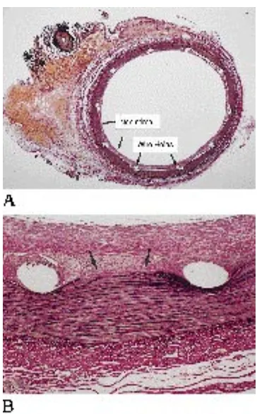
Longitudinal sectioning with removal of half the arterial wall revealed fibrous connective tis-sue filling the pores of the stent (Fig 3). Cross sections through the fistula (section 3) also demonstrated dense (white) connective tissue within the fistula hole, causing complete sepa-ration of the arterial and venous lumens (Fig 4). There was a small organized thrombus that was attached to the fistula site by a small stalk.
The neointima was comparatively uniform in thickness around the arterial lumen (Fig 5). All specimens through the stent revealed fragmen-tation (disruption) of the internal elastic lamina caused by outward pressure of the stent wires (Fig 6). The area between the artery and vein consisted of fibrous connective tissue with fibro-Fig 1. A, Arteriogram. The catheter lies within the carotid artery proximal to the fis-tula (arrow). The jugular vein (arrowheads) lies lateral to the carotid artery.
B, The stent lies within the carotid and across the fistula hole.
C, Arteriogram immediate after stent placement. Fistula remains patent.
[image:3.612.56.381.98.464.2]blasts, macrophages, and giant cells (Fig 7). Vascular spaces of various sizes were present in this area (Fig 7). The increased amount of ma-ture collagen and the decreased cellularity along the fistula margins suggested that the margins of the fistula were thrombosed first. The connective tissue within the fistula on the arterial side was organized in such a fashion that the spindloid cells (fibroblasts) were oriented parallel to the endothelial surface of the artery, whereas the cells within the thrombus of the
vein were much more haphazard. The signifi-cance of this lies in the body’s apparent attempt to form a more organized, properly oriented border around the artery.
Discussion
[image:4.612.237.552.97.398.2]The dog was chosen for our experiment be-cause it is one of the best experimental animals for studying the ingrowth of fibrous tissue through the wall of the porous graft. Sauvage et al implanted 1084 porous Dacron arterial grafts in experimental animals (dogs, pigs, and calves) (13). Six-centimeter grafts were used to bridge defects created by excision of 5- to 6-cm lengths of descending thoracic aorta of the dog, growing pig, mini pig, and calf. Transinterstices ingrowth refers to the movement of fibrous tis-sue through the wall of the porous graft. Transinterstices ingrowth occurred quickly in the pigs and calves, but more slowly in the dogs. In general, they postulate that the longer it takes for a prosthesis to heal in an experimental animal, the more probable it is that the data from that animal will be relevant in humans. Arterial prostheses heal much slower in humans than in any experimental animal thus far tested. For this reason, they prefer to use mongrel dogs Fig 2. The illustration depicts the
se-quential changes at the fistula after stent placement. A, Before stent placement, blood/contrast is shunted from the carotid artery (open arrows) through the fistula (small arrow) and into the jugular vein (large arrows). B, Immediately after stent placement, the fistula remains open. C, Eight weeks after stent placement, the blood/contrast is no longer shunted from the carotid because the fistula is closed. The carotid remains patent.
[image:4.612.58.297.559.698.2]as models because of their slower healing rate, which approximates the healing rate in humans more closely than that of other animals.
Hemorrhage within and adjacent to the stent was seen in our experiment (Fig 3). Based on information from coagulation and fibrinolytic assays, the incidents of delayed hemorrhage in the interstices of an arterial prosthesis are much higher in dogs than in humans (13). This is thought to be caused by a highly active fibrino-lytic system associated with insufficient re-serves of fibrinogen. Plasmin, the active fibrino-lytic enzyme, often is detectable in dog blood, but seldom in human blood. Fibrinogen levels in humans do not change substantially after place-ment of an intraarterial graft, as opposed to in dogs. Fibrinogen levels were tested in dogs re-ceiving micronet arterial grafts: a dramatic in-crease in fibrinogen level was seen in the first 4
to 6 postoperative days, followed by a large decrease (13). This decrease was followed shortly by evidence of major transinterstices hemorrhage in many cases.
[image:5.612.59.529.99.500.2]of the vessel lumen; however, lyophilized su-tured vein graft and autologous vein attached with cyanoacrylate led to an early and complete thrombosis of the stented vessel 1 week after implantation because of immunologic rejection and distinct foreign-body reaction, respectively. Proliferation of neointima and endothelium over the stent wires is a well-documented phe-nomenon. This neointima/endothelial layer pro-vides a smooth, nonthrombogenic surface. In our experience, this proliferative tissue re-sponse may be sufficient to cover partially or completely the pores of a stent (14).
Schatz et al studied the acute and chronic biological changes caused by balloon-expand-able intracoronary stents in the adult mongrel dog (11). They placed 20 stainless steel stents in the coronary arteries of 20 dogs. Angiogra-phy was performed at 1, 3, 6, and 12 months. The animals were killed in groups of three at 1, 3, 8, and 32 weeks for gross, light, and
elec-tronmicroscopic analysis. All stents were
patent, and there was no evidence of myocar-dial infarction. The stent was covered initially by a thin layer of thrombus that was later replaced
by neointimal muscular proliferation that
reached maximal thickness by 8 weeks. His-topathologic analysis at 1 week revealed that the struts of the stent had compressed the arte-rial wall; and the indentations were filled with thrombus material largely composed of red cells and fibrin covered by immature endothe-Fig 5. A, Cross section through carotid artery (approximately
4 mm from the fistula) after removal of the stent wires. The neointima lies deep to the stent wires (holes) and encircles the inner lumen of the artery (Verhoeff’s van Giesson’s elastic stain, magnification 33.3).B, The stent wires (holes) depressed the elastic lamina. Note the fragmentation (disruption) of the internal elastic lamina (arrows) caused by the outward pressure of the stents wires (Verhoeff’s van Giesson’s elastic stain, magnification
[image:6.612.86.268.98.391.2]333).
Fig 6. Cross section through the fistula. The area between the artery and vein con-sists of fibrous connective tissue with fibro-blasts and macrophages. Vascular spaces (arrows) of various sizes lie within the fi-brous connective tissue filling the fistula hole (Verhoeff’s van Giesson’s elastic stain, mag-nification36.6).
[image:6.612.232.555.498.737.2]lium. In the 3-week specimen, the neointima covering the stent was more cellular, and
he-mosiderin-laden macrophages partially
re-placed the underlying thrombus. At 8 weeks, the degree of intimal hyperplasia peaked be-cause of fibroblast proliferation, so that intimal thickness was greatest; the amount of underly-ing residual thrombus was minimal. By 32 weeks, the neointima was thinner and appeared less cellular. Schatz’s results indicated that the early thrombus covering the stent wires is re-placed ultimately by a thin layer of neointima that is grossly indistinguishable from that of ad-jacent nonstented artery.
Palmaz et al demonstrated total early endo-thelialization of intravascular stents in 1 to 3 weeks in the peripheral vessels (15–17). Exces-sive intimal hyperplasia was a rare finding. The biological effects of vascular implants are re-lated to phenomena that occur at the interface between the host tissue and the foreign implant material (10, 18). The series of events after stent placement facilitates the growth of endo-thelial cells over the surface of the stent (19). When observed on cross sections of the stented arterial wall, replacement of thrombus by fibro-muscular intima occurred first around the stent struts and then expanded eccentrically. Fibro-myocytes in direct contact with the metal sur-face develop physical attachment. It has been suggested that the attachment occurs between the cell membrane and the metal oxide layer through an intervening proteinaceous film. Eight weeks after stent placement, when throm-bus has been replaced by fibromuscular tissue, neovasculature is present and is more dense around stent struts than near the pores between the struts.
At times this proliferative tissue reaction can lead to significant narrowing of the intraluminal diameter of the vessel; however, stent-related stenosis was not present in our specimens. Ste-nosis of the stented vessel is related to cellular proliferation within the vessel wall, stimulated by interactions between platelets adherent to damaged intima, endothelial cells, and smooth muscle cells (20, 21). Endovascular place-ments of stents may induce this process either by outward pressure of the stent, as seen with
the self-expandable stents (Wallstent and
Gianturco Z stent), or as a result of inflation of a delivery balloon (Palmaz, Strecker, Gianturco-Robin). Intimal fibrocellular tissue growth is a known phenomenon of implantable stents.
Wa-khloo et al compared balloon-expandable tan-talum stents and self-expanding nitinol stents for the treatment of carotid aneurysms in Labra-dor dogs (22). The stents consisted of elastic, loosely connected woven loops made of bio-compatible tantalum or nitinol filaments, 100 mm thick. Nine-month angiographic follow-up revealed maximal stenosis of the stented vessel segment of up to 40% after placement of the tantalum stent; however, no more than 15% ste-nosis followed placement of the nitinol stents. Significantly greater intimal fibrocellular tissue growth surrounded a tantalum filament when compared with the nitinol filaments. The stent-induced stenosis was usually located at the dis-tal rather than the proximal portion of the stented segment. This was appreciated 3 weeks after implantation of the stent and persisted over 6 months. Histologic examination revealed thick intimal fibrocellular proliferative tissue surrounding the tantalum filaments (300 to 350 mm). The nitinol filaments were covered with a smooth and regular neointimal layer of approx-imately 150 mm. Few hemosiderin-laden mac-rophages were present in the fibrotically thick-ened intimal wall underlying either type of stent, and the media of the stented vessels revealed minor signs of atrophy. The proposed theory is that intimal-media injury caused by dilating the stent toward the arterial wall may induce this fibroproliferative reaction. The greater prolifer-ative reaction in the vessels containing the bal-loon-expandable tantalum stent versus the self-expanding nitinol prosthesis may be caused by the intimal-medial injury induced by the dilating balloon.
re-sponsible for a 20% to 25% luminal narrowing in all vessels. This proliferative response was as-sociated with multiple disruptions of the internal elastic membrane and occasional intimal splits with associated intimal hemorrhage.
Plugging of the stent pores with neointimal tissue seems more likely if the density of the woven wire network is greater and the pores between the wire filaments are smaller. For this reason, we chose the Wallstent for our experi-ment. It is constructed with 20 wires, 0.0035-in diameter; the pore dimension is 1.17 mm in length by 1.53 mm in width; the surface area covered by the stent wire is 17.67%, with the surface free area (open space) being 82.33%. This results in a pore density of 97.6 pores per square centimeter at 5-mm diameter. In com-parison, the balloon-expandable tantalum stent has a pore density of 34.7 pores per square centimeter at 5-mm diameter; the self-expand-ing nitinol stent, made of heat-treated nickel-titanium alloy, has a density of 62.4 pores per square centimeter at 5-mm diameter (22). The greater pore density of the Wallstent results in smaller pores, with a greater number of wire segments per given length, and thus provides a more favorable substructure upon which orga-nized fibrous tissue can proliferate.
The purpose of this study was not to deter-mine whether stents may act as a source of emboli. The study of thromboembolic disease caused by stent implantation was beyond the scope of this work. The possibility of stents act-ing as a potential source of emboli exists. En-dothelialization of stent wires several weeks af-ter implantation has been documented (15, 16). This endothelium provides a smooth, protec-tive, nonthrombogenic surface. Previous studies have suggested that stent-induced thrombus formation can be avoided successfully with sys-temic anticoagulation (23, 24).
An endovascular stent constructed for neu-rointerventional applications has not yet been developed. The 7F introducer catheter with af-fixed stent used in our study would be much too stiff to negotiate the curvature of the cervical/ intracranial carotid artery of humans. A minified version, perhaps 4F, with great flexibility and trackability, would need to be developed before stents could be considered seriously for the treatment of intracranial vascular disease.
In conclusion, the proliferation of fibrous tis-sue, collagen, and endothelium is a known phe-nomenon after intravascular implantation of a
vascular graft or stent. Placement of a tubular woven wire mesh (stent) across a fistula hole provides the foundation for this fibrous tissue process to occur. The result was either com-plete or near-comcom-plete closure of the fistulous communication between the carotid artery and jugular vein. Although preliminary, these results may stimulate interest in further development in stent technology, with particular focus on neu-rointerventional applications.
References
1. Lasjaunias P, Bernstein A.Surgical Neuroangiography. Heidel-berg, Germany: Springer-Verlag; 1987;2
2. Marks M, Dake M, Steinberg G, et al. Stent placement for arterial and venous cerebrovascular disease: preliminary experience. Ra-diology1994;191:441– 446
3. Palmaz J, Laborde J, Rivera F, Encarnacion C, Lutz J, Moss J. Stenting of the iliac arteries with the Palmaz stent: experience from a multicenter trial.Cardiovasc Intervent Radiol1992;15:291–297 4. Zollikofer CL, Antonucci F, Pfyffer M, et al. Arterial stent place-ment with use of the Wallstent: midterm results of clinical experi-ence.Radiology1991;179:449 – 456
5. Vorweck D, Gunther R. Stent placement in iliac arterial lesions: three years of clinical experience with the Wallstent.Cardiovasc Intervent Radiol1992;15:285–290
6. Strecker E, Liermann D, Barth K, et al. Expandable tubular stents for treatment of arterial occlusive diseases: experimental and clin-ical results.Radiology1990;175:97–102
7. Dotter C, Buschmann R, McKinney M, et al. Transluminal expand-able nitinol coil stent grafting: preliminary report.Radiology1983; 147:259 –260
8. Duprat G, Wright K, Charnsangavej C, et al. Flexible balloon-expanded stent for small vessels.Radiology1987;162:276 –278 9. Duprat G, Wright K, Charnsangevej C, et al. Self-expanding
me-tallic stents for small vessels: an experimental evaluation. Radiol-ogy1987;162:469 – 472
10. Palmaz J. Intravascular stents: tissue-stent interactions and de-sign considerations.AJR Am J Roentgenol1993;160:613– 618 11. Schatz R, Palmaz J, Tio F, et al. Balloon-expandable
intracoro-nary stents in the adult dog.Circulation1987;76:450 – 457 12. Minor E, Ehringer H, Ahmadi R, et al. Platelet deposition at
an-gioplasty sites and its relation to restenosis in human iliac and femoropopliteal arteries.Radiology1989;170:767–772 13. Sauvage L, Berger K, Wood S, et al. Interspecies healing of porous
arterial prostheses.Arch Surg1974;109:698 –705
14. Geremia G, Haklin M, Brennecke L. Embolization of experimen-tally created aneurysms with intravascular stent devices.AJNR Am J Neuroradiol1994;15:1223–1231
15. Palmaz J, Sibbitt R, Tio F, Reuter S, Peters J, Garcia F. Expand-able intraluminal vascular grafts: a feasibility study. Surgery 1986;99:199
16. Palmaz J. Balloon-expandable intravascular stent. AJR Am J Roentgenol150:1263–1269
17 Palmaz J, Kopp D, Hayashi H, et al. Normal and stenotic renal arteries: experimental balloon-expandable intraluminal stenting. Radiology1987;164:705–708
19. Flugelman M, Virmani R, Leon M, et al. Genetically engineered endothelial cells remain adherent and viable after stent develop-ment and exposure to flow in vitro.Circ Res1992;70:348 –354 20. Szikora I, Guterman L, Wells K, et al. Combined use of stents and
coils to treat experimental wide-necked carotid aneurysms: pre-liminary results.AJNR Am J Neuroradiol1994;15:1091–1102 21. Serruys P, Strauss B, van Beusekom H, van der Giessen W.
Stenting of coronary arteries: has a modern Pandora’s box been opened?J Am Coll Cardiol1991;17:143B–154B
22. Wakhloo A, Schellhammer F, de Vries J, et al. Self-expanding and balloon-expandable stents in the treatment of carotid aneurysms: an experimental study in a canine model.AJNR Am J Neuroradiol 1994;15:493–502
23. Carozza JP, Richard PE, Levine MJ, et al. Angiographic and clin-ical outcome of intracoronary stenting: immediate and long term results from a large single-center experience.J Am Coll Cardiol 1992;20:328 –337
24. Schatz RA. A view of vascular stents.Circulation1989;79:445– 457