Noriaki Tomura, Manabu Hashimoto, Ryuji Sashi, Hiroko Hirano, Mitsuru Kobayashi, Yoshinori Hirano, Kimihiko Satoh, Jiro Watarai, and Masayoshi Kowada
PURPOSE: To determine the efficacy of superselective angio-CT in the diagnosis of astrocytoma. METHODS: Nineteen patients with astrocytomas had superselective angio-CT before chemother-apeutic agents were administered via superselective intraarterial infusion. CT was performed after contrast material was delivered through a microcatheter, which had been advanced into the feeding arteries of the tumor. Superselective angio-CT scans were compared with digital subtraction angiograms, conventional contrast-enhanced CT scans, and contrast-enhanced T1-weighted MR images. RESULTS: Superselective angio-CT scans depicted contrast enhancement of the tumor in all of the patients and the medullary veins of the tumors in 32% of the patients. Digital subtraction angiograms showed tumor stains in 68% of the patients and the medullary veins in only 5%. Conventional CT scans and MR images showed contrast enhancement of the tumor in 89% of the patients. Superselective angio-CT scans confirmed the proper position of the catheter tip for the infusion of a chemotherapeutic agent. CONCLUSIONS: Superselective angio-CT can be used to depict contrast enhancement of tumors and the vascular structures that are characteristic of astrocytomas.
Index terms: Astrocytoma; Brain neoplasms, computed tomography; Computed tomography, technique
AJNR Am J Neuroradiol17:1073–1080, June 1996
Arterial angio– computed tomography (an-gio-CT), performed with contrast administration directly into arteries through a catheter, has been used in the diagnosis of various lesions in the liver (1–3), lung (2), and other organs (4 – 6). As previously reported, arterial angio-CT can depict vascular structures of tumors, which, because arteries and veins in the lesions can be seen with better contrast resolution, is useful in the differential diagnosis (1, 6). The advent of microcatheters (7) has made possible the use of superselective catheterization for such intravas-cular procedures as embolization of arterio-venous malformations and aneurysms, and thrombolytic therapy.
For the treatment of brain tumors, direct in-traarterial infusion of chemotherapeutic agents is known to have pharmacokinetic advantages (8 –11). In our institution, patients with gliomas have been treated with a combination of radia-tion and superselective intraarterial infusion of chemotherapeutic agents. To confirm the distri-bution of these drugs, we performed arterial an-gio-CT with superselective administration of contrast material, a procedure that we have named superselective angio-CT.
Superselective angio-CT scans showed the distribution of contrast material as well as new findings concerning the vascular structure of the brain tumors. The purpose of this study was to compare superselective angio-CT scans with digital subtraction angiograms, conventional enhanced CT scans, and magnetic resonance (MR) images to determine the efficacy of super-selective angio-CT in the depiction of tumors.
Subjects and Methods
Between March 1993 and November 1994, superselec-tive angio-CT was performed in 19 patients with
astrocy-Received July 25, 1995; accepted after revision December 18. From the Departments of Radiology (N.T., M.H., R.S., H.H., M.Kob., Y.H., K.S., J.W.) and Neurosurgical Service (M. Kow.), Akita (Japan) University School of Medicine.
Address reprint requests to Noriaki Tomura, MD, Department of Radi-ology, Akita University School of Medicine, 1–1–1 Hondo, Akita City, Akita, 010 Japan.
AJNR 17:1073–1080, June 1996 0195-6108/96/1706 –1073
qAmerican Society of Neuroradiology
tomas who expected to be treated with superselective in-traarterial infusion of 1-(4-amino-2-methyl pyrimidine-5-yl)-methyl-3-(2-chloroethyl)-3-nitrosourea hydrochloride (ACNU) (12). Informed consent was obtained from the patients and/or their families before the examinations.
All tumors were proved histologically by biopsy or sur-gery. Four astrocytomas were grade II, 10 were grade III, and 5 were grade IV. Superselective angio-CT was per-formed before superselective infusion of ACNU. The pa-tients underwent transfemoral catheterization with a 6F catheter (PU; Toray, Tokyo, Japan). The catheter was placed in either the cervical internal carotid artery or the cervical segment of a vertebral artery. After intraarterial digital subtraction angiography with the PU catheter, a Tracker-18 catheter (Target Therapeutics, Los Angeles, Calif) was passed coaxially inside the PU catheter and advanced into the feeding arteries of the tumor. Hepa-rinized saline was administered through the PU and Track-er-18 catheters. Once the Tracker catheter was in the proper position, digital subtraction angiography was per-formed with a hand injection of diluted contrast material. Patients were then moved to the CT suite.
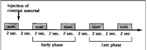
Axial CT of the whole brain was first performed parallel to the orbitomeatal plane without contrast administration. A section including tumor was scanned before contrast injection, and then four contiguous scans at the same section were obtained with contrast injection into the feed-ing arteries. The scannfeed-ing protocol is illustrated in Figure 1. A 9800 CT scanner was used, with 2-second scanning time, 170 mAs, and 10-mm collimation. These 10-mm-thick sections were chosen to gain an improved signal-to-noise ratio.
For superselective angio-CT, it is necessary to dilute the contrast material before superselective intraarterial in-jection, owing to the possibility of blood-brain barrier dis-ruption; therefore, 0.5 mL of iopamidol diluted with 2.5 mL of saline was administered by hand injection at a rate of 1 mL/s. When necessary, subtraction images with an edge enhancement were obtained by using the Data View Sys-tem (GE Medical SysSys-tems, Milwaukee, Wis). If superselec-tive angio-CT did not show proper positioning of the infu-sion catheter, the catheter tip was withdrawn proximally and the imaging procedure was repeated. After comple-tion of the CT studies, the patient was returned to the angiography suite, where the position of the tip of the catheter was confirmed. ACNU was administered in a dose of 50 to 80 mg, dissolved in sterile water in a concentration
of 2.5 mg/mL, with use of a mechanical injector at a rate of 80 mL/h.
The tip of the Tracker catheter was in the horizontal portion of the middle cerebral artery in 4 patients, in the insular portion of the middle cerebral artery in 12, in the opercular portion of the middle cerebral artery in 1, in the horizontal portion of the anterior cerebral artery in 1, in the superior cerebellar artery in 1, and in the posterior cerebral artery in 1.
All patients had conventional enhanced CT and con-trast-enhanced T1-weighted MR imaging within 2 weeks before the superselective angio-CT study. Conventional enhanced CT was performed parallel to the orbitomeatal plane with 2-second scanning time, 170 mAs, and 10-mm collimation. Enhanced T1-weighted MR images were ac-quired on a 1.5-T unit. Four-millimeter-thick sections with a 2-mm section gap were obtained parallel to the plane including the anterior and posterior commissures, using a spin-echo sequence (400 – 600/20 – 40/2 [repetition time/ echo time/excitations]).
To evaluate tumor stain (contrast enhancement) and the presence of medullary veins (13, 14) in the tumor, we retrospectively compared superselective angio-CT scans with conventional digital subtraction angiograms obtained via infusion through the guiding catheter, superselective digital subtraction angiograms obtained via infusion through the Tracker-18 catheter, conventional enhanced CT scans, and contrast-enhanced T1-weighted MR im-ages. Scans were examined conjointly by more than three observers, including a neuroradiologist. Evaluation of the appearance of tumor stain, the contrast enhancement of the tumor, and the medullary veins was by consensus.
Results
[image:2.612.57.299.87.170.2]gio-CT showed contrast enhancement only of tumor tissue (Fig 4). In 6 patients, superselec-tive angio-CT clearly showed irregular tumor vessels draining into the enlarged medullary veins and the subependymal veins of the lateral ventricle (Figs 2 and 3). The enlarged medul-lary veins did not appear in normal tissue. Those veins seemed to appear early; however, it was not evident because we could not differen-tiate between the arterial and the venous phases.
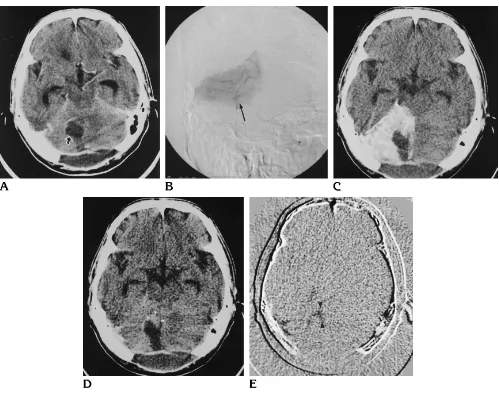
In a patient with astrocytoma in the parahip-pocampal gyrus (case 10, Fig 5), superselec-tive angio-CT with contrast injection into the posterior cerebral artery ensured proper posi-tioning of the catheter tip for the infusion of ACNU. Superselective angio-CT was also useful in identifying a feeding artery in another patient
(case 5) with astrocytoma in the left frontal lobe. In no patient did the catheter tip retract or move during transfer to the CT scanner or upon return to the angiography suite. No significant complication resulted from catheterization or contrast administration.
Discussion
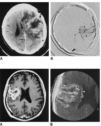
Arterial angio-CT is known to be a useful diagnostic tool for tumors of the liver (1–3), because it affords excellent contrast resolution for differentiating normal from abnormal atten-uation areas. Angio-CT can be performed either with an intravenous bolus infusion of contrast material or via direct intraarterial administration through a catheter. The former is a well-known Fig 3. Case 8: 38-year-old man with grade II astrocytoma.
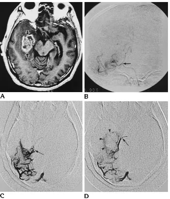
[image:3.612.58.392.81.495.2]Contrast-enhanced T1-weighted MR image (A) shows tumor in right frontal and temporal lobe (arrowheads); superselec-tive angio-CT scan (B) with contrast ad-ministration into the insular portion of the right middle cerebral artery shows en-larged abnormal vessels (arrows) in the tumor, which are presumably medullary veins draining into the subependymal vein (arrowheads) of the lateral ventricle.
Fig 2. Case 3: 51-year-old man with residual grade IV astrocytoma after sur-gery.
noninvasive technique for detecting vascular le-sions, such as aneurysms (15–17). Arterial an-gio-CT has been widely used in detecting liver metastasis (3). Angio-CT with contrast admin-istration into the superior mesenteric artery can show a small tumor in the liver not discernible by conventional enhanced CT. Angio-CT has also been used to identify the feeding artery of liver tumors before embolization (2).
Encouraging results of intraarterial infusion of chemotherapeutic agents have been reported (8 –11). It has been shown, for example, that intraarterial infusion of carmustine (BCNU) to the brain resulted in a considerably higher drug level in the infused areas than that attained with intravenous delivery. Intraarterial delivery of carmustine to brain tumors might also be achieved in higher concentrations. However, to avoid toxic reactions to the eye as a complica-tion of internal carotid arterial infusion of
che-motherapeutic agents, the catheter should be introduced into the supraclinoid or a more distal segment (18, 19).
With the advent of microcatheters, supers-elective catheterization has become an easy and safe procedure (7). In our institution, pa-tients with glioma have been treated with a combination of radiation and superselective in-traarterial infusion of ACNU (12). Initially, we attempted superselective angio-CT to confirm the distribution of contrast material through the catheter tip before infusion of ACNU. We found that superselective angio-CT scans showed contrast enhancement of the tumor even in cases in which no such enhancement was seen on conventional CT scans or contrast-enhanced T1-weighted MR images. However, this finding might be the result of increased contrast reso-lution caused by administration of contrast ma-terial directly into the feeding arteries.
Comparison of superselective angio-CT with other imaging techniques
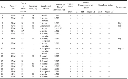
Case Age, y/ Sex
Grade of Astro-cytoma
Radiation dose, Gy
Location of Tumor
Location of Tip of Microcatheter
Tumor Stain
Contrast Enhancement of
Tumor
Medullary Veins
Comments
DSA CT MR Angio-CT DSA Angio-CT
1 69/M III* 28 L frontal L M2 1 1 1 1 2 2
2 50/M III 60 L frontal,
parietal
L M2 1 1 1 1 2 2
3 51/M IV 60 L frontal L M2 1 1 1 1 2 1 Fig 2
4 52/M III 60 Cerebellum R SCA 2 2 2 1 2 2 Fig 4
5 30/M IV 60 L frontal L A1, M1 1 1 1 1 2 2 †
6 41/F III* . . . L frontal L M2 1 1 1 1 2 2
7 52/F III* . . . R frontal,
parietal
R M2 1 1 1 1 2 2
8 38/M II* 60 R frontal,
temporal
R M2 1 1 1 1 2 1 Fig 3
9 57/M III . . . L frontal,
parietal
L M2 1 1 1 1 2 2
10 46/M II* . . . R temporal,
parietal
R PCA 2 1 1 1 2 2 Fig 5†
11 43/F II* . . . L frontal L M3 2 1 1 1 2 2
12 49/M III 60 R basal
ganglia
R M2 2 2 2 1 2 2
13 65/M IV . . . R frontal R M2 1 1 1 1 1 1
14 38/M III 60 R frontal R M1 2 1 1 1 2 1
15 19/M II* 28 L frontal L M2 1 1 1 1 2 2
16 26/F III . . . R frontal R M1 1 1 1 1 2 2
17 41/M III . . . R temporal R M1 1 1 1 1 2 2
18 51/F IV 60 L frontal L M2 2 1 1 1 2 1
19 64/M IV 24 R frontal R M2 1 1 1 1 2 1
Note.—1indicates detectable;2, not detectable; DSA, digital subtraction angiography; M1, horizontal portion of the middle cerebral artery; M2, insular portion of the middle cerebral artery; M3, opercular portion of the middle cerebral artery; A1, horizontal portion of the anterior cerebral artery; PCA, posterior cerebral artery; and SCA, superior cerebellar artery.
* Histology was proved at biopsy.
[image:4.612.59.558.99.409.2]Enlarged medullary veins are usually found in two pathologic processes: malignant infiltrating gliomas and arteriovenous malformations (20). They may also be seen in other lesions of the white matter, such as metastasis, malignant lymphoma, cerebral hemorrhage, inflammatory diseases, and venous thrombosis (20). Huang and Wolf (21) reported that in approximately 10% of their patients with glioblastoma multi-forme, no vascular abnormalities appeared other than enlargement of the deep medullary veins, and they concluded that streaming within the deep medullary veins was a reliable sign of glioblastoma multiforme. Hooshmand et al (22)
[image:5.612.58.557.82.475.2]noted that the deep medullary veins were seen in low-grade astrocytomas where there was central necrosis. In a case of astrocytoma grade II in our series, the enlarged medullary veins were seen on superselective angio-CT scans. These veins appeared early, as they were drain-ing arteriovenous shuntdrain-ing in the tumor. Scatliff et al (23) studied the vascular structure of glio-blastomas on postmortem specimens and re-ported four major types of vascular compo-nents: type I, cortical and transcortical (medullary) arteries draining into the enlarged medullary veins and subependymal veins; type II, gray matter hypervascularity consisting of Fig 4. Case 4: 52-year-old man with grade III astrocytoma in the cerebellar vermis.
neocapillary circulation and vasodilatation of cortical arteries and veins; type 3, irregular neo-capillary formation around necrotic areas; and type 4, a rounded capillary network around ne-crotic area or in viable tumor. In our series, cases 3 (Fig 2), 8 (Fig 3), 13, and 14 seem to fit the type I category, and this was clearly seen on the superselective angio-CT scans. These en-larged medullary veins were seen on digital sub-traction angiograms that had an increased spa-tial resolution. Until now, this radiologic finding could be seen only on conventional or digital subtraction angiograms. Superselective an-gio-CT made it possible to depict this angio-graphic finding by CT.
Superselective angio-CT scans seem similar to microangiographic sections obtained by postmortem injections. In investigations of the vascular structure of tumors, this new technique is superior to digital subtraction angiography,
conventional enhanced CT, and contrast-en-hanced T1-weighted MR imaging. The type of vascular structure reported by Scatliff et al (23) is thought to be characteristic of glioma.
[image:6.612.224.558.84.475.2]Superselective angio-CT has also proved useful in identifying feeding arteries that were unclear on conventional or digital subtraction angiograms. In 2 patients (cases 5 and 10) in our series, only superselective angio-CT scans showed the major feeding artery of the tumor. This indicated that superselective angio-CT is valuable at the time of intraarterial infusion of chemotherapeutic agents and will be feasible for evaluation of feeding arteries before chemo-therapy. Moreover, this technique is useful for determining the proper position of a catheter tip prior to infusion of chemotherapeutic agents (Fig 5). The distribution of contrast material seen on the superselective angio-CT scan, how-ever, may not be representative of the depiction Fig 5. Case 10: 46-year-old man with
grade II astrocytoma.
of ACNU distribution, because the infusion rate of contrast material is quite different from that of ACNU; moreover, contrast material has a higher specific gravity than drugs. To assess the exact distribution of intraarterial cerebral che-motherapeutic agents, single-photon emission CT (SPECT) performed with intraarterial ad-ministration of technetium-99m hexamethyl-propyleneamine oxime (HMPAO) has recently been proposed (24). Aoki et al (24) studied drug distribution with 99mTc HMPAO SPECT and reported that uniform distribution could be obtained by manual pulsatile injection (24, 25). The technique presented here, while attrac-tive for the depiction of the character and vas-cularity of tumors, can be safely performed with modern digital subtraction angiographic equip-ment, mounted on a C-arm, and with high-res-olution fluoroscopy and road-mapping capabil-ities. Moving patients to a CT suite is necessary for superselective angio-CT; therefore, new equipment combining angiography and CT is expected. We encountered no complications re-sulting from catheterization and injection of contrast materials. Although this study is pre-liminary, our findings suggest that implementa-tion of superselective angio-CT might facilitate improved survival rates for patients with brain tumors. Our results should encourage further evaluation of the effectiveness and safety of su-perselective angio-CT.
In conclusion, we performed superselective angio-CT in patients with astrocytomas imme-diately before superselective intraarterial che-motherapy. Superselective angio-CT scans de-picted contrast enhancement of the tumors in all patients and made it possible to see the enlarged medullary veins that are characteristic of gliomas. This technique can be used to iden-tify feeding arteries that are unclear on angio-grams as well as to determine the proper posi-tion of a catheter tip before injecposi-tion of chemotherapeutic agents.
Acknowledgments
Takashi Hatayama and his team of technologists kindly performed the CT examinations. We thank Dr Aryeh Stoll-man (Department of Radiology, Mt Sinai [NY] School of Medicine) for his help in editing this paper.
References
1. Takayasu K, Muramatsu Y, Moriyama N, et al. Focal nodular hyperplasia of the liver: arterial angio-CT and microangiography. J Comput Assist Tomogr1992;16:212–215
2. Rieber A, Brambs HJ, Gorich J, Kauffmann GW. Intraarterielles Angio-CT als Perfusionskontrolle vor intraarteieller Chemothera-pie bei Bronchi al-, Mamma- und Lebertumoren.Rofo Fortschr Geb Rontgenstr Neuen Bildgeb Verfahr1991;154:514 –517 3. Safi F, Roscher R, Bittner R, Beger HG. The clinical relevance of
tumor marker CEA, CA 19 –9 in regional chemotherapy of hepatic metastases of colorectal carcinoma.Int J Biol Markers1988;3: 101–106
4. Gorich J, Hasan I, Sittek H, et al. Superselektive intraarterielle Chemotherapie bei Mammakarzinomen.Radiologe1993;33:308 – 312
5. Hirai Y, Kaku S, Nakayama K, et al. Significance of preoperative diagnosis of endometrial carcinoma by angio-CT.Nippon Sanka Fujinka Gakkai Zasshi1988;40:373–378
6. Moriyama N. CT diagnosis of hepatic and pancreatic cancers.Gan No Rinsho1983;29:1169 –1174
7. Kikuchi Y, Strother CM, Boyer M. New catheter for endovascular interventional procedures.Radiology1987;165:870 – 871 8. Feun LG, Wallace S, Stewart DJ, et al. Intracarotid infusion of
cis-diamminedichloroplatinum in the treatment of recurrent ma-lignant brain tumors.Cancer1984;54:794 –799
9. Greenberg HS, Ensminger WD, Seeger JF, et al. Intraarterial BCNU chemotherapy for the treatment of malignant gliomas of the central nervous system: a preliminary report.Cancer Treat Rep 1981;65:803– 810
10. Yamada K, Bremer AM, West CR, Ghoorah J, Park HC, Takita H. Intraarterial BCNU therapy in the treatment of metastatic brain tumor from lung carcinoma.Cancer1979;44:2000 –2007 11. Mahaley MS Jr, Hipp SW, Dropcho EJ, et al. Intracarotid cisplatin
chemotherapy for recurrent gliomas.J Neurosurg1989;70:371– 378
12. Yamashita J, Handa H, Tokuriki Y, et al. Intraarterial ACNU ther-apy for malignant brain tumors: experimental studies and prelim-inary clinical results.J Neurosurg1983;59:424 – 430
13. Stein RL, Rosenbaum AE. Normal deep cerebral venous system. In: Newton TH, Potts DG, eds.Radiology of the Skull and Brain.St Louis, Mo: Mosby; 1974:1904 –1910
14. Huang YP. Veins of the white matter of the cerebral hemisphere. In: Salamon G, Huang YP, eds.Radiologic Anatomy of the Brain. Berlin, Germany: Springer-Verlag; 1976:216 –217
15. Fukui K, Sadamoto K, Sakai S. Development of the new cerebral angio-CT technique and magnetic resonance angio- imaging. Acta Radiol Suppl1986;369:67– 68
16. Leonardi M, Biasizzo E, Calabro A. Applications of dynamic-CT, or angio-CT in neuroradiology: a disappointing experience. Com-put Radiol1985;9:29 –36
17. Schmid UD, Steiger HJ, Huber P. Accuracy of high resolution computed tomography in direct diagnosis of cerebral aneurysm. Neuroradiology1987;29:152–159
18. Gebarski SS, Greenberg HS, Gabrielsen TO, Vine AK. Orbital angiographic changes after intracarotid BCNU chemotherapy. AJNR Am J Neuroradiol1984;5:55–58
19. Defer G, Fauchon F, Schaison M, Chiras J, Brunet P. Visual toxicity following intraarterial chemotherapy with hydroxyethyl-CNU in patients with malignant gliomas.Neuroradiology1991; 33:432– 437
21. Huang YP, Wolf BS. Veins of the white matter of the cerebral hemispheres (the medullary veins): diagnostic importance in ca-rotid angiography.AJR Am J Roentgenol1964;92:739 –755 22. Hooshmand I, Rosenbaum AE, Stein RL. Radiographic anatomy
of normal cerebral deep medullary veins: criteria for distinguish-ing them from their abnormal counterparts.Neuroradiology1974; 7:75– 84
23. Scatliff JH, Radcliffe WB, Pittman HH, Park CH. Vascular structure of glioblastomas.AJR Am J Roentgenol1969;105:795– 805
24. Aoki S, Terada H, Kosuda S, et al. Supraophthalmic chemother-apy with long tapered catheter: distribution evaluated with intraar-terial and intravenous Tc-99m HMPAO. Radiology 1993; 188:347–350