Case Report
First reported case of a squamous cell carcinoma
arising in the duodenum in a patient with
Lynch syndrome
Ali I Amjad1, Aatur D Singhi2, Edward P Balaban3, Beth Dudley4, Randall E Brand4, Nathan Bahary1,5
1Division of Hematology Oncology, Department of Medicine, University of Pittsburgh, PA, USA; 2Department of
Pathology, University of Pittsburgh, PA, USA; 3Penn State Hershey Cancer Institute, Hershey, PA, USA; 4Division of
Gastroenterology, Hepatology and Nutrition, Department of Medicine, University of Pittsburgh, PA, USA; 5
Depart-ment of Molecular Genetics and DevelopDepart-mental Biology, University of Pittsburgh School of Medicine, Pittsburgh, PA, USA
Received November 25, 2014; Accepted November 26, 2014; Epub December 1, 2014; Published December 15,
2014
Abstract: A 58 y/o male with Lynch syndrome, who was diagnosed with a squamous cell carcinoma (SCC) arising in the duodenum, is described. Previous malignancies included two metachronous colorectal adenocarcinomas, and a known family history of Lynch syndrome associated with deletion of exons 8-15 of the MSH2 gene. Analysis of his small bowel SCC revealed loss of MSH2 and MSH6 protein expression, suggesting a pathogenic role of the germ-line deletion. While small bowel adenocarcinomas have previously been reported in Lynch syndrome, to our knowledge
this is the first report of Lynch syndrome-associated squamous histology. As patients with Lynch syndrome live
longer with early detection and treatment of their cancers, unusual sites and histology of previously unreported cancers may emerge. It is also important to recognize variant histologies that otherwise might not prompt pursuing a diagnosis of Lynch syndrome in the appropriate clinical setting.
Keywords: Hereditary non-polyposis colorectal cancer, Lynch syndrome, squamous cell carcinoma, small bowel cancers, duodenal cancers
Case presentation
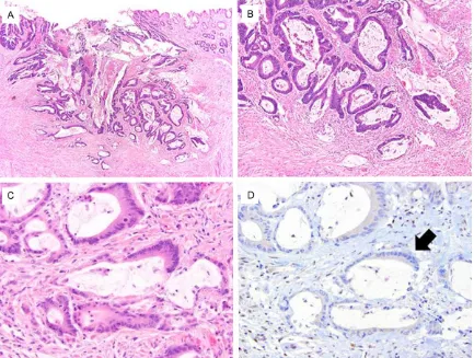
A 58-year-old Caucasian male presented with two days of nausea and vomiting from an upper gastrointestinal tract obstruction. Imaging and endoscopy showed a mass arising in the duo-denum (Figure 1). A biopsy of this mass revea- led squamous cell carcinoma. He underwent resection of his duodenum with a distal gas-trectomy and Billroth II gastrojejunostomy. Pathology revealed an invasive, moderately dif-ferentiated keratinizing squamous cell carcino-ma (Figure 2A-C) with a focal glandular differ-entiation measuring 11 cm. The tumor trans- murally invaded the duodenal wall with involve-ment of the subserosa. There was angiolym-phatic invasion but no perineural invasion. The tumor was immunoreactive for the squamous cell markers p40 and CK5/6, and demonstrat-ed very focal positivity with mucicarmine. One of 17 lymph nodes was positive for carcinoma.
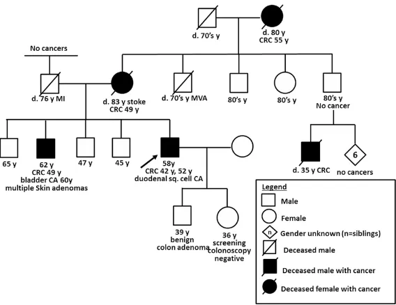
His ancestry was notable for German-Scottish descent. His mother and maternal grandmoth-er both wgrandmoth-ere successfully treated for colorectal cancer at the ages of 49 and 55 years, respec-tively. His mother also had a hysterectomy for unclear reasons. His brother was diagnosed with colorectal cancer at the age of 49 years and later with a bladder cancer at the age of 60 years. His brother was also treated for multiple skin adenomas over his lifetime. One of his maternal first cousins died at the age of 35 years from metastatic colorectal cancer (Figure 3). There was no history of cancers on the paternal side of the family.
Because his histology was with a squamous cell carcinoma, we elected to adjuvantly treat him with three monthly cycles of infusional 5-fluorouracil in combination with cisplatin. He experienced no complications and is currently following an individualized plan of surveillance with annual upper gastrointestinal endoscopy and small bowel capsule endoscopy.
Discussion
Lynch syndrome, also known as hereditary non-polyposis colorectal cancer (HNPCC), is the most common form of hereditary colorectal cancer. It is characterized by an autosomal dominant inheritance pattern with incomplete penetrance and accounts for 2-5% of all colorectal cancers [1]. Patients with HNPCC are also at an excess risk of extracolonic malignan-cies including endometrial, ovarian, stomach, small bowel, pancreas, hepatobiliary, brain and urothelial cancers. Some patients with HNPCC have the Muir-Torre variant and present with
recurrent sebaceous adenomas, sebaceous carcinomas and/or multiple keratoacanthomas [2]. Lynch syndrome is caused by germ-line defects in mismatch repair genes, with MLH1
and MSH2 accounting for approximately ninety percent of identified mutations [1].
We report a case of a patient with known Lynch syndrome who was diagnosed with a squamous cell carcinoma arising in the duodenum and previously treated for two metachronous colo- rectal adenocarcinomas. The patient’s inherit-ed cancer prinherit-edisposition was due to a previ-ously identified germ-line MSH2 deletion of exons 8-15. His duodenal squamous cell carci-noma showed loss of MSH2 and MSH6 by immunohistochemistry, suggesting a patho-genic role for his MSH2 germ-line mutation in this tumor. To the best of our knowledge, our patient seems to be the first report of a duode-nal squamous cell carcinoma, since Love and Lynch first recognized Lynch syndrome-associ-ated small bowel cancers in the late 1980s [3, 4].
Given the glandular differentiation of the lining of the gastrointestinal tract, pathogenesis of squamous cell cancers remains elusive. One may speculate that these cancers arise from pre-existing adenomas and adenocarcinomas, as historically evidenced by areas of squamous differentiation sometimes seen in gastrointes-tinal adenocarcinomas.[5] We retrieved and reanalyzed the two prior colorectal adenocarci-noma specimens and found no squamous fea-tures (Figures 4 and 5). However, there was focal glandular differentiation of the squamous carcinoma specimen from the duodenum. Another proposed theory is squamous meta-plasia of pluripotent basal cells which then undergo malignant transformation to form squamous cell cancers [6]. Whether this finding relates to mismatch repair defect in Lynch Syndrome is unknown.
[image:2.612.89.289.70.224.2]We performed an extensive literature search to review published data on HNPCC-associated small bowel cancers (Table 1). In the largest reported cohort of Lynch syndrome patients (from German and Dutch national registries), small bowel cancers comprised 2.5% patients (54 of 2,118), more commonly affecting males (HR 2.5; p 0.002) with a cumulative lifetime risk of 12.0% (95% CI 5.7 to 19.3). MSH2 gene mutation was not associated with higher inci-dence of small bowel cancers (compared with
MSH6 and MLH1 HR 1.0; p = 0.993) [7]. Histology of the small bowel cancers were not reported in this cohort. Another large study (reporting proven and probable muta-tion carriers from Denmark, Holland, Finland and USA) quotes a frequency of small bowel cancers of 0.93% (56 of 6,041 patients), more likely in males (HR 3.1, p = 0.0002) with a lifetime risk of 4.3% (95% CI 3.1-6.0). The small bowel cancers included ade-nocarcinoma and neuroendo-crine tumors. No squamous cell histology was reported [8]. Other smaller retrospec-tive studies have quoted a
Figure 2. Histopathology of the patient’s duodenal tumor demonstrated (A-C) moderately-differentiated keratinizing squamous cell carcinoma with focal glandular differentiation. Immunohistochemical stains for MSH2 (D) and MSH6 showed loss of nuclear expression (arrow), while the surrounding stroma had preserved staining.
[image:3.612.91.375.461.680.2]lifetime cumulative incidence of small bowel cancers ranging from 2.5% to 7.2% with none resembling the histology of our patient [9-11].
The largest dedicated series of small bowel cancers in Lynch syndrome was published by Park et al in 2006 [12]. This was a
[image:4.612.91.524.72.399.2]question-Figure 4. The patient’s first cancer was diagnosed at the age of 42 years and located within the rectum. The histo -pathology (A-C) was consistent with an invasive moderately-differentiated adenocarcinoma with mucinous features that extended through the mucosa, submucosa and into the muscularis propria. Immunohistochemical stains dem-onstrate the tumor had loss of both MSH2 (D, arrow) and MSH6.
[image:4.612.91.525.471.635.2]Table 1. Review of clinical characteristics of patients from published case series of small bowel cancers associated with Lynch syndrome Characteristics HNPCC-associated Small Bowel Cancer (SBC) Case Series Current patient
Study Identification Park et al. (2006) [12] Rodriguez-Bigas et al. (1998) [13] Schulmann et al. (2005) [14] Amjad et al. (2015) Source of patient (s) Questionnaires mailed to 55
mem-bers of InSiGHT* from 21 countries Questionnaires mailed to members of ICG-HNPCC+ from 6 countries
Patient data retrieved from
German HNPCC Consortium University of Pittsburgh Cancer Institute Patient Demographics
No. of patients 85 42 31
No. of Small bowel cancers 90 49 32 + 1 adenoma
Males 51/85 (60.0%) 32/42 (76.2%) 22/31 (70.9%) Male
Age at diagnosis, years Mean 48 (range 11-81) Mean 49 (range 25-88) Mean 44 (range 15-73) 58
Met Amsterdam criteria 60/85 (70.6%) 34/43 (80.9%) 16/31 (51.6%) Yes (Met Amsterdam I criteria)
Disease Characteristics
Histology Squamous cell carcinoma (first scientific report
to the best of our knowledge)
Adenocarcinoma 74/90 (82.2%) 41/49 (83.7%) 24/33 (72.7%) + 1 adenoma
Carcinoid 2/90 (2.2%) 3/49 (6.1%) 1/33 (3.0%)
Not specified 14/90 (15.6) 5/49 (10.2%) 7/33 (21.2%)
Squamous cell carcinoma None reported None reported None reported
Location
Unknown 18/90 (20.0%) 2/49 (4.1%) 3/33 (9.1%)
Known 72/90 (80.0%) 47/49 (95.9%) 30/33 (90.9%)
Duodenum 31/72 (43.1%) 17/47 (36.2%) 16/30 (53.3%) Duodenum
Jejunum 31/72 (43.1%) 18/47 (38.3%) 10/30 (33.3%)
Ileum 10/72 (13.9%) 12/47 (25.5%) 4/30 (13.3%)
Synchronous None reported 7/42 patients (16.6%) 1/31 patient Capsule endoscopy negative for synchronous
SBC. Patient will be offered surveillance Metachronous 4/85 patients (4.7%) One patient had
2 metachronous SBCs
4/42 patients (9.5%) None reported
Other HNPCC-associated cancers (Information available for 41 patients) (Information available for 42 patients)
Not reported
Total number of other cancers 59 77 Not reported
Colorectal 51 in 27 patients 59 in 25 patients 2 prior colorectal cancers
Endometrial 5 NR
Renal pelvis/ureter 4 NR
Ovarian 3 NR
Pancreatic 1 NR
SBC as the first HNPCC-associated cancer 14/41 patients (34.1%) 24/42 patients (57.1%) 14/31 (45.1%) No
Genetics All patients were known mutation
carriers (inclusion criteria) No. of patients who were mutation carriers unknown 27 patients underwent genetic testing Mutations in mismatch repair genes 69 different germ-line mutations
identified
MSH2 34/69 (49.3%) in 38 patients 6 patients 9 patients Exon deletion 8-15 in MSH2
Truncation 28/34 (82.4%)
MLH1 31/69 (44.9%) in 42 patients 9 patients 16 patients
Truncation 25/31 (80.6%)
Missense 6/31 (19.4%)
MSH6 3/69 (4.3%) in 3 patients 2 patients
Truncation 2/3
Missense 1/3
PMS2 1/69 (1.4%) in 2 patients
naire-based survey with collaboration of mem-bers of the International Society for Gastro- intestinal Hereditary Tumors from 21 countries. They identified 85 patients with 90 small bowel cancers with characteristics as reported in Table 1. One interesting finding from this study was that in patients with small bowel cancers and MSH2 mismatch repair gene mutations, the distribution of the mutations within the gene was significantly different as compared to their HNPCC controls. There was an increased frequency of mutations in the Walker-A region, comprising codons 626-733, (26.5% versus 2.8%, p < 0.001) and fewer mutations in the MutL homologue interaction domain (2.9 ver-sus 19.9%, p = 0.019). Our patient’s large dele-tion encompasses the Walker-A region and part of the MutL homologue interaction domain. Two other similar studies by Rodriguez-Bigas et al and Schulmann et al are summarized in Table 1 [13, 14]. We were especially interested in the reported tumor histology and none of the small bowel cancers were of squamous cell origin. Conclusion
Based on our review of literature, this is the first report of squamous cell carcinoma arising in the duodenum in a patient with Lynch syn-drome. As patients with Lynch syndrome live longer with early detection and treatment of their cancers, unusual sites and histology of previously unreported cancers may emerge, as is illustrated by this case. It is also important to recognize alternative histologies that otherwise might not prompt pursuing a diagnosis of Lynch syndrome in the appropriate clinical scenario. Disclosure of conflict of interest
None to disclose.
Address correspondence to: Dr.Ali I Amjad, Division of Hematology Oncology, Department of Medicine, University of Pittsburgh, PA, USA. Tel: 412-648-6413; Fax: 412-648-6579; E-mail: amjadai@upmc. edu
References
[1] Lynch HT and de la Chapelle A. Hereditary colorectal cancer. N Engl J Med 2003; 348: 919-932.
[2] Ponti G and Ponz de Leon M. Muir-Torre syn-drome. Lancet Oncol 2005; 6: 980-987. [3] Love RR. Small bowel cancers, B-cell lymphatic
leukemia, and six primary cancers with
metas-tases and prolonged survival in the cancer family syndrome of Lynch. Cancer 1985; 55: 499-502.
[4] Lynch HT, Smyrk TC, Lynch PM, Lanspa SJ, Bo-man BM, Ens J, Lynch JF, Strayhorn P, Carmody T and Cristofaro G. Adenocarcinoma of the small bowel in lynch syndrome II. Cancer 1989; 64: 2178-2183.
[5] Williams GT, Blackshaw AJ and Morson BC. Squamous carcinoma of the colorectum and its genesis. J Pathol 1979; 129: 139-147. [6] Cabrera A and Pickren JW. Squamous
meta-plasia and squamous-cell carcinoma of the rectosigmoid. Dis Colon Rectum 1967; 10: 288-297.
[7] Engel C, Loeffler M, Steinke V, Rahner N, Holin -ski-Feder E, Dietmaier W, Schackert HK, Goer-gens H, von Knebel Doeberitz M, Goecke TO, Schmiegel W, Buettner R, Moeslein G, Lette-boer TG, Gomez Garcia E, Hes FJ, Hoogerbrug-ge N, Menko FH, van Os TA, Sijmons RH,
Wag-ner A, Kluijt I, Propping P and Vasen HF. Risks
of less common cancers in proven mutation carriers with lynch syndrome. J Clin Oncol 2012; 30: 4409-4415.
[8] Watson P, Vasen HF, Mecklin JP, Bernstein I,
Aarnio M, Jarvinen HJ, Myrhoj T, Sunde L, Wi-jnen JT and Lynch HT. The risk of extra-colonic, extra-endometrial cancer in the Lynch syn-drome. Int J Cancer 2008; 123: 444-449. [9] Barrow E, Robinson L, Alduaij W, Shenton A,
Clancy T, Lalloo F, Hill J and Evans DG. Cumula-tive lifetime incidence of extracolonic cancers in Lynch syndrome: a report of 121 families with proven mutations. Clin Genet 2009; 75: 141-149.
[10] Vasen HF, Stormorken A, Menko FH, Nagen
-gast FM, Kleibeuker JH, Griffioen G, Taal BG,
Moller P and Wijnen JT. MSH2 mutation carri-ers are at higher risk of cancer than MLH1 mu-tation carriers: a study of hereditary nonpolyp-osis colorectal cancer families. J Clin Oncol 2001; 19: 4074-4080.
[11] Vasen HF, Morreau H and Nortier JW. Is breast
cancer part of the tumor spectrum of heredi-tary nonpolyposis colorectal cancer? Am J Hum Genet 2001; 68: 1533-1535.
[12] Park JG, Kim DW, Hong CW, Nam BH, Shin YK, Hong SH, Kim IJ, Lim SB, Aronson M, Bisgaard ML, Brown GJ, Burn J, Chow E, Conrad P, Doug-las F, Dunlop M, Ford J, Greenblatt MS, Heikki J, Heinimann K, Lynch EL, Macrae F, McKinnon
[13] Rodriguez-Bigas MA, Vasen HF, Lynch HT, Wat -son P, Myrhoj T, Jarvinen HJ, Mecklin JP, Mac-rae F, St John DJ, Bertario L, Fidalgo P, Madlen-sky L and Rozen P. Characteristics of small bowel carcinoma in hereditary nonpolyposis colorectal carcinoma. International Collabora-tive Group on HNPCC. Cancer 1998; 83: 240-244.
[14] Schulmann K, Brasch FE, Kunstmann E, Engel
C, Pagenstecher C, Vogelsang H, Kruger S, Vo
-gel T, Knaebel HP, Ruschoff J, Hahn SA,
Kneb-el-Doeberitz MV, Moeslein G, Meltzer SJ,