Original Article
Distinct different sensitivity of Treg and Th17 cells to
Fas-mediated apoptosis signaling in patients with acute
coronary syndrome
Qing Li1*, Yiping Wang2*, Yi Wang3,4,5, Qing Zhou3,4,5, Ke Chen6, Yuan Min Wang7, Wei Wei4,5, Yuan Wang3,4,5 1The central laboratory of Medical research center in the Affiliated Anhui Provincial Hospital of Anhui Medical University, Hefei, Anhui230001, PR China; 2The Centre for Transplantation and Renal Research, Western Clinical
School, University of Sydney, Westmead, NSW Australia; 3Laboratory of Molecular Biology and Department of Bio
-chemistry, Anhui Medical University, Hefei, Anhui230032, PR China; 4Key Laboratory of Gene Resource Utilization for Severe Disease (Anhui Medical University), Ministry of Education and Anhui Province, Hefei, Anhui230032, PR China; 5Institute of Clinical Pharmacology, Anhui Medical University, Hefei, Anhui230032, PR China; 6De -partment of Cardiovascular Disease, The Affiliated Anhui Provincial Hospital of Anhui Medical University, Hefei, Anhui230001, PR China; 7Centre for Kidney Research, Children’s Hospital at Westmead, Sydney, NSW, Australia. *These authors contributed equally to this work.
Received November 12, 2012; Accepted December 11, 2012; Epub January 15, 2013; Published February 1, 2013
Abstract: Objective: An imbalance in CD4+CD25+ regulatory T (Treg) cells and Th17 cells has been found to correlate to occurrence of acute coronary syndrome [ACS, including unstable angina (UA) and acute myocardial infarction (AMI)]. However, the mechanisms of Th17/Treg imbalance in ACS patients are still unclear. The purpose of this study is to investigate the possibility of differences in sensitivity of Th17 and Tregs to Fas-mediated apoptosis which could lead to Th17/Treg imbalance in ACS patients. Methods: We examined the apoptosis of Th17 and Treg cells,
apop-tosis-related Fas/Fas ligand(FasL) pathway, and inflammatory markers in patients with AMI, UA, stable angina (SA) and controls by Flow cytometry and ELISA. Then we analysed the correlation of inflammatory markers and sFasL to
Treg apoptosis, and the effect of anti-FasL antibody on Treg apoptois in vitro. Results: Our study demonstrated that
apoptotic Tregs, Fas and FasL expression, Caspase-3 activity of Tregs were significantly higher in ACS patients than
those in NCA and SA patients (all P < 0.05). The percentage of apoptotic Tregs is positively correlated with the levels
of inflammatory markers and sFasL. In vitro incubation of peripheral blood mononuclear cells from ACS patients
with anti-FasL antibody resulted in a markedly reduction of apoptotic Treg cells. However, there were no significant
differences in apoptotic Th17 cells and in Fas and FasL expression for Th17 cells between the four groups (all P >0.05). Conclusions: Tregs, but not Th17 cells, become apoptotic through Fas/FasL pathway, which contributed to reduction of Tregs leading to an imbalance between Th17 and Treg cells. This could be the mechanism underlying Th17/Treg imbalance and occurrence of ACS.
Keywords: Apoptosis, Fas, Fas ligand, T helper 17, regulatory T cells, acute coronary syndrome
Introduction
Atherosclerosis (AS) is a chronic inflammatory
disease in which immune response plays a vital role. Various immune cells, particularly CD4+ T-helper cells (Th), participate in pathogenesis of atherogenesis [1, 2]. It has been demonstrat-ed that Th cells play an important role in coro-nary plaque rupture resulting in occurence of acute coronary syndrome (ACS), including
unstable angina (UA) and acute myocardial infarction (AMI) [3].
Recently, CD4+CD25+ regulatory T cells (Treg) and Th17 cells have been described as two new subsets in Th cells. Treg cells have important effects on the maintenance of immune homeo-stasis by contact-dependent suppression and
the ability to release anti-inflammatory cyto
-forming growth factor (TGF)-β [4]. Th17 cells
exert an important role in the pathogenesis of
many autoimmune diseases and inflammatory
conditions [5].
Previous studies from our group and others, both Treg cells and Th17 cells have shown to be involved in pathogenesis of atherosclerosis and ACS, and Th17/Treg imbalance appear to be critical in the development of AS, especially ACS [6, 7]. However, few studies have explored the mechanisms of Th17/Treg imbalance in AS and ACS patients.
Apoptosis is an active process of cell death involving the sequential activation of a series of caspases after appropriate stimulation [8]. Apoptosis can also be induced by receptor-mediated or mitochondrial-receptor-mediated pathways [9]. Fas and Fas ligand (FasL) are important proapoptotic proteins, which belong to the TNFR family [10], and play essential roles in many human autoimmune diseases [11]. Activation-induced cell death (AICD) is a major mechanism of peripheral T cell apoptosis in which Fas/FasL pathway plays an important role [12, 13].
Human Treg have been shown to express Fas and FasL [14]. Whether Treg are susceptible to Fas-mediated apoptosis in disease is contro-versial[15-17]. Recent studies have shown that Th17 cells are less sensitive than Th1 cells to Fas-mediated apoptosis [18]. The role of the Fas/Fas ligand mediated apoptosis in Th17/
Treg imbalance is unknown. In this study, we
examined the apoptosis of Th17 and Treg cells and their sensitivity to Fas/FasL pathway in ACS patients, and determined the differences in sensitivity of Th17 and Tregs to Fas-mediated apoptosis which may contribute to Th17/Treg imbalance and ACS occurrence.
Methods
Patients
The study was approved by the institutional review boards of Anhui Medical University and
conforms with the declaration of Helsinki. All
patients gave written informed consent prior enrolment into the study.
Patients at Anhui provincial hospital who under-went diagnostic catheterization between July 2008 and June 2010 (105 males and 60
females) were examined. Patients were
classi-fied into four groups: Group 1, AMI patients (23
males and 15 females, mean age 57.8±16.7
years). Myocardial infarction was confirmed by definite (>2mm) ST-segment elevations in at least two consecutive leads and significant rise of creatine kinase MB and troponin I levels.
Group 2, UA patients (24 male and 16 female,
mean age 63.9±12.4). UA was defined as chest pain at rest accompanied by definite ischaemic
electrocardiographic changes (ST-segment changes and/or T-wave inversions). Group 3, SA patients (28 male and 14 female, mean age 60.6±11.8). SA was diagnosed by typical exer-tional chest discomfort associated with downsloping or horizontal ST-segment depres-sion >1mm in an exercise test. Group 4, sub-jects with normal coronary arteries (NCA). Control subjects were selected on the basis of a recent angiography showing NCA (30 male and 15 female, mean age 56.7±15.9). Patients with ACS and stable angina had similar extent of coronary atherosclerosis. There were no evi-dent differences between the four groups with regard to age.
No patient had been treated with anti-inflam -matory drugs and/or immunosuppressive agents. None had thromboembolism, dissemi-nated intravascular coagulation, advanced liver disease, renal failure, malignant disease, other
inflammatory disease, chronic-immune-mediat
-ed disorders, valvular heart disease, atrial fibril
-lation or pacemaker.
Blood samples and cell preparation
Blood samples were obtained from all the
patients in a fasting state on the following morning after admission. The time interval between symptom onset and blood sampling
was less than 24 h in all cases. PB (5-10 ml)
was collected from 45 NCA individuals and 120 patients. All samples were anti-coagulated with heparin and examined within 4 h. Peripheral
blood mononuclear cells (PBMCs) were pre -pared by Ficoll density gradient for analysis of
flow cytometry (FCM). Serum was obtained after centrifugation and stored at −80°C for the
measurement of soluble Fas Ligand (sFasL),
inflammation markers and other Biochemical
indicators.
culture medium (RPMI 1640 supplemented
with 100 U/ml penicillin and 100μg/ml strepto -mycin, 2mM glutamine and with 10%
heat-inac-tivated fetal calf serum, Gibco BRL). The cell
suspension was transferred to each well of 24-well plates. Cultures were stimulated with phorbol myristate acetate (PMA, 25 ng/ml) plus
ionomycin (1μg/ml) for 4 h, in the presence of monensin (1.7μg/ml, all from Alexis Biochemicals, San Diego, CA) The incubator was set at 37 °C under a 5% CO2 environment.
After culture, the contents of the well were transferred to 5ml sterile tubes. The cells were then centrifuged at 1200 rpm for 5 min. For the
analysis of Treg, 100 μl of PBMCs (106) was added to tubes for further staining.
Cell staining for annexinV: apoptosis analysis
in Treg and Th17 cells
Apoptotic cells were identified by staining for AnnexinV with a commercially available kit (Annexin V-FITC Apoptosis Detection Kit I, BD
Pharmingen, San Diego, CA, USA). For Treg
apoptosis analysis, aliquots (100μL) of mono
-nuclear cells were incubated with 20μL of
APC-anti-CD4, PerCP-anti-CD25 and PE-cy7-anti-CD127 for 15 min at room temperature. Cells
were then washed with PBS and suspended in 100μL binding buffer (BD Pharmingen). After incubation with 5μL FITC-AnnexinV for 15 min
at room temperature, cells were resuspended
in 400μL binding buffer and analyzed by flow cytometry (FCM) using FACS Aria II flow cytom
-eter with BD FACSDiva Software (Becton Dickinson, San Jose, CA, USA).
For Th17 apoptosis analysis, the cells were incubated with APC-conjugated anti-CD4
(13B8.2 clone; Beckman Coulter-Immunotech, Marseille, France) at 4°C for 15 min. Cells were then washed with PBS and incubated with 5 μL
FITC-AnnexinV for 15 min at room temperature.
After fixation and permeabilization according to
the manufacturer’s instructions, cells were stained with PerCP-conjugated anti-IL-17A
(ebio64DEC17 clone, eBioscience).
In order to evaluate the influence of in vitro
stimulation, fixation/permeabilization on cell
apoptosis, CD4+ T cell apoptosis was examined among groups under pre-stimulation,
post-stimulation, and post-fixation/permeabilization
before apoptosis analysis of Th17.
Fas and FasL expression in Treg and Th17 cells
As for Treg analysis, cell surface staining was performed by the use of
isothiocyanate(FITC)-conjugated anti-Fas (DX2 clone, eBioscience,
San Diego, CA, USA),
phycoerythrin(PE)-conju-gated anti-FasL (DOK-1 clone, eBioscience),
APC-conjugated anti-CD4, peridinin chlorophy II
protein (PerCP) conjugated anti-CD25 (B1.49.9 clone, eBioscience), PE-cy7-conjugated anti-CD127 (ebioRDR5 clone, eBioscience).
[image:3.612.90.526.81.254.2]Antibodies and appropriate isotype controls were incubated with cells for 30 min at room Table 1. Patient characteristics
Item NCA(n=45) SA(n=42) UA(n=40) AMI(n=38)
Age 56.7±15.9 60.6±11.8 63.9±12.4 57.8±16.7
Sex (male/female) 30/15 28/14 24/16 23/15
CAD extent (n×vessels) 0 2.0±0.7 1.9±0.5 2.1±0.9
Hypertension n(%) 18(40.0) 19(45.2) 21(52.5) 19(50.0)
Smoking rate n (%) 10(22.2) 11(26.2) 12(30.0) 10 (26.3)
Obesity, n (%) 7(15.6) 8(19.0) 7(17.5) 6(15.8)
FBG (mmol/L) 4.95±0.52 5.57±0.84* 5.52±0.89* 5.61±0.93*
TC (mmol/L) 4.39±0.67 4.61±0.82 5.10±0.78#† 5.26±0.89#‡
TG (mmol/L) 1.14±0.45 1.25±0.57 1.68±0.59*† 1.86±0.53#†
HDL-C(mmol/L) 1.30±0.19 1.27±0.23 1.06±0.25 1.12±0.28
LDL-C(mmol/L) 2.51±0.38 2.70±0.41 3.03±0.59* 3.27±0.74#†
VLDL-C(mmol/L) 0.49±0.15 0.45±0.17 0.60±0.23 0.56±0.21
Values are expressed as mean±SD. AMI: acute myocardial infarction; UA: unstable angina; SA: stable angina; NCA: subjects with normal coronary arteries; CAD: coronary artery disease. FBG: fasting blood glucose; TC: total cholesterol; TG: total triglyceride;
temperature in the dark. Cells were then washed in phosphate buffered solution (PBS).
For Th17 analysis, the cells were incubated with FITC-conjugated anti-Fas, PE-conjugated
anti-FasL, APC-conjugated anti-CD4 at 4°C for 15 min, cells were fixed and permeabilized
according to the manufacturer’s instructions after culture, then were stained with PerCP-conjugated anti-IL-17A (ebio64DEC17 clone,
eBioscience) after fixation and permeabiliza -tion according to the manufacturer’s instruc-tions. Stained cells were assessed by FCM. The frequency of Fas and FasL in Treg (CD4+CD25+CD127low)and Th17(CD4+IL17+)T cells was expressed as a percentage of Treg or Th17 by sequential gating on lymphocytes, CD4+ T cells, Treg or Th17 cells.
Measurement of caspase-3 activity in Treg
cells
Intracellular caspase-3 activity in Treg cells was detected by active Caspase-3 apoptosis Kit
(Becton Dickinson Pharmingen,). Purified PBMCs (1×10⁶ cells/mL) were stained with APC-conjugated CD4, PE-conjugated
anti-CD25(B1.49.9 clone, eBioscience), PerCP-conjugated anti-CD127(ebioRDR5 clone, eBio
-science) in the dark at room temperature for 30 min, then cells were fixed and permeabilized
according to the manufacturer’s instructions
after washing in PBS. At last, cells were stained
with FITC-conjugated monoclonal anti-active Caspase-3 antibody for 30 min, and FACS anal-ysis was performed using the FACS Aria II.
Detection of sFasL, blood Biochemistry and inflammatory markers
The levels of sFasL in serum were examined by
enzyme-linked immunosorbent assay (ELISA).
According to the manufacture’s instructions
(sFasL ELISA kits, from R&D Systems), and measured at 450 nm on Biocell HT1 ELISA
microplate reader. The minimal detectable con-centrations were 2.66 pg/ml. Intra-assay and
inter-assay coefficients of variation for ELISA
were <5%. All samples were measured in duplicate.
Blood sugar and lipids were determined by the enzymatic method. Inflammatory factors, high
sensitive C-reactive protein (hsCRP) and Lipoprotein (a) [Lp(a)] was measured by immu-noturbidimetric method. All the assays were conducted on an Olympus AU2700 biochemical autoanalyzer (Olympus, Janpan).
Analysis of the effect of anti-FasL Ab on Treg
apoptosis
PBMCs (1.0 × 10⁶ cells/ml) from AMI subjects (n = 5) were incubated with culture medium (RPMI 1640 supplemented with 10%
heat-inac-tivated fetal calf serum), including 2μg/ml anti -FasL (Mouse IgG1, San Diego, CA, USA) for 48 h in vitro. After incubation, the apoptosis of AnnexinV+ Treg cells was measured using FCM. Statistical analysis
Values were expressed as mean ±standard
[image:4.612.91.519.81.232.2]deviation (SD) in the text and figures. Data were
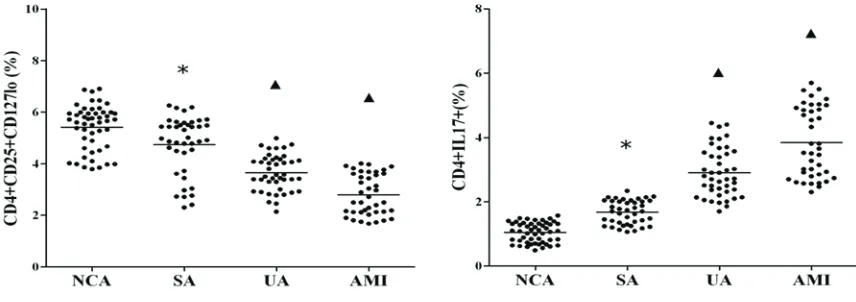
Figure 1.The frequencies of CD4+CD25+CD127low Treg and CD4+IL-17+Th17 cells in ACS patients. *P<0.01 vs. NCA;
Figure 2. Apoptosis of Treg and Th17 cells in each group. A. Comparison of apoptotic Treg and Th17 cells in the four groups. *P<0.01 vs. NCA and SA, ▲P<0.05 vs. NCA, ♦P<0.05 vs. UA. B. Flow cytometry plots for apoptotic Treg
cells (gated by CD4+CD25+CD127low cells) in each group. C. Flow cytometry plots for apoptotic Th17 cells (gated by
CD4+IL17+ cells) in each group. D. CD4+T cells apoptosis among groups at pre-stimulation, post-stimulation, and
analyzed by using statistical software (SPSS 11.0, LEAD Technologies, Inc., Chicago, IL,
USA). Statistical significance for the difference
in different groups was assessed by one-way
analysis of variance (ANOVA). If significance was found, Bonferroni test was performed for
post-hoc analysis to detect the difference among groups when equal variance were assumed, while Dunnett’s C test were per-formed when equal variance were not assumed. Spearman’s correlation was used as a test of correlation between two continuous variables. A probable value of P<0.05 was considered to
be statistically significant.
Results
Patient characteristics
Table 1 shows the characteristics of patients.
There were no significant differences in age,
gender, coronary artery disease (CAD) extent,
hypertension, smoking rate, obesity, fasting blood glucose (FBG), high-density
lipoprotein-cholesterol (HDL-C), and very low-density lipo-protein-cholesterol (VLDL-C) concentrations among patients with AMI, UA and SA. The Levels
of TC and TG in AMI and UA groups were signifi -cantly higher than those in SA group and NCA controls (P<0.05, P<0.01 respectively).
Besides, the Levels of low density lipoprotein cholesterol (LDL-C) and FBG in AMI and UA groups were also significantly higher than those
in NCA controls. (P<0.05, P<0.01 respectively).
Apoptosis in Treg and Th17 cells
[image:7.612.97.522.143.418.2]As shown in Figure 1, the frequencies of CD4+ CD25+ CD127low Treg cells were significantly lower in the patients with AMI and UA as com-pared with the NCA group and SA patients (P<0.01), while Treg frequencies in the SA Figure 3. Expression of Fas, FasL and active Caspase-3 in Treg and Th17 cells for each group. A. Comparison of Fas and FasL in Tregs and Th17 among the four groups. ▲P<0.01 vs. NCA and SA, *P<0.05 vs. NCA and SA, #P<0.05 vs. NCA, ♦P<0.01 vs.UA. B. Comparison of active Caspase-3 in Tregs among the 4 groups. ▲P<0.01 vs. NCA and SA,
♦P<0.05 vs. UA, *P<0.05 vs. NCA. C. The flow cytometry plots about Fas, FasL and active Caspase-3expression in Tregs (gated by CD4+CD25+CD127low cells) from the 4 groups.
Figure 4. Correlation of Treg apoptosis to inflammatory markers and sFasL. A. Comparison of CRP, Lp(a) and sFasL among the four groups. ▲P<0.01 vs. NCA and SA, *P<0.05 vs. NCA. B. Spearman correlation of Treg apoptosis to
groups were also markedly lower than those of
the NCA group (P<0.05). On the contrary, the frequencies of CD4+IL-17+ Th17 cells were evi-dently higher in patients with AMI and UA than those with NCA and SA (P<0.01), while there was also obvious difference between the SA and NCA group (P<0.01).
In addition, AnnexinV+ apoptotic Tregs in AMI and UA groups were significantly higher than
those in NCA and SA patients (P<0.01),
Apoptotic Tregs in SA group were significantly
higher than those in NCA patients, while
Apoptotic Tregs in AMI group were also strik -ingly higher than those in UA patients (P<0.05)
. Moreover, There were no significant differenc -es in apoptotic Th17 cells between the four groups (all P>0.05, Figure 2A-C).
In the control test of apoptosis analysis in Th17 cells, CD4+ T cells apoptosis was not signifi -cantly increased in the post-stimulation group
or the post-fixation/permeabilization group in
comparison to the pre-stimulation group (P>0.05, Figure 2D).
Expression of Fas and FasL in Treg and Th17 cells.
As shown in Figure 3A, the levels of Fas and
FasL expression for Tregs were markedly higher
in the AMI and UA groups than those in the SA and NCA group (P<0.01, P<0.05 respectively). In addition, Fas expression for Treg cells in the
SA group was significantly higher than that in
Treg cells
Caspase-3 activity of Treg cells in the UA and
AMI groups was significantly higher than that in
NCA and SA groups (P<0.01, P<0.05 respec-tively); In addition, Caspase-3 activity for Treg
cells in the AMI group was significantly higher
than that in the UA group, and in the SA group
caspase-3 activity was also markedly higher
than that in NCA group (both P<0.05) (Figure 3B-C).
Correlation of Treg apoptosis to inflammatory markers and sFasL
The concentrations of hsCRP, Lp(a) and sFasL
in the AMI and UA groups was significantly
increased compared with SA and NCA groups (P<0.01 for all). In addition, all the three
indica-tors were significantly increased in SA group
compared to the NCA group (P< 0.05) (Figure 4A).
For the four groups, inflammatory markers and sFasL had significant correlation with Treg
apoptosis. The apoptotic rate of Tregs positively correlated with the concentrations of hsCRP, Lp(a) and sFasL (P<0.01 and r=0.826,0.727, 0.872 respectively, Figure 4B).
Anti-FasL antibody reduce Treg apoptosis in ACS patients in vitro
AnnexinV+ apoptotic Treg cells in UA and AMI patients incubated with anti-FasL were mark -edly decreased in comparison to the group Figure 5.Treg apoptosis in the four groups in vitro treatment of PBMC with anti-FasL
Ab for 48h. *P<0.05 vs. no-anti-FasL group, #P>0.05 vs. no-anti-FasL group.
the NCA group, and Fas and FasL expression for Treg cells in the AMI
group was also signifi -cantly higher than those in the UA group (P<0.01 respectively). At the same time, FasL expres-sion for Tregs in SA and NCA groups was not
sig-nificantly different (P>0. 05). Interestingly, there
were no significant dif -ferences in Fas and FasL expression for Th17 cells among the four groups (all P>0.05).
[image:8.612.94.403.73.248.2]without incubation with anti-FasL after 48 h incubation in vitro (both P <0.05). Apoptotic Treg cells in NCA and SA patients incubated with anti-FasL were slightly decreased, but this
is no significant between anti-FasL group and
non-anti-FasL group (both P >0.05, Figure 5). Discussion
Treg and Th17 cells play important role in
inflammation, and could participate in plaque
destabilization and the onset of ACS. Our
previ-ous study demonstrated a significant increase of Th17 and related cytokines, and a decline of Treg and related cytokines in ACS patients com -pared to SA and NCA patients [6]. This abnor-mality could possibly cause Th17/Treg imbal-ance. In this study, we explored the mechanism underlying Th17/Treg imbalance in ACS patients by examining apoptosis and the Fas/Fas ligand
pathway. ACS patients exhibited a remarkable
increase in apoptotic rate, Fas/FasL expression and Caspase-3 activity of Tregs. The levels of hsCRP, Lp(a) and sFasL in ACS patients also increased. In addition, the apoptotic rate of Tregs was positively correlated with the concen-trations of hsCRP, Lp(a) and sFasL in serum. At the same time, Treg apoptosis in ACS patients
is decreased markedly by Anti-FasL Ab in vitro.
However, there were no marked changes in
apoptotic rate and Fas/FasL expression of Th17 cells among the four groups. Therefore,
our findings demonstrated, for the first time,
that apoptosis and Fas/Fas Ligand pathway contributes to the decrease of Tregs, not Th17, resulting in Th17/Treg imbalance and the onset of ACS.
Apoptosis plays an important part in lympho-cyte development and regulation of immune response. Apoptosis disorders, both interrupt-ed as well as acceleratinterrupt-ed apoptosis, were dem-onstrated as central defects in immune func-tion, and could lead to a range of diseases [19]. Gritzapis et al [20] have demonstrated that the decrease in intratumor Tregs was due to apop-tosis induced by cell contact with FasL+ Th. In other studies [18, 21], Th17 cells are remark -ably less susceptible to apoptosis than Th1. However, apoptosis of Treg and Th17 in ACS patients has not yet been reported.
In our study, ACS patients exhibited remarkable
rise in apoptotic Tregs. Apoptotic Tregs in SA
patients were significantly increased compared
to those in NCA group, and those in AMI patients were also apparently elevated over those in UA
patients. However, there were no significant dif -ferences in apoptotic Th17 cells between the four groups. Increased apoptosis of Treg cells may lead to relative reduction of Treg expres-sion, and Th17/Treg imbalance. To exclude the
effects of simulation and fixation/permeabiliza -tion on the results of Th17 apoptosis, CD4+ T cell apoptosis was examined pre-stimulation,
post-stimulation, and post-fixation/permeabili -zation before Th17 apoptosis analysis. We found that the apoptotic rates of CD4+ T cells were not markedly altered in the three groups,
then concluded that the process had no effects on Th17 apoptosis.
The Fas/FasL pathway plays an important role in T cell apoptosis [12, 13].Fas are activated by their natural ligands FasL. The combination of Fas and FasL on cell membrane as a trimeric complex triggers the activation cascade of cas-pases [22, 23]. Cascas-pases are a family of cyste-ine proteases in the downstream section of the Fas/FasL pathway that are necessary for the execution of apoptosis. Among them, cas-pase-3 is considered to be a major executioner protease and plays a crucial role in the trans-duction of apoptotic signals. Activated cas-pase-3 can interact with a large number of tar-gets within an affected cell to bring about its disintegration and apoptotic bodies formation [24, 25].
A study by Bossowska et al [26] has shown that
a high expression of Fas and FasL on lympho-cytes occurs in ACS patients. Furthermore, the results of Venet et al [27] indicate that Tregs act on monocytes by inhibiting their LPS-induced survival through a proapoptotic mech-anism involving the Fas/FasL pathway. Other studies also suggest that the resistance of Th17 cells to is due to lower production of FasL, as compared to Th1 cells[18]. However, little
was previously known about the sensitivity of
Th17 and Treg to Fas-mediated apoptosis in ACS patients.
between Th17 and Treg cells, leading to plaque destabilization and the onset of ACS. We need to further prove our conclusion in a large scale of the population, and ongoing efforts should be made to explore whether other pathways is related to Treg apoptosis and Th17/Treg imbal-ance. Our study provides a new insight into Th17/Treg imbalance, may be useful to devise novel strategies for research on the pathogen-esis and treatment of ACS.
Acknowledgments
This study was supported by National Natural Science Foundation of China (No. 30971226), Natural Science Youth Foundation of Anhui province of China (No. 11040606Q08), Natural Science Key Foundation of Anhui Universities of China (No. KJ2011A166), and Anhui Medical University Research Foundation(No. 2010X- KJ083).
We thank Dr. Helen Williams from Vascular Biology Research Centre, Department of
Surgery, the University of Sydney at Westmead Hospital for her assistance with grammar and for her valuable comments on the manuscript. Disclosure
The authors declare no conflict of interest.
Address correspondence to: Dr. Yuan Wang,
Biochemistry & Molecular Biology, Anhui Medical
University, Hefei 230032, Anhui Province, PR China. Phone: +86-551-5161140; Fax: +86-551-5161140; E-mail: wangyuan@ahmu.edu.cn; Or: Dr. Qing Li, The central laboratory of Medical research center in Anhui Provincial Hospital, Hefei 230001, Anhui Province, PR China. Phone: +86 -551-2283574; Fax: +86-551-2283574; E-mail: liqing-2001@163.com
References
[1] Shoenfeld Y, Sherer Y, Harats D.
Artherosclero-sis as an infectious, inflammatory and autoim -mune disease. Trends Immunol 2001; 22: 293-5.
[2] Zeman K. Atherosclerosis and infection? Vnitr
Lek 2006; 52: 823-6.
[3] Hansson GK. Inflammation, atherosclerosis,
and coronary artery disease. N Engl J Med 2005; 352: 1685-95.
[4] Sakaguchi S, Sakaguchi N, Shimizu J, Yamaza
-ki S, Sa-kihama T, Itoh M, Kuniyasu Y, Nomura T, Toda M, Takahashi T. Immunologic tolerance
higher than apoptotic rates of Tregs for ACS patients, which is due to activation of cas-pase-3 prior to exposure of phosphatidylserine during apoptosis [28], so detection of cas-pase-3 activation in the cytoplasm may contrib-ute to future prediction of apoptosis. Interestingly, the Fas/FasL pathway did not play
a role in Th17 expression in ACS patients. By
multiple analysis, Th17 cells expressed low level of Fas and FasL indicating no effect of Fas-mediated apoptosis on Th17 cells.
Here we found that Tregs, but not Th17 are sen-sitive to Fas-mediated apoptosis which may be the reason why Th17/Treg imbalance occurrs in ACS. We found that cell apoptosis and Fas-FasL apoptotic pathways could be responsible for the alteration of the fragile balance between Treg and Th17 cells, which leads to increases
the risk of plaque rupture and ACS.
Apoptotic Tregs incubated with anti FasL were decreased in ACS patients incubated with
anti-FasL. These results not only confirmed that
Fas/FasL pathway is essential to Treg apopto-sis, but also suggested that targeting Treg apoptosis may be a therapeutic strategy to maintain Th17/Treg balance in ACS, further leading to plaque stabilization. Consequently,
blocking the Fas/FasL pathway and inhibiting Treg apoptotic processes could be beneficial to
ACS. Treatments using anti-FasL, including cas-pase inhibitors, may show good results in terms of prevention and cure for ACS. However, this
strategy needs to be verified in vivo by animal models.
The Th17/Treg imbalance is related to the
nature of an inflammatory response. The levels
of hsCRP, Lp(a) and sFasL in ACS patients
increased significantly, and their levels in SA
patients also elevated in comparison to NCA group. Furthermore, there was a positive asso-ciation between Treg apoptosis and the con-centration of inflammation markers as well as sFasL, which also seemed to be linked directly to the degree of myocardial damage and plaque destabilization.
Conclusions
Our findings show that Tregs, but not Th17 cells, give rise to significant apoptosis in ACS
maintained by CD25+CD4+regulatory T cells: their common role in controlling autoimmunity, umor immunity, and transplantation tolerance. Immunol Rev 2001; 182: 18-32.
[5] Miossec P, Korn T, Kuchroo VK. Interleukin-17
and type 17 helper T cells. N Engl J Med 2009; 361: 888-98.
[6] Li Q, Wang Y, Chen K, Zhou Q, Wei W, Wang Y, and Wang Y. The role of oxidized low-density
li-poprotein in breaking peripheral Th17/Treg
balance in patients with acute coronary
syn-drome. Biochem Biophys Res Commun 2010;
394: 836-42.
[7] Cheng X, Liao YH, Ge H, Li B, Zhang J, Yuan J,
Wang M, Liu Y, Guo Z, Chen J, Zhang J, Zhang L. The Th17/Treg imbalance in patients with acute coronary syndrome. Clin Immunol 2008; 127: 89-97.
[8] hompson CB. Apoptosis in the pathogenesis
and treatment of disease. Science 1995; 267: 1456-62.
[9] Zhang H, Xu Q, Krajewski S, Krajewska M, Xie Z, Fuess S, Kitada S, Paulowski K, Godzik A, Reed JC. BAR: an apoptosis regulator at the in
-tersection of caspases and Bcl-2 family pro -teins. Proc Natl Acad Sci 2000; 97: 2597-602. [10] Griffith TS, Brunner T, Fletcher SM, Green DR,
Ferguson FA. Fas ligand-induced apoptosis as a mechanism of immune privilege. Science 1995; 270: 1189-92.
[11] Suvannavejh GC, Dal Canto MC, Matis LA, Mil-ler SD. Fas-mediated apoptosis in clinical re-missions of relapsing experimental autoim-mune encephalomyelitis. J Clin Invest 2000; 105: 223-31.
[12] Zhang Y, Xu G, Zhang L, Roberts AI, Shi Y. Th17 cells undergo Fas-mediated activation-induced cell death independent of IFN-r. J Immunol 2008; 181: 190-6.
[13] Wei Y, Chen K, Sharp GC, Yagita H, Braley-Mul -len H. Expression and regulation of Fas and
Fas ligand on thyrocytes and infiltrating cells
during induction and resolution of granuloma-tous experimental autoimmune thyroiditis. J Immunol 2001; 167: 6678–86.
[14] Strauss L, Bergmann C, Szczepanski M, Good -ing W, Johnson JT, Whiteside TL. A unique sub-set of CD4+CD25highFoxp3+ T cells secreting
IL-10 and TGF-β mediates suppression in the
tumor microenvironment. Clin Cancer Res 2007; 13: 4345-54.
[15] Grossman WJ, Verbsky JW, Barchet W, Colonna M, Atkinson JP, Ley TJ. Human T regulatory
cells can use the perforin pathway to cause au-tologous target cell death. Immunity 2004; 21: 589-601.
[16] Janssens W, Carlier V, Wu B, VanderElst L, Jac -quemin MG, Saint-Remy JM. CD4+CD25+ T
cells lyse antigen-presenting B cells by Fas-Fas
ligand interaction in an epitope-specific man -ner. J Immunol 2003; 171: 4604-2.
[17] Fritzsching B, Oberle N, Eberhardt N, Quick S, Haas J, Wildemann B, Krammer PH, Suri-Payer
E. In contrast to effector T cells, CD4+CD25+Foxp3+ regulatory T cells are highly susceptible to CD95 ligand- but not to TCR-mediated cell death. J Immunol 2005; 175: 32–6.
[18] Fang Y, Yu S, Ellis JS, Sharav T, Braley-Mullen
H. Comparison of sensitivity of Th1, Th2, and Th17 cells to Fas-mediated apoptosis. J
Leu-koc Biol 2010; 87: 1019-28.
[19] Szuster-Ciesielska A, Daniluk J, Bojarska-Ju
-nak A. Apoptosis of blood mononuclear cells in alcoholic liver cirrhosis. The influence of in vi -tro ethanol treatment and zinc supplementa-tion. Toxicology 2005; 212: 124-34.
[20] Gritzapis AD, Voutsas IF, Lekka E, Papamichail M, Baxevanis CN. Peptide vaccination breaks
tolerance to HER-2/neu by generating
vaccine-specific FasL(+) CD4(+) T cells: first evidence
for intratumor apoptotic regulatory T cells. Cancer Res 2010; 70: 2686-96.
[21] Tan C, Ramaswamy M, Shi G, Vistica BP, Siegel RM, Gery I. Inflammation-inducing Th1 and
Th17 cells differ in their expression patterns of apoptosis-related molecules. Cell Immunol 2011; 271: 210-3.
[22] Krueger A, Fas SC, Baumann S, Krammer PH.
The role of CD95 in the regulation of peripheral T2 cell apoptosis. Immunol Rev 2003; 193: 58-69.
[23] Brunner T, Wasem C, Torgler R, Cima I, Jakob S,
Corazza N. Fas (CD95/Apo-1) ligand regulation in T cell homeostasis, cell-mediated cytotoxici-ty and immune pathology. Semin Immunol 2003; 15: 167-76.
[24] Creagh EM, Conroy H, Martin SJ. Caspase-acti-vation pathways in apoptosis and immunity. Immunol Rev 2003; 193: 10-21.
[25] Riedl SJ, Shi Y. Molecular mechanisms of cas-pase regulation during apoptosis. Nat Rev Mol
Cell Biol 2004; 5: 897-907.
[26] Bossowska A, Bossowski A, Galar B. Analysis of apoptotic markers Fas/FasL (CD95/CD95L)
expression on the lymphocytes in patients with acute coronary syndrome. Kardiol Pol 2007; 65: 883-89.
[27] Venet F, Pachot A, Debard AL, Bohe J, Bienvenu
J, Lepape A, Powell WS, Monneret G. Human CD4+CD25+ regulatory T lymphocytes inhibit li-popolysaccharide-induced monocyte survival through a Fas/Fas ligand-dependent mecha-nism. J Immunol 2006; 177: 6540-7.
[28] Bonomini M, Dottori S, Amoroso L, Arduini A,