Original Article
UCH-L1 involved in regulating the degradation of EGFR
and promoting malignant properties in
drug-resistant breast cancer
Yiting Jin1, Wei Zhang1, Jiawen Xu3, Hongying Wang1,Zijing Zhang1, Chengyu Chu1, Xiuping Liu2,3, Qiang Zou1
1Department of General Surgery, Huashan Hospital, Fudan University, Shanghai, P. R. China; 2Department of Pa-thology, The Fifth People’s Hospital of Shanghai, Fudan University, Shanghai, P. R. China; 3Department of Pathol-ogy, School of Basic Medical Sciences, Fudan University, Shanghai, P. R. China
Received August 28, 2015; Accepted September 28, 2015; Epub October 1, 2015; Published October 15, 2015
Abstract: Ubiquitin carboxy terminal hydrolase-L1 (UCHL1) belongs to the UCH proteases family that deubiquitinates ubiquitin-protein conjugates in the ubiquitin-proteasome system. Our previous research showed that UCH-L1 and EGFR could regulate the expression of P-gp, CD147 and MMPs in multi-drug resistance (MDR) breast cancer cells, respectively. But it is still unclear whether direct regulation exists between the UCH-L1 and EGFR in MDR breast cancer. In order to clarify this, MDR human breast carcinoma cell line MCF7/Adr, that expresses relatively high UCH-L1, and its parental cell line MCF7, that expresses relatively low UCH-L1, were chosen for this study. We added ubiquitin proteasome inhibitor MG-132 into the culture of MCF7/Adr cells and transfected pIRES2-UCH-L1-EGFP plasmid into MCF7 cells, respectively. Using quantitative real-time polymerase chain reaction and western blot analyses, we found accompanying over-expression of UCH-L1, EGFR was up-regulated in both MCF7/ADR and MCF7 cells. Preliminary results indicated the degradation of EGFR might be regulated by ubiquitin level. So we speculated that up-regulated UCH-L1 could promote expression level of EGFR, thereby enhance the invasion and metastasis abilities of tumor cells. Moreover, to further explore the role of UCH-L1 and EGFR, we investigated the expression of UCH-L1, EGFR and P-gp in 65 local advanced breast cancer cases by immunohistochemistry assay. The result showed that the patients not responding to chemotherapy had higher UCH-L1, EGFR and P-gp expression levels and more lymph nodes metastasis. The Kaplan-Meier survival analysis showed that the patients with elevated UCH-L1 expression after chemotherapy presented shorter overall survival and disease free survival times than those with down-regulated or unchanged expression of UCH-L1. Our findings suggest that UCH-L1 may be an indicator of chemotherapy-response and poor-survival in breast cancer. UCH-L1 might be an appropriate target for improving chemo-resistant breast cancer therapy.
Keywords: UCH-L1, EGFR, breast cancer, multidrug resistance
Introduction
UCH-L1 is one member of the ubiquitin carboxy terminal hydrolase (UCH) family, which is selec-tively expressed in the testis/ovary and brain [1-3]. Recently some studies showed that UCH-L1 was abnormally expressed in some tumors, and correlated with cancer cell differentiation, metastasis and multi-drug resistance (MDR) [4-7]. In our past assays, we found UCH-L1 not only regulated the expression of P-gp, CD147 and MMPs, but also the ubiquitination and deg-radation of P-gp and CD147 in MDR breast can-cer cells. P-gp and CD147 were ubiquitinated, and modification of ubiquitin was important for
their degradation. We also found that EGFR could promote malignant properties of MDR breast cancer cells via up-regulating CD147, MMPs [8]. So we pondered the linkage between UCH-L1 and EGFR.
pathway was proposed. It is supposed that up-regulated UCH-L1 could suppress the degrada-tion of EGFR via ubiquitin-proteasome pathway, and then enhance the expression of P-gp, CD147 and MMPs, which directly resulted in promoting malignant properties of MDR breast cancer cells. This might provide further insight into the mechanism underlying the linkage between drug resistance and tumor metasta- sis.
Materials and methods
Cell culture
The MDR breast cell line MCF7/Adr was cul-tured in RPMI-1640 medium (Gibco-BRL, Karlsruhe, Germany), and the parental cell line, MCF7, was cultured in DMEM (high glucose; Gibco-BRL) supplemented with 0.01 mg/mL bovine insulin (Sigma, St. Louis, MO, USA). All cell culture media contained 10% foetal bovine serum (FBS; PAA Laboratories, Linz, Austria), 100 U/ml penicillin and 100 μg/mL streptomy-cin. The cells were cultured at 37°C in a humidi-fied atmosphere containing 5% CO2. For stable MDR1 gene expression, the MCF7/Adr cells were maintained in the presence of Adriamycin (Sigma). Ubiquitin proteasome inhibitor MG-132 was purchased from Sigma.
were cultured at 37°C for another 42 h till har-vested. To detect the transfection efficiency, the fluorescent microscopy (Zeiss, Oberkochen, Germany) was used to detect the green fluores-cent expression of the EGFP-labelled cells.
Reverse transcription and quantitative real-time polymerase chain reaction
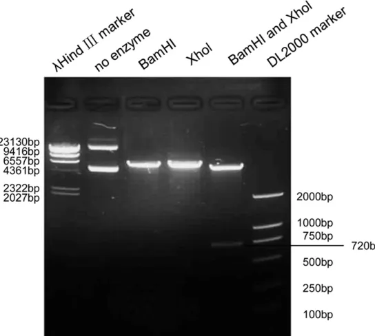
[image:2.612.94.366.75.319.2]Total cellular RNA was extracted using the Tripure isolation reagent (Sangon, Shanghai, China). The RNA samples were subjected to reverse transcription (RT) with 2 μg of RNA, Oligo (dT)18, dNTP, and reaction buffer supplied with M-MLV reverse transcriptase (Promega, Madison, WI, USA). Real-time polymerase chain reaction (PCR) reactions were then performed in 20 μL of solution with 2 μg of cDNA, 1 mM of each forward and reverse primer and 2× SYBR green mix (Takara, Shuzo, Kyoto, Japan). Changes in the mRNA expression level were calculated following normalisation to the glycer-aldehyde-3-phosphate dehydrogenase (GAP- DH) mRNA level. Relative gene expression was determined by the fluorescence intensity ratio of the target gene to GAPDH. The primers used in the real-time PCR reactions were designed based on information from the human genome database. The following are the primers used for the specific amplification of GAPDH and Figure 1. pIRES2-UCH-L1-EGFP plasmid was identified by digestion with
re-striction enzymes.
Plasmid transfection
UCH-L1: GAPDH forward primer: 5’-AAC GGA TTT GGT CGT ATT G-3’, and reverse primer: 5’-GGA AGA TGG TGA TGG GAT T-3’; UCH-L1 for-ward primer: 5’-GCC AAT GTC GGG TAG ATG-3’,
[image:3.612.95.522.75.220.2]and reverse primer: 5’-CAA AGT CCC TCC CAC AGA-3’; EGFR forward primer: 5’-CCA AGG CAC GAG TAA CAA GC-3’, and reverse primer: 5’-CCA AAT TCC CAA GGA CCA CC-3’.
Figure 2. Ubiquitin proteasome inhibitor MG-132 blocked the degradation of EGFR in MCF7/Adr cells. after co-culture with ubiquitin proteasome inhibitorMG-132, in MCF7/Adr cells, significantly elevated expressions of EGFR were found in both protein and mRNA levels. A. EGFR protein level was assessed by western blot. B. EGFR mRNA level was assessed by real-time PCR. Bar graphs represent mean ± SEM of three independent experiments. *P < 0.05 vs. control cells. Results are representative of three similar experiments.
[image:3.612.88.520.304.584.2]Western blot
The cells were collected and lysed in a modified RIPA buffer [50 mM Tris (pH 7.8), 150 mM NaCl,
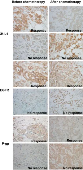
[image:4.612.107.375.75.618.2]every 3 weeks for 4 cycles) at our institution between 2009 and 2011 to be included in this study. All patients had received a radical mas-tectomy after 4 cycles of chemotherapy. Figure 4. Expression of UCH-L1, EGFR and P-gp before and after
chemo-therapy was measured by immunohistochemistry in patients’ tissues. Rep-resentative photographs of HE staining of UCH-L1, EGFR and P-gp of differ-ent groups are shown. (Original magnification 200×).
5 mM EDTA, 15 mM MgCl2, 1% NP-40, 0.5% sodium deoxy-cholate, 1 mM DTT, and 20 mM N-ethylmaleimide] con-taining 1 Complete Protease Inhibitor Cocktail tablet (Ro- che Molecular Biochemical, Indianapolis, IN, USA) per 50 mL of buffer. Total cell lysate (50 μg of protein) was resolved using standard sodium dodec-yl sulphate polyacrdodec-ylamide gel electrophoresis (SDS-PAGE) and electrophoretically trans-ferred onto polyvinylidene flu-oride (PVDF) membranes (Mi- llipore, Bedford, MA, USA). The membranes were blocked in 5% non-fat milk for 1 h at room temperature and then incu-bated overnight with primary antibodies against UCH-L1 (Chemicon, Temecula, CA, USA), EGFR (BD Biosciences, USA), and β-actin (Sigma) at 4°C. The membranes were then incubated for 1 h at 4°C with the appropriate HRP-conjugated secondary anti-bodies (Invitrogen, Carlsbad, CA, USA). Detection of the pro-tein expression levels using enzyme-linked chemilumines-cence (ECL; Pierce, Rockford, USA) was performed accord-ing to the manufacturer’s protocol.
Patient and tissue specimens
After approval from our institu-tional review board, we select-ed 65 cases of local advancselect-ed breast cancer from a cohort of women who had undergone a core-needle biopsy (patholo-gy-confirmed breast invasive ductal carcinoma) and neo-adjuvant chemotherapy (pacli-taxel 175 mg/m2 (Bristol-My-
According to the Japanese Breast Cancer Society (JBCS) criteria for the histological evalu-ation of the therapeutic response after neo-adjuvant chemotherapy [13], the cohort was divided into three groups, including pathologi-cal complete response (PCR, n = 13), partial response (n = 27) and no response (n = 25). Tissue specimens before and after chemother-apy were conserved for testing.
We ensure that the work described has been carried out in accordance with The Code of Ethics of the World Medical Association (Declaration of Helsinki) for experiments involv-ing humans. The privacy rights of human sub-jects must always be observed.
Immunohistochemistry
Paraffin-embedded tissue samples from 18 murine tumours and 65 mammary carcinomas were prepared. Briefly, the slides were dehy-drated in xylene and graded alcohols. Antigen retrieval of EGFR was performed with protein-ase K at room temperature for 20 min. UCH-L1 and P-gp antigen retrieval were performed with 0.05 M TBS buffer and 0.01 M citrate buffer at pH 6.0 at 95°C for 20 min, respectively. Then, the slides were incubated with diluted primary antibody (anti-EGFR, 1:100 dilution; anti-UCH-L1, 1:50 dilution; anti-P-gp, 1:50 dilution) for 12 h, followed by incubation with a biotinylated
[image:5.612.87.524.96.163.2]polyclonal Ab (BD), a UCH-L1 rabbit polyclonal Ab (Chemicon) and a P-gp mouse monoclonal Ab (Calbiochem, USA). The positive controls for UCH-L1 and P-gp included sections of formalin-fixed, paraffin-embedded human brain tissues as indicated in the instruction manual, and the positive controls for EGFR included sections of FFPE human breast carcinoma tissues. The negative controls were incubated with an immunoglobulin fraction in place of the poly-clonal primary Ab in the positive tissues men-tioned above. The saturation and intensity of the immunostained cells were evaluated over three visual fields, at a power of 400× under a light microscope (Carl Zeiss, Gottingen, Germany). For the statistical analysis, accord-ing to the study by Han et al. [14] and our previ-ous work (unpublished data), the total staining of EGFR, UCH-L1 and P-gp was scored based on the intensity and percentage of cells with EGFR, UCH-L1 and P-gp cytoplasmic staining using the following scale: score 0, negative staining for all of the tumour cells; score 1, neg-ative/weak staining for all of the tumour cells or moderate staining in less than 10% of the cells; score 2, moderate staining in more than 30% but less than 70% of the tumour cells or strong staining within 10% of the tumour cells; score 3, moderate staining in more than 70%, or strong staining in more than 30% but less than 70% of all the tumour cells; and score 4, strong Table 1. Statistical quantification and comparison of IHC scores of the variation of UCH-L1, EGFR and P-gp before and after chemotherapy
UCH-L1 EGFR P-gp
Before After P value Before After P value Before After P value PCR (13 cases) 2.15 N/A N/A 0.63 N/A N/A 0.5 N/A N/A Response (27 cases) 1.852 1.704 0.404 0.593 0.556 0.866 0.926 0.778 0.46 No response (25 cases) 1.84 2.44 0.001* 0.72 1.16 0.046* 1.12 1.6 0.037*
*P<0.05.
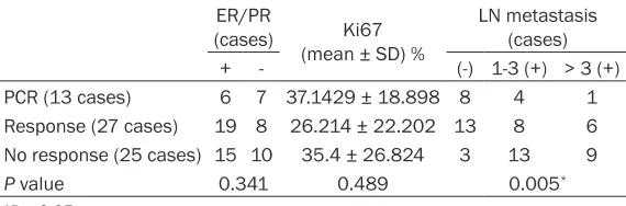
Table 2. Statistical quantification and comparison of the expres-sion of ER/PR and Ki67 and the status of LN metastasis after chemotherapy
ER/PR
(cases) (mean ± SD) %Ki67 LN metastasis (cases) + - (-) 1-3 (+) > 3 (+) PCR (13 cases) 6 7 37.1429 ± 18.898 8 4 1 Response (27 cases) 19 8 26.214 ± 22.202 13 8 6 No response (25 cases) 15 10 35.4 ± 26.824 3 13 9
P value 0.341 0.489 0.005*
*P < 0.05.
[image:5.612.91.376.230.324.2]staining in more than 70% of all the tumour cells. The reproducibility of EGFR, UCH-L1 and P-gp staining was examined between 2 labora-tories (by 2 independent pathologists) using 2 different primary antibodies.
Statistical analysis
Statistics were calculated using SPSS 19.0 software. All experiments were repeated at least three times, and the results are present-ed as the mean ± standard errors (SEM). The differences were analysed by ANOVA and Student›s t-test. Pearson’s correlation coeffi-cients were used to determine whether two prognosis-related factors correlated with each other. Kaplan-Meier survival analysis was used to estimate the prognostic relevance, and the survival difference between groups was assessed by the log-rank test. The statistical significance was determined at P < 0.05 (two-tailed).
Results
Ubiquitin proteasome inhibitor MG-132 can block the degradation of EGFR in MCF7/Adr cells
In our past findings, we resolved that EGFR could regulate the expression of P-gp and
CD147, and ubiquitin proteasome pathway also played an important role in the degradation of P-gp and CD147. So it is wondered whether EGFR is also regulated by ubiquitin proteasome pathway in MDR breast cancer cells. After co-culture with ubiquitin proteasome inhibitor MG-132, in MCF7/Adr cells, significantly elevat-ed expressions of EGFR were found in both pro-teins and mRNA levels (Figure 2A, 2B). It sug-gested that the block of ubiquitin proteasome pathway could affect the degradation of EGFR in MDR breast cancer cells.
pIRES2-UCH-L1-EGFP plasmid caused specific
and effective up-regulation of UCH-L1 and EGFR expression in MCF7 cells
[image:6.612.92.521.73.276.2]Our previous study has found both UCH-L1 and EGFR were involved in regulating the invasion/ metastasis abilities of MDR breast cancer. So we wondered about the relationship between UCH-L1 and EGFR. For this purpose, we trans-fected pIRES2-UCH-L1-EGFP plasmid into MCF7 cells which expressing UCH-L1 in a rela-tively low level. The transfection efficiencies were initially evaluated using immunofluores-cence analysis as shown in Figure 3A. In addi-tion, real-time PCR and western blot analysis showed elevated expression of UCH-L1 in MCF7 cells, which resulted in up-regulation of EGFR (Figure 3B, 3C).
UCH-L1 and EGFR are involved in MDR and metastasis process in chemo-resistant breast cancer tissue samples
65 local advanced breast cancer (LABC) cases undergoing neoadjuvant chemotherapy with the P-gp substrates, paclitaxel and epirubicin, were chosen for this study. The expression of UCH-L1, EGFR and P-gp were assayed by immu-nohistochemical staining (Figure 4). According to the chemo-response, the patients were divided into 3 groups: pathological complete response (PCR; n = 13), partial response (n = 27) and no response (n = 25). The elevated expression of UCH-L1, EGFR and P-gp after chemotherapy were observed only in the no-response group (P < 0.05; Table 1), which also exhibited an increase in lymph node metasta-sis (P < 0.01; Table 2). However, in the partial-response group, the expression of UCH-L1, EGFR and P-gp were decreased, and fewer lymph node metastases were found. There was no statistic correlation between UCH-L1 and ER, PR and Ki67, so was EGFR and P-gp (all P > 0.05; Table 2). In the PCR group, due to tumour missing after chemotherapy, the differential comparison before and after chemotherapy couldn’t be conducted. The above results reveal that UCH-L1 and EGFR may be involved in the progression of promoting malignant properties in MDR breast cancer.
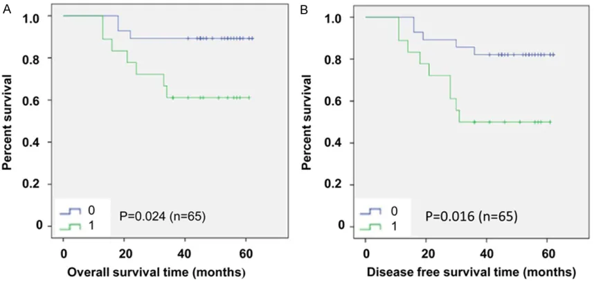
A 65-case survival analysis reveals a correla-tion between up-regulacorrela-tion of UCH-L1 and shorter OS/DFS in MDR patients
The 65 patients were divided to 2 groups according to the status of UCH-L1 after chemo-therapy. One group presented up-regulated expression of UCH-L1, while the other group presented down-regulated or unchanged expression of UCH-L1. The Kaplan-Meier sur-vival analysis showed that there was a correla-tion between overexpressed UCH-L1 after che-motherapy and shorter overall survival (OS) time (P < 0.05; Figure 5A) and shorter disease-free survival (DFS) time (P < 0.05; Figure 5B). It further suggested that UCH-L1 might be involved in chemotherapy resistance and poor survival.
Discussion
Ubiquitin proteasome system (UPS) partici-pates in the turnover of many key regulatory
proteins and in the development of cancer. UPS has three parts: ubiquitin, ubiquitin ligase and 26S proteolytic enzyme complex. The target protein is covalently connected to ubiquitin via ubiquitin ligase, and then is identified and hydrolyzed by 26S proteolytic enzyme complex [15]. UCH-L1 belongs to the UCH protease fam-ily that deubiquitinates ubiquitin-protein conju-gates in the UPS [16]. It has a dual function: a hydrolase activity that removes small COOH-terminal ubiquitin to generate an ubiquitin monomer and a dimerization-dependent ubiq-uitin ligase activity [17]. So UCH-L1 can obstruct the degradation process of the target protein. UCH-L1 over-expression has been observed in breast cancer [4]. In this study, two closely associated mammary carcinoma cell lines, MCF7 and MCF7/Adr, were chosen for compari-son. Our previous experiments indicated that MCF7/Adr expressed much higher UCH-L1 than MCF7 cells [7, 18, 19]. Furthermore, the expres-sion of UCH-L1 promoted coincided with up-regulation of MDR1 gene and increase of inva-sion abilities in MCF7 cells [21]. Through the role of deubiquitination, over-expressed UCH-L1 may block the degradation of P-gp and CD147, and then enhance expression of MMPs in MCF7/Adr cells. EGFR is also reported to play a very crucial part in enhancing CD147 and MMPs expression to establish favorable condi-tions for cells migration/invasion in MDR breast cancer cells [8]. EGFR is mostly reported degrading by autophagy-lysosome pathway, but the process can be affected by the regula-tion of ubiquitin.
deubiquitinat-ing enzyme and inhibits the conjugatdeubiquitinat-ing of the ubiquitin monomer and EGFR, which could fur-ther block the internalization and degradation of EGFR.
Our previous studies have shown both UCH-L1 and EGFR were involved in enhancing malig-nant properties in MDR breast cancer cells [8, 21]. However, there was no relevant clinical study reported. In our study, 65 local advanced breast cancer patients were conducted to veri-fy the above findings. We found the patients not responding to chemotherapy had higher UCH-L1, EGFR and P-gp expression levels and more lymph nodes metastasis, which indicating po- orer prognosis. Just the opposite, the patients who have responsed to chemotherapy present-ed decreasing UCH-L1, EGFR and P-gp expres-sion and less lymph nodes metastasis. The Kaplan-Meier survival analyses showed that the patients with elevated UCH-L1 expression after chemotherapy presented shorter OS and DFS time than those with down-regulated or unchanged expression of UCH-L1. From the clinical perspective, this work revealed that high UCH-L1 and EGFR were involved in enhanc-ing chemo-resistance and metastasis ability in breast cancer patients and lead to poorer survival.
Glogowska et al have reported that the combi-nation of existing ErbB inhibitors and the UCH-L1 gene silencing effect of specific exon 23-encoded peptides may be a novel treatment for patients with aggressive ErbB1/2-depen- dent cancers [22]. This result is similar to sug-gest that UCH-L1 and EGFR have cooperation in promoting cancer malignant properties. In summary, our findings indicate that UCH-L1 plays a crucial role in modulating the degrada-tion of EGFR and promoting malignant proper-ties in MDR breast cancer. UCH-L1 might be a novel target for improving EGFR-related chemo-resistant breast cancer therapy.
Acknowledgements
This work was supported by grants from the National Nature Science Foundation of China (No. 30872971) and Shanghai Municipal Science and Technology Commission (No. 134119a8502). We thank the members of our laboratory for helpful discussions.
Disclosure of conflict of interest
None.
Address correspondence to: Dr. Qiang Zou, De- partment of General Surgery, Huashan Hospital, Fudan University, 12 Middle Wulumuqi Road, Sh- anghai 200040, P. R. China. Tel: 86-21-52889355; Fax: 86-21-62525470; E-mail: zouqiangzq@aliyun. com; Dr. Xiuping Liu, Department of Pathology, The Fifth People’s Hospital of Shanghai, Fudan University, 128 Ruili Road, Minhang, Shanghai 200240, P. R. China. Tel: 86-21-54237528; Fax: 86-21-54237596; E-mail: [email protected]
References
[1] Kwon J, Wang YL, Setsuie R, Sekiguchi S, Sak-urai M, Sato Y, Lee WW, Ishii Y, Kyuwa S, Noda M, Wada K, Yoshikawa Y. Developmental regu-lation of ubiquitin C-terminal hydrolase iso-zyme expression during spermatogenesis in mice. Biol Reprod 2004; 71: 515-21.
[2] Sekiguchi S, Yoshikawa Y, Tanaka S, Kwon J, Ishii Y, Kyuwa S, Wada K, Nakamura S, Taka-hashi K. Immunohistochemical analysis of pro-tein gene product 9.5, a ubiquitin carboxyl-ter-minal hydrolase, during placental and embryonic development in the mouse. Exp Anim 2003; 52: 365-9.
[3] Wilkinson KD, Lee KM, Deshpande S, Duerk-sen-Hughes P, Boss JM, Pohl J. The neuron-specific protein PGP 9.5 is a ubiquitin carboxyl-terminal hydrolase. Science 1989; 246: 670-3. [4] Miyoshi Y, Nakayama S, Torikoshi Y, Tanaka S,
Ishihara H, Taguchi T, Tamaki Y, Noguchi S. High expression of ubiquitin carboxy-terminal hydrolase-L1 and -L3 mRNA predicts early re-currence in patients with invasive breast can-cer. Cancer Sci 2006; 97: 523-9.
[5] Yamazaki T, Hibi K, Takase T, Tezel E, Nakaya-ma H, Kasai Y, Ito K, AkiyaNakaya-ma S, Nagasaka T, Nakao A. PGP9.5 as a marker for invasive colorectal cancer. Clin Cancer Res 2002; 8: 192-5.
[6] Tezel E, Hibi K, Nagasaka T, Nakao A. PGP9.5 as a prognostic factor in pancreatic cancer. Clin Cancer Res 2000; 6: 4764-7.
[7] Yang JM. Emerging roles of deubiquitinating enzymes in human cancer. Acta Pharmacol Sin 2007; 28: 1325-30.
[8] Xu JW, Li QQ, Tao LL, Cheng YY, Yu J, Chen Q, Liu XP, Xu ZD. Involvement of EGFR in promo-tion of malignant properties in multidrug resis-tant breast cancer cells. Int J Oncol 2011; 39: 1501-9.
stability and function of the epithelial Na + channel (ENaC) by ubiquitination. Eur Mol Biol Organ J 1997; 16: 6325-36.
[10] Hicke L, Riezman H. Ubiquitination of a yeast plasma membrane receptor signals its ligand-stimulated endocytosis. Cell 1996; 84: 277-87.
[11] Zhong D, Ru Y, Wang Q, Zhang J, Zhang J, Wei J, Wu J, Yao L, Li X, Li X. Chimeric ubiquitin li-gases inhibit non-small cell lung cancer via negative modulation of EGFR signaling. Can-cer Lett 2015; 359: 57-64.
[12] Glogowska A, Stetefeld J, Weber E, Ghavami S, Hoang-Vu C, Klonisch T. Epidermal growth fac-tor cytoplasmic domain affects ErbB protein degradation by the lysosomal and ubiquitin-proteasome pathway in human cancer cells. Neoplasia 2012; 14: 396-409.
[13] Horii R, Honma N, Ogiya A, Kozuka Y, Fukuda T, Yoshida M, Ohsumi S, Mukai H; Japanese Breast Cancer Society. The Japanese Breast Cancer Society Clinical Practice Guideline for pathological diagnosis of breast cancer. Breast Cancer 2015; 22: 59-65.
[14] Han HJ, Russo J, Kohwi Y, Kohwi-Shigematsu T. SATB1 reprogrammes gene expression to pro-mote breast tumor growth and metastasis. Na-ture 2008; 4: 329-32.
[15] Larsen CN, Krantz BA, Wilkinson KD. Substrate specificity of deubiquitinating enzymes: ubiqui-tin C-terminal hydrolases. Biochemistry 1998; 37: 3358-68.
[16] Ohkawa K, Asakura T, Takada K, Sawai T, Hashizume Y, Okawa Y, Yanaihara N. Calpain inhibitor causes accumulation of ubiquitinated P-glycoprotein at the cell surface: possible role of calpain in P-glycoprotein turnover. Int J On-col 1999; 15: 677-86.
[17] Liu Y, Fallon L, Lashuel HA, Liu Z, Lansbury PT Jr. The UCH-L1 gene encodes two opposing en-zymatic activities that affect alpha-synuclein degradation and Parkinson’s disease suscep-tibility. Cell 2002; 111: 209-18.
[18] Li QQ, Wang WJ, Xu JD, Cao XX, Chen Q, Yang JM, Xu ZD. Involvement of CD147 in regulation of multidrug resistance to P-gp substrate drugs and in vitro invasion in breast cancer cells. Cancer Sci 2007; 98: 1064-9.
[19] Li QQ, Wang WJ, Xu JD, Cao XX, Chen Q, Yang JM, Xu ZD. Up-regulation of CD147 and matrix metalloproteinase-2, -9 induced by P-glycopro-tein substrates in multidrug resistant breast cancer cells. Cancer Sci 2007; 98: 1767-74. [20] Wang WJ, Li QQ, Xu JD, Cao XX, Li HX, Tang F,
Chen Q, Yang JM, Xu ZD, Liu XP. Over-expres-sion of ubiquitin carboxy terminal hydrolase-L1 induces apoptosis in breast cancer cells. Int J Oncol 2008; 33: 1037-45.
[21] Wang WJ, Li QQ, Xu JD, Cao XX, Li HX, Tang F, Chen Q, Yang JM, Xu ZD, Liu XP. Interaction be-tween CD147 and P-glycoprotein and their regulation by ubiquitination in breast cancer cells. Chemotherapy 2008; 54: 291-301.